X-ray computer tomography apparatus

a computer and tomography technology, applied in applications, instruments, nuclear engineering, etc., can solve problems such as cost increase, and achieve the effects of reducing cost, increasing convenience, and widening the rang

Active Publication Date: 2009-02-03
MORITA MFG CO LTD
View PDF8 Cites 41 Cited by
  • Summary
  • Abstract
  • Description
  • Claims
  • Application Information

AI Technical Summary

Benefits of technology

[0020]According to the X-ray imaging apparatus (claim 1) of the present invention, a panoramic imaging mode or an offset scan / CT mode can be arbitrarily selected by the imaging mode selecting means of selecting the panoramic imaging mode or the offset scan / CT mode. It is thereby possible to perform panoramic imaging and offset scan / CT using a costless small two-dimensional X-ray sensor, and as necessary or upon request, it is possible to continuously acquire a panoramic image and an offset scan / CT image which are optimum for treatment. Accordingly, when an approximate position of an region of interest is obtained by panoramic imaging having a large imaging region and an imaging position is then set to perform the offset scan / CT on the region of interest, it is possible to perform CT in a larger range than a conventional range, so as to perform CT in a large range while holding down cost.
[0021]According to the X-ray imaging apparatus (claim 2) of the present invention, since the offset scan / CT mode and the normal scan / CT mode are selected as necessary, the convenience in CT increases, and hence with the use of CT, it is possible to acquire a CT image with even higher resolution.
[0022]According to the X-ray imaging apparatus (claim 3) of the present invention, a slit corresponding to the panoramic imaging mode or the CT mode is selectively arranged, and it is thereby possible to obtain the optimum X-ray beam required for each imaging mode.
[0023]According to the X-ray imaging apparatus (claim 4) of the present invention, the first or the second slit is adjusted to use only an X-ray inclined at a certain angle from the X-ray irradiation central axis, and it is thereby possible to efficiently obtain clear X-ray image data.
[0024]According to the X-ray imaging apparatus (claim 5, 6) of the present invention, the driving means is provided which moves the rotational central axis of the central arm in accordance with either the panoramic imaging mode or the CT mode selected by the imaging mode selecting means of selecting either mode, and it is thereby possible to obtain an arbitrary locus of the rotating means that is optimum for imaging.

Problems solved by technology

There has been a problem with a conventional X-ray CT apparatus for jaw / face imaging in dentistry and the like in that, since the normal scan / CT method is a dominant method, in the case of performing CT by detecting a range not smaller than a visual field detectable by a two-dimensional X-ray sensor, a large-sized two-dimensional X-ray sensor is required, which causes a cost increase.

Method used


Image

  • X-ray computer tomography apparatus
  • X-ray computer tomography apparatus
  • X-ray computer tomography apparatus

Examples


Embodiment Construction

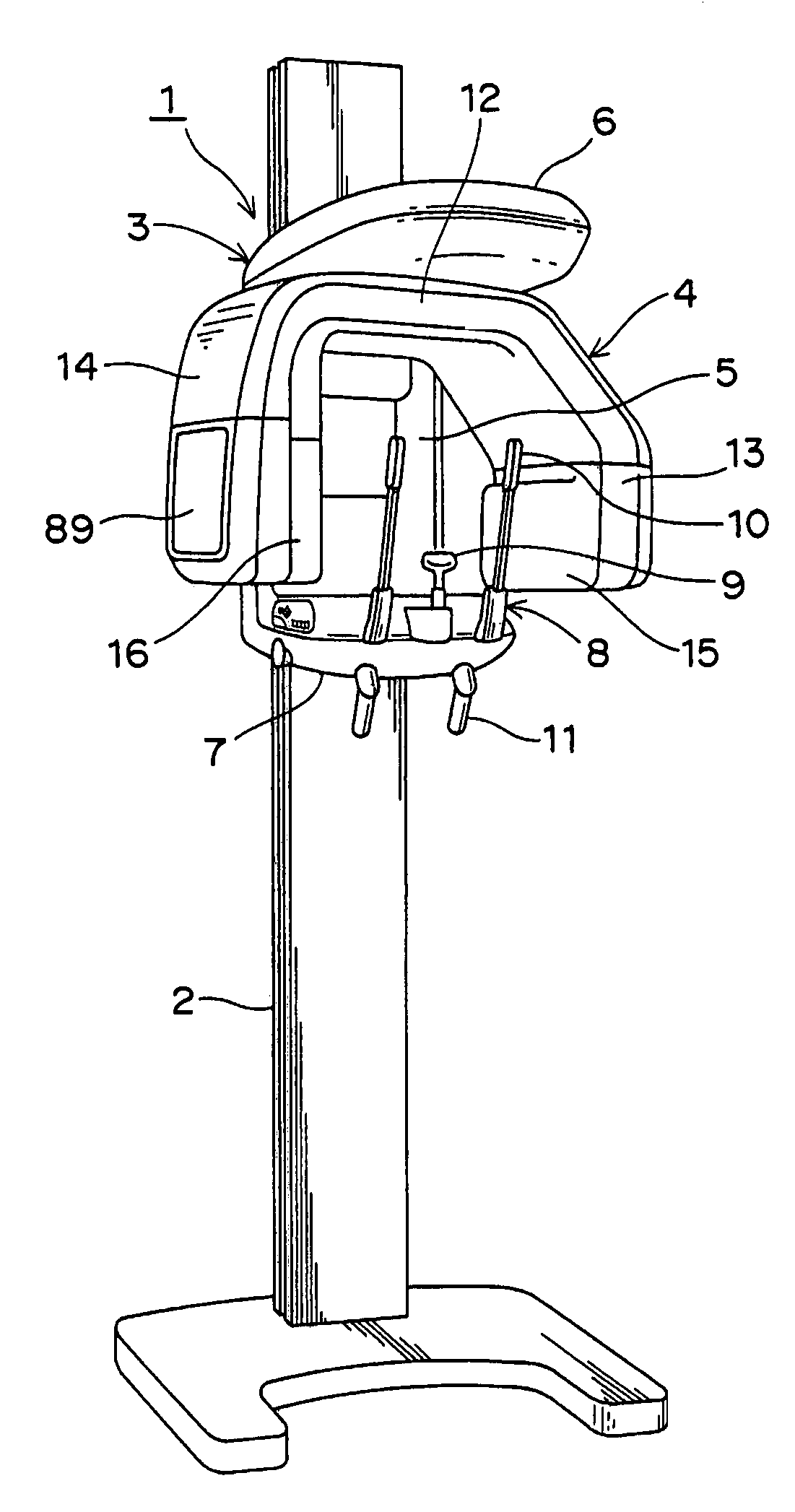
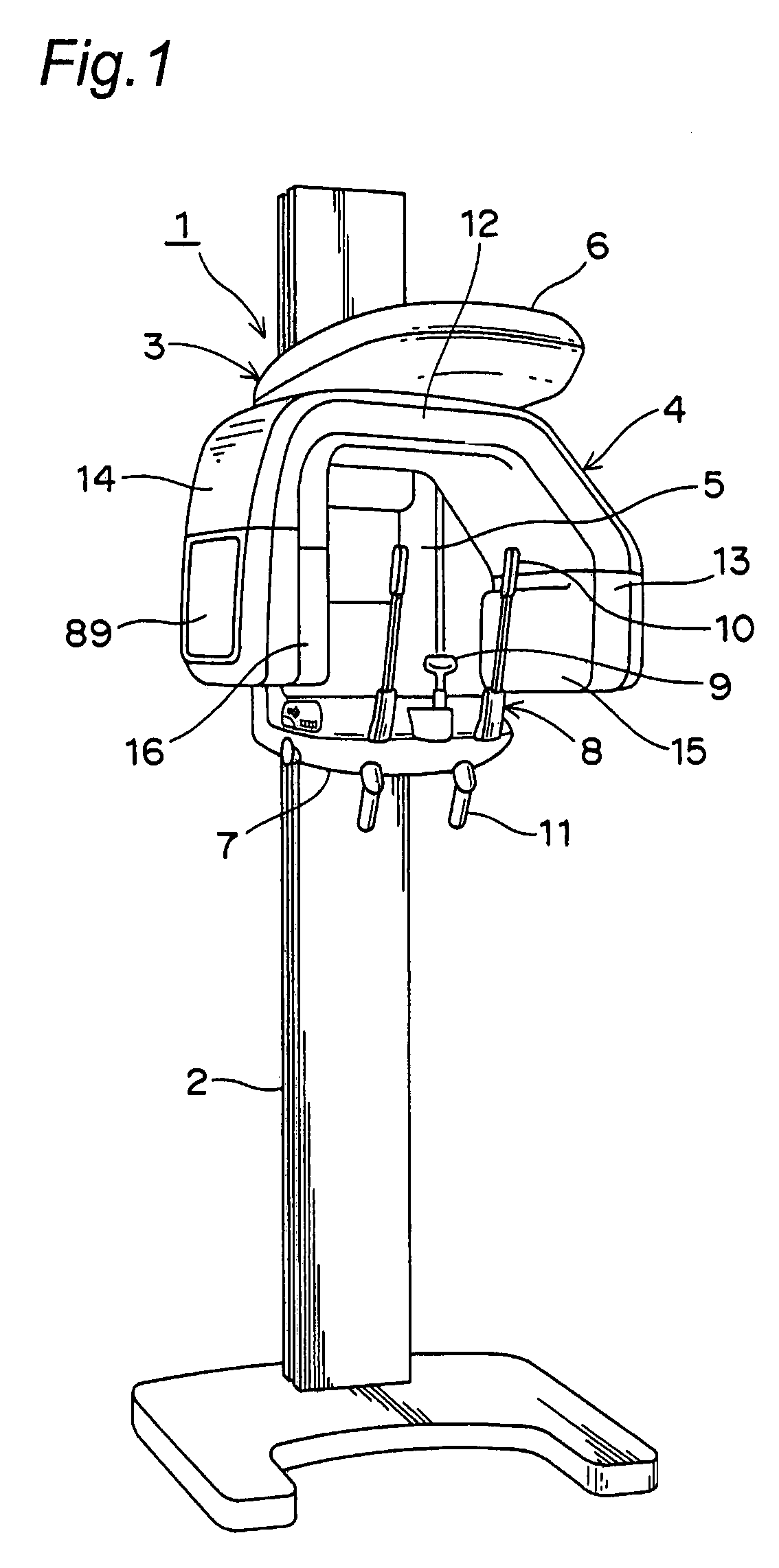
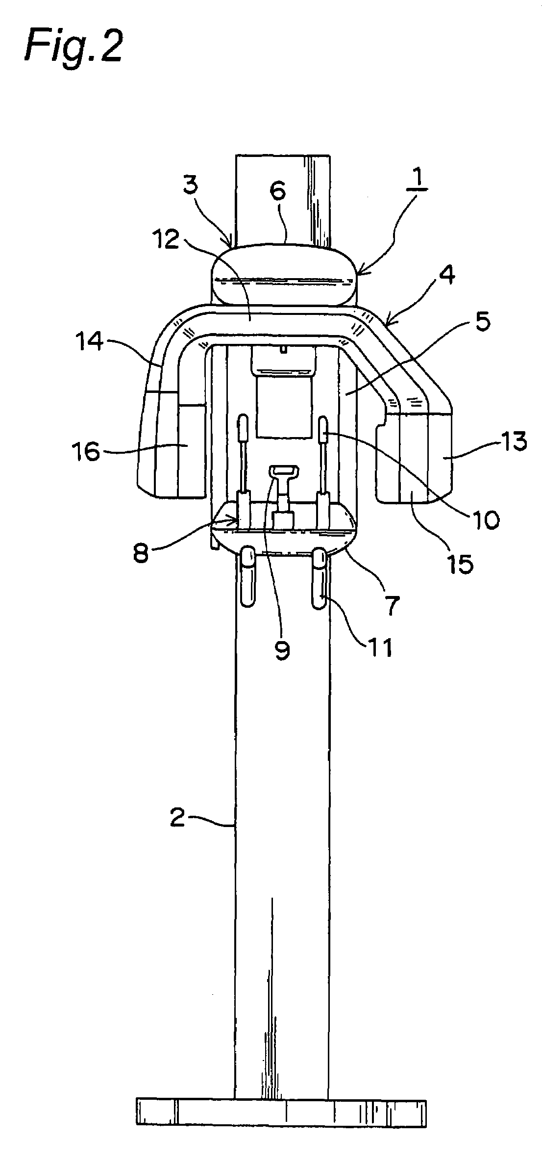
[0044]With reference to the attached drawings, several embodiments of the X-ray imaging apparatus according to the present invention will be described below. It is to be noted that, although terms that mean a specific direction or a place (e.g. “upper”, “lower”, “left”, “right” and other terms including those terms) are used in the following description, these terms are used for the purpose of facilitating visual understanding of configurations represented in the drawings, and should not be used for defining a technical range of the invention.

[0045]FIGS. 1 to 5 show respective appearances of an X-ray CT apparatus according to one embodiment of the present invention. The X-ray CT apparatus is capable of performing three-dimensional computer X-ray tomography (Computer Tomography: hereinafter referred to as “CT”) in addition to a variety of imaging which are conventionally widely known in the dentistry field (e.g. panoramic imaging, linear tomography, linear scan imaging, scanogram ima...


PUM


Abstract

There is provided a new X-ray imaging apparatus by the use of which X-ray CT and panoramic imaging can be effectively performed. In an X-ray imaging apparatus which rotates an X-ray generating section and an X-ray detecting section around an imaging object arranged between these X-ray generating section and the X-ray detecting section and also detects in the X-ray detecting section X-rays having been radiated from the X-ray generating section and transmitted through the imaging object to form an X-ray image, a panoramic imaging mode in which the X-ray generating section and the X-ray detecting section are driven to form a panoramic image of the imaging object and an offset scan / CT mode in which the X-ray generating section and the X-ray detecting section are driven to form a tomographic image of the imaging object are set, so as to selectively execute these two imaging modes. Consequently, the panoramic imaging mode and the offset scan / CT mode can be arbitrarily selected, thereby allowing formation of an X-ray imaged image that is optimum for treatment.

Description

BACKGROUND OF THE INVENTION[0001]1. Field of the Invention[0002]The present invention relates to an X-ray computer tomography (CT) apparatus for dentistry or a jaw / face region. In particular, the present invention relates to an X-ray CT apparatus capable of performing local X-ray CT on a certain tooth or the like as an region of interest in dentistry or the jaw / face region, and performing panoramic X-ray imaging on a curved cross section of a dental arch, a jaw joint, and the like.[0003]2. Description of the Related Art[0004]The Japanese Patent Publication No. 10-225455 (A) discloses an X-ray CT apparatus for dental diagnosis. This X-ray CT apparatus is one having a local X-ray computer tomography (hereinafter referred to as “CT”) along with a panoramic X-ray imaging mode. This X-ray CT apparatus is capable of selecting the X-ray CT mode and the panoramic X-ray imaging mode, and characterized as follows: when the X-ray CT mode is selected, a rotational central axis is fixed onto a c...

Claims


Application Information

Patent Timeline
Patent Type & Authority Patents(United States)
IPC IPC(8): A61B6/00A61B6/14
CPCA61B6/14
Inventor SUZUKIYOSHIMURA, TAKAHIROHONJO, MAKOTO
Owner MORITA MFG CO LTD
Why Eureka
Try Eureka