Abdominopelvic region surgical training model

a training model and abdominopelvic technology, applied in educational models, instruments, educational appliances, etc., can solve the problems of urinary incontinence, bladder instability, and damage to the urethral sphincter or loss of support of the urethral sphincter

Inactive Publication Date: 2009-06-30
BOSTON SCI SCIMED INC
View PDF25 Cites 111 Cited by
  • Summary
  • Abstract
  • Description
  • Claims
  • Application Information

AI Technical Summary

Problems solved by technology

One cause for this condition is damage to the urethral sphincter or loss of support of the urethral sphincter, such as can occur in males after prostatectomy or following radiation treatment, or that can occur due to pelvic accidents and aging related deterioration of muscle and connective tissue supporting the urethra.
Other causes of male incontinence include bladder instability, over-flowing incontinence and fistulas.
Weakening and elongation of the pubourethral ligaments and the arcus tendineus fascia pelvis, weakening of the endopelvic fascia and pubourethral prolapse of the anterior vaginal wall, and their complex interaction with intraabdominal forces are all suspected to play a role in the loss of pelvic support for the urethra and subsequent hypermobility to an unnaturally low non-anatomic position, leading to urinary incontinence.
Cadavers are not always readily available for the initial training, and repeated training is not convenient.
Moreover, it is not possible to visualize the advancement of the needle tip within the cadaver body, and it is not possible it assess whether damage has been done to internal organs, vessels, and nerves in the abdominopelvic region without destructive autopsy at the site.
Similar problems are encountered in training to perform other procedures involving blindly advancing a needle or other surgical instrument around the internal organs and alongside bones, nerves, and blood vessels to effect other treatments and implantations of implantable medical devices in the abdominopelvic region.

Method used


Image

  • Abdominopelvic region surgical training model
  • Abdominopelvic region surgical training model
  • Abdominopelvic region surgical training model

Examples


Embodiment Construction

[0046]In the following detailed description, references are made to illustrative embodiments of methods and apparatus for carrying out the invention. It is understood that other embodiments can be utilized without departing from the scope of the invention.

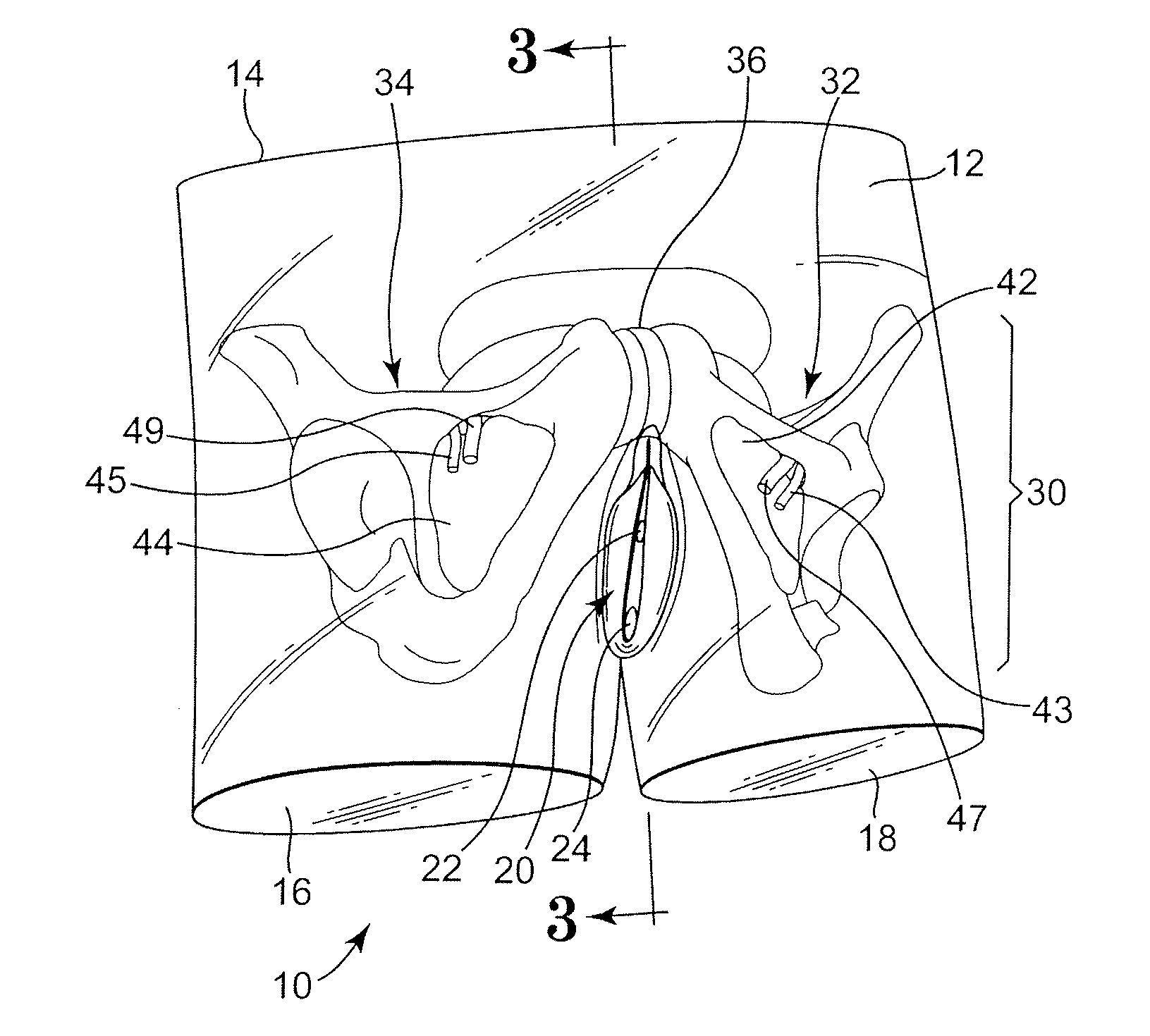
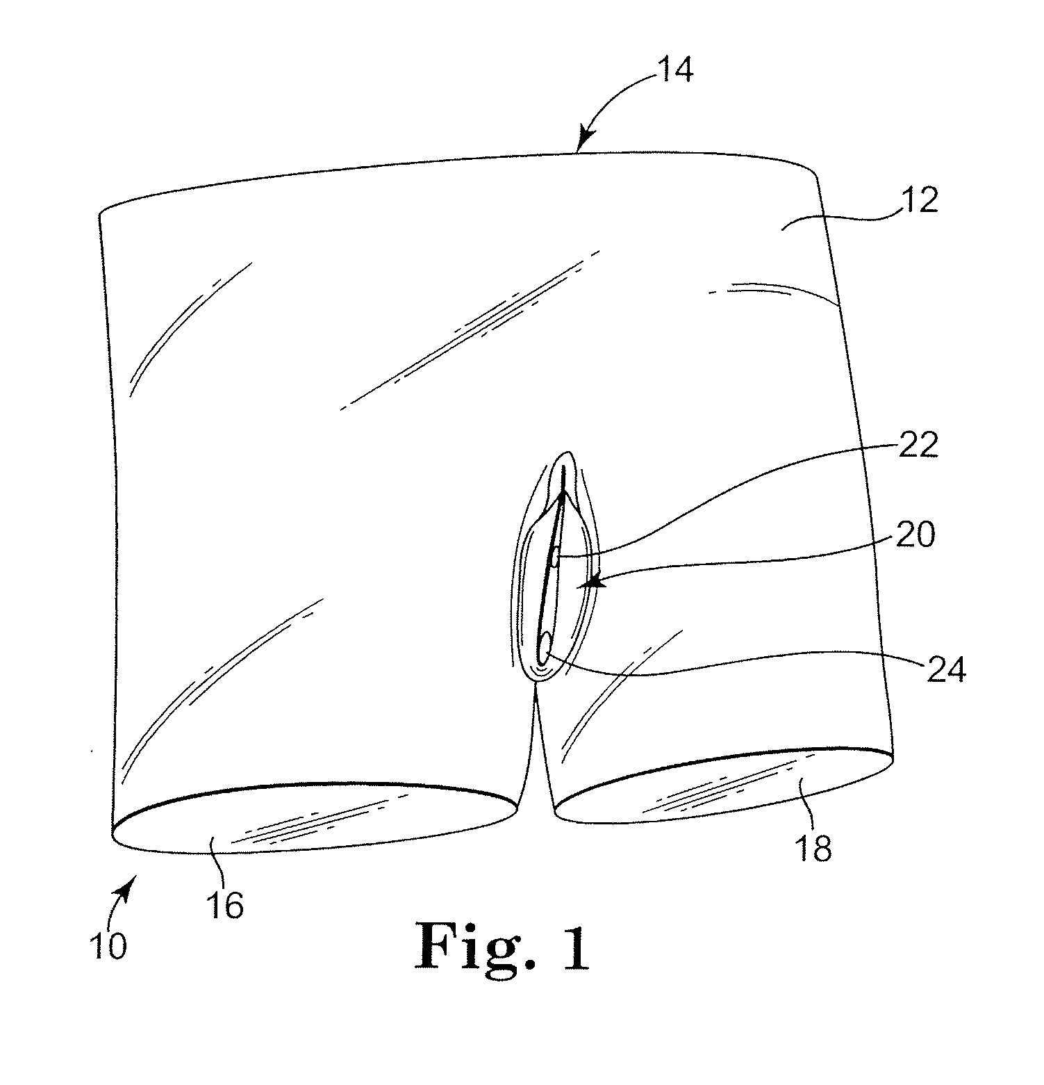
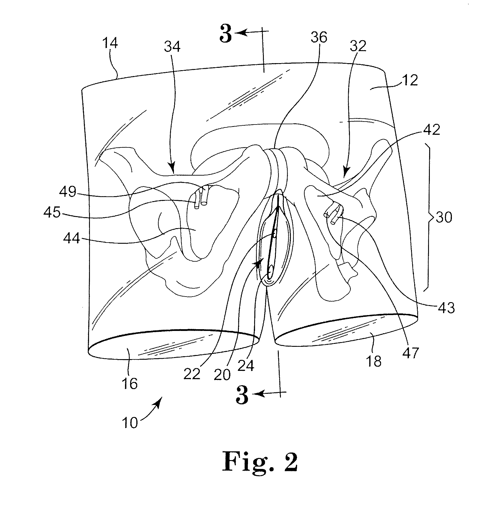
[0047]In FIG. 1, a first preferred embodiment of a female training model 10 of the female abdominopelvic region that is covered or bounded by a shell or “skin”12 and that extends between a model abdominal end 14 and model leg ends 16 and 18. The skin 12 is shaped to conform to representative external genitalia 20, and the external genitalia model may represent the urethral orifice 22 and vaginal orifice 24. In one preferred embodiment, the urethral orifice 22 and vaginal orifice 24 are coupled to internal representations or models of the urethra 52 and vagina 54 (shown in FIG. 3). Furthermore, the external female genitalia 20 may be modeled to represent a vaginal incision made to expose a section of the urethra 52 (FIG. 3) between ...


PUM


Abstract

Surgical training models of the abdominopelvic region of male and female human patient's bodies to assist in training an operator in microsurgical techniques are disclosed. The surgical training models may be used in training in implantation of implantable medical devices, e.g., in the performance of surgical procedures using particular sling implantation tools to implant a urethral sling to support the urethra or bladder to treat incontinence.

Description

[0001]This application claims priority to U.S. Provisional Application No. 60 / 747,112, filed May 12, 2006, the entire content of which is incorporated herein by reference.RELATED APPLICATION[0002]This application is related to commonly assigned Provisional Application No. 60 / 704,563, filed Aug. 2, 2005, for ABDOMINOPELVIC REGION MALE ANATOMIC MODEL in the name of Adam Gullickson et al.FIELD[0003]The present invention pertains to a surgical training model of the abdominopelvic region of a human patient's body to assist in training an operator in implantation of implantable medical devices, e.g., in the performance of surgical procedures using particular sling implantation tools to implant a urethral sling to support the urethra or bladder to treat incontinence.BACKGROUND[0004]Models of the entire or particular regions or systems of the human anatomy are commonly employed in the general education and training of medical personnel as well as specialized training of specialists, surgeon...

Claims


Application Information

Patent Timeline
Patent Type & Authority Patents(United States)
IPC IPC(8): G09B23/28
CPCG09B23/281
Inventor ARNAL, KEVIN R.HAUSCHILD, SIDNEY F.
Owner BOSTON SCI SCIMED INC
Why Eureka
Try Eureka