Protective cap for arthroscopic instruments

a protective cap and arthroscopic technology, applied in the field of arthroscopic surgical instruments, can solve the problems of shortening the useful life of the prosthetic, affecting the patient's recovery, and requiring additional, painful surgeries to correct the problem, so as to prevent accidental damage to the arthroscope

Active Publication Date: 2009-06-30
PRISTINE SURGICAL LLC
View PDF25 Cites 108 Cited by
  • Summary
  • Abstract
  • Description
  • Claims
  • Application Information

AI Technical Summary

Benefits of technology

[0006]The methods and devices shown below provide for a protective cap that is placed over the distal portion of an arthroscope. The cap is made of a transparent, yet durable material that prevents accidental damage to the arthroscope caused by trimming instruments or impacts with hard tissue within the surgical field. Holes may be placed in the cap to provide for the inflow and outflow of fluids from the cap. One or more lenses or filters may be provided within the cap to adjust the field of view as seen through the arthroscope.

Problems solved by technology

The rigid cannula can also become stuck between bones or cartilage during a procedure.
A rigid cannula can also damage metal prosthetics used to replace joints, resulting in a shortening of the useful life of the prosthetic and forcing the patient to undergo additional, painful surgeries to correct the problem.
An additional problem associated with arthroscopic surgery is maintaining a clear surgical field during surgery.
Blood and debris can cloud the field, impairing a surgeon's ability to visualize tissue.
A further problem associated with arthroscopic surgery is accidental damage to the arthroscope.
The arthroscope is often damaged if the working end of a trimming instrument accidentally strikes the sensitive optical components on the distal portion of the arthroscope.
The arthroscope may also be damaged if the arthroscope becomes stuck between bones, cartilage or other tissue and excessive force must be used to free the arthroscope.
Arthroscopes are expensive, costing thousands of dollars, so accidental damage to arthroscopes is a significant cost problem.
A damaged arthroscope could cause delays during surgery and broken pieces of the arthroscope could be deposited in the surgical field.
Both situations are harmful to the patient.

Method used


Image

  • Protective cap for arthroscopic instruments
  • Protective cap for arthroscopic instruments
  • Protective cap for arthroscopic instruments

Examples


Embodiment Construction

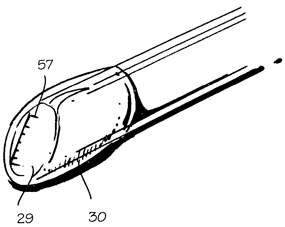
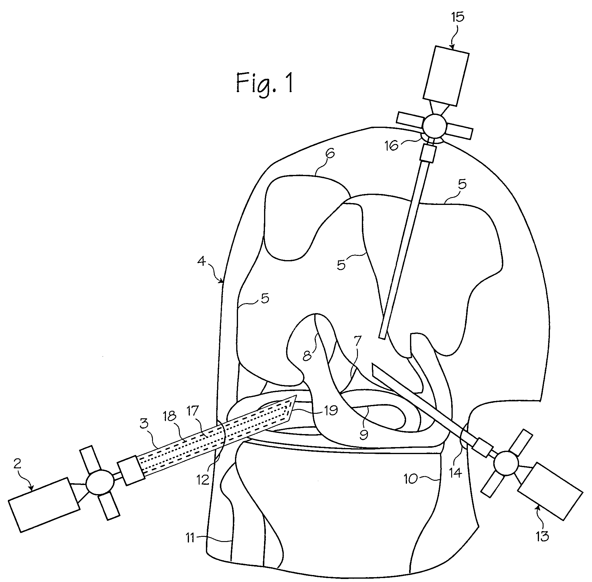
[0019]FIG. 1 shows a method of performing arthroscopic surgery on a patient using an arthroscopic instrument 2 sheathed in an atraumatic introducer sheath 3. (The various parts of the arthroscope are shown in phantom to indicate their positions inside the sheath.) Various anatomical landmarks in a patient's knee 4 are shown for reference, including the femur 5, patella 6, posterior cruciate ligament 7, anterior cruciate ligament 8, meniscus 9, tibia 10 and fibula 11. During surgery, the surgeon introduces the arthroscope 2 into the knee via a first incision 12 in order to visualize the surgical field. A trimming instrument 13 is introduced through a second incision 14 to remove or trim tissue that the surgeon determines should be removed or trimmed. Optionally, an irrigating instrument 15 may be introduced through a third incision 16 in order to irrigate the surgical field and thereby maintain a clear view. As illustrated below, a combined arthroscope and inflow / outflow atraumatic s...


PUM


Abstract

A protective cap for use on arthroscopic instruments and sheaths disposed over arthroscopic instruments. The cap protects the distal end of the instrument from accidental damage caused by other instruments used during a surgical procedure. The portion of the cap that covers a view port on an arthroscope is transparent in order to allow a user to see through the instrument and through the cap.

Description

FIELD OF THE INVENTIONS[0001]The inventions described below relate the field of arthroscopic surgical instruments.BACKGROUND OF THE INVENTIONS[0002]Arthroscopic surgery involves using optical instruments, such as an arthroscope, to visualize an operating field inside or near a joint of a patient. The same instrument or other instruments may be used to perform a surgical procedure in the operating field. Common instruments used in addition to the arthroscope include a trimming instrument for cutting tissue and an irrigation instrument for irrigating the surgical field. Each of the instruments requires its own incision to be introduced into the surgical field; thus, many surgeons prefer to use only a trimming instrument and an arthroscope during arthroscopic surgical procedures.[0003]Arthroscopes are fragile in relation to the forces applied during arthroscopic surgery, so a rigid cannula is placed over the arthroscope to reinforce it. The distal end of the rigid cannula is pointed, u...

Claims


Application Information

Patent Timeline
Patent Type & Authority Patents(United States)
IPC IPC(8): A61B1/00
CPCA61B1/00135A61B1/00137A61B1/00142A61B1/317A61B1/00096A61B1/00101A61B1/00177A61B1/00183A61B1/00089A61B1/012
Inventor KUCKLICK, THEODORE R.
Owner PRISTINE SURGICAL LLC
Why Eureka
Try Eureka