Anti-IGF-1R antibodies and uses thereof

a technology of igf-1r and antibodies, applied in the field of anti-igf-1r antibodies, can solve the problems of increasing the risk of developing cancer in individuals with higher than normal circulating igf levels, inhibiting igf-1r function, and providing encouraging but limited success

Active Publication Date: 2009-11-03
BIOGEN MA INC
View PDF14 Cites 25 Cited by
  • Summary
  • Abstract
  • Description
  • Claims
  • Application Information

AI Technical Summary

Benefits of technology

[0533]Whether or not IGF-1R-specific antibodies or immunospecific fragments thereof disclosed herein are used in a conjugated or unconjugated form, it will be appreciated that a major advantage of the present invention is the ability to use these molecules in myelosuppressed patients, especially those who are undergoing, or have undergone, adjunct therapies such as radiotherapy or chemotherapy. That is, the beneficial delivery profile (i.e. relatively short serum dwell time, high binding affinity and enhanced localization) of the molecules makes them particularly useful for treating patients that have reduced red marrow reserves and are sensitive to myelotoxicity. In this regard, the unique delivery profile of the molecules make them very effective for the administration of radiolabeled conjugates to myelosuppressed cancer patients. As such, the IGF-1R-specific antibodies or immunospecific fragments thereof disclosed herein are useful in a conjugated or unconjugated form in patients that have previously undergone adjunct therapies such as external beam radiation or chemotherapy. In other preferred embodiments, binding molecules, e.g., binding polypeptides, e.g., IGF-1R-specific antibodies or immunospecific fragments thereof (again in a conjugated or unconjugated form) may be used in a combined therapeutic regimen with chemotherapeutic agents. Those skilled in the art will appreciate that such therapeutic regimens may comprise the sequential, simultaneous, concurrent or coextensive administration of the disclosed antibodies or other binding molecules and one or more chemotherapeutic agents. Particularly preferred embodiments of this aspect of the invention will comprise the administration of a radiolabeled binding polypeptide.
[0534]While IGF-1R-specific antibodies or immunospecific fragments thereof may be administered as described immediately above, it must be emphasized that in other embodiments conjugated and unconjugated binding molecules may be administered to otherwise healthy patients as a first line therapeutic agent. In such embodiments binding molecules may be administered to patients having normal or average red marrow reserves and / or to patients that have not, and are not, undergoing adjunct therapies such as external beam radiation or chemotherapy.
[0535]However, as discussed above, selected embodiments of the invention comprise the administration of IGF-1R-specific antibodies or immunospecific fragments thereof to myelosuppressed patients or in combination or conjunction with one or more adjunct therapies such as radiotherapy or chemotherapy (i.e. a combined therapeutic regimen). As used herein, the administration of IGF-1R-specific antibodies or immunospecific fragments thereof in conjunction or combination with an adjunct therapy means the sequential, simultaneous, coextensive, concurrent, concomitant or contemporaneous administration or application of the therapy and the disclosed binding molecules. Those skilled in the art will appreciate that the administration or application of the various components of the combined therapeutic regimen may be timed to enhance the overall effectiveness of the treatment. For example, chemotherapeutic agents could be administered in standard, well known courses of treatment followed within a few weeks by radioimmunoconjugates described herein. Conversely, cytotoxin-conjugated binding molecules could be administered intravenously followed by tumor localized external beam radiation. In yet other embodiments, binding molecules may be administered concurrently with one or more selected chemotherapeutic agents in a single office visit. A skilled artisan (e.g. an experienced oncologist) would be readily be able to discern effective combined therapeutic regimens without undue experimentation based on the selected adjunct therapy and the teachings of the instant specification.
[0536]In this regard it will be appreciated that the combination of a binding molecule (with or without cytotoxin) and the chemotherapeutic agent may be administered in any order and within any time frame that provides a therapeutic benefit to the patient. That is, the chemotherapeutic agent and IGF-1R-specific antibody or immunospecific fragment thereof, may be administered in any order or concurrently. In selected embodiments IGF-IR-specific antibodies or immunospecific fragments thereof of the present invention will be administered to patients that have previously undergone chemotherapy. In yet other embodiments, IGF-1R-specific antibodies or immunospecific fragments thereof of the present invention will be administered substantially simultaneously or concurrently with the chemotherapeutic treatment. For example, the patient may be given the binding molecule while undergoing a course of chemotherapy. In preferred embodiments the binding molecule will be administered within 1 year of any chemotherapeutic agent or treatment. In other preferred embodiments the polypeptide will be administered within 10, 8, 6, 4, or 2 months of any chemotherapeutic agent or treatment. In still other preferred embodiments the binding molecule will be administered within 4, 3, 2 or 1 week of any chemotherapeutic agent or treatment. In yet other embodiments the binding molecule will be administered within 5, 4, 3, 2 or 1 days of the selected chemotherapeutic agent or treatment. It will further be appreciated that the two agents or treatments may be administered to the patient within a matter of hours or minutes (i.e. substantially simultaneously).
[0537]Moreover, in accordance with the present invention a myelosuppressed patient shall be held to mean any patient exhibiting lowered blood counts. Those skilled in the art will appreciate that there are several blood count parameters conventionally used as clinical indicators of myelosuppression and one can easily measure the extent to which myelosuppression is occurring in a patient. Examples of art accepted myelosuppression measurements are the Absolute Neutrophil Count (ANC) or platelet count. Such myelosuppression or partial myeloablation may be a result of various biochemical disorders or diseases or, more likely, as the result of prior chemotherapy or radiotherapy. In this respect, those skilled in the art will appreciate that patients who have undergone traditional chemotherapy typically exhibit reduced red marrow reserves. As discussed above, such subjects often cannot be treated using optimal levels of cytotoxin (i.e. radionuclides) due to unacceptable side effects such as anemia or immunosuppression that result in increased mortality or morbidity.
[0538]More specifically conjugated or unconjugated IGF-1R-specific antibodies or immunospecific fragments thereof of the present invention may be used to effectively treat patients having ANCs lower than about 2000 / mm3 or platelet counts lower than about 150,000 / mm3. More preferably IGF-1R-specific antibodies or immunospecific fragments thereof of the present invention may be used to treat patients having ANCs of less than about 1500 / mm3, less than about 1000 / mm3 or even more preferably less than about 500 / mm3. Similarly, IGF-1R-specific antibodies or immunospecific fragments thereof of the present invention may be used to treat patients having a platelet count of less than about 75,000 / mm3, less than about 50,000 / mm3 or even less than about 10,000 / mm3. In a more general sense, those skilled in the art will easily be able to determine when a patient is myelosuppressed using government implemented guidelines and procedures.

Problems solved by technology

Experimental approaches undertaken to inhibit IGF-1R function in tumors have provided encouraging but limited success, and their effectiveness in treating cancer is yet to be determined in the clinic.
Individuals with higher than normal circulating IGF levels have increased risk for developing cancer.

Method used


Image

  • Anti-IGF-1R antibodies and uses thereof
  • Anti-IGF-1R antibodies and uses thereof
  • Anti-IGF-1R antibodies and uses thereof

Examples


example 1

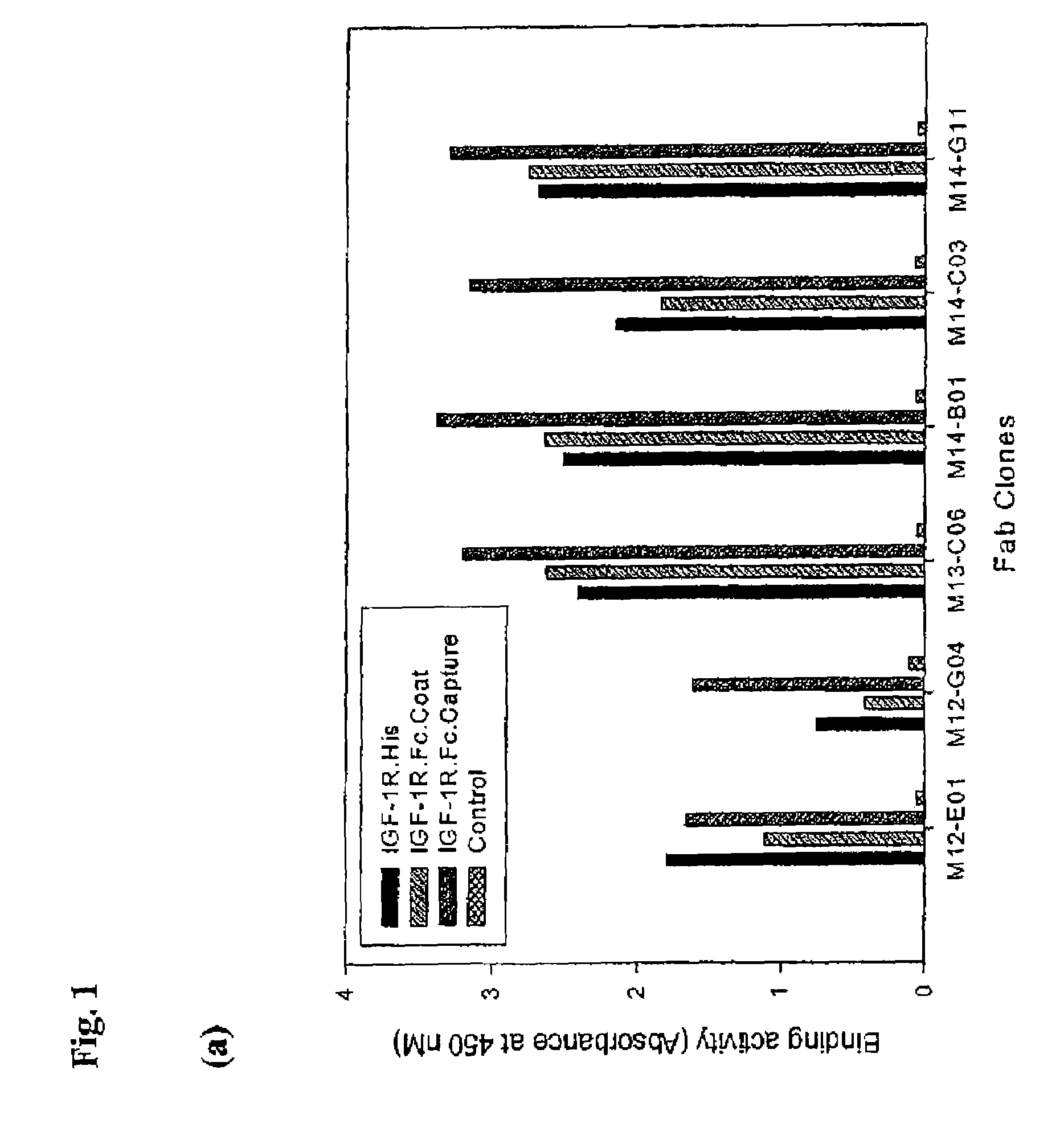
Selection of IGF-1R Specific Fabs from Phage Libraries

[0559]Recombinant human IGF-1R ectodomain was used to screen a human naïve phagemid Fab library containing 3.5×10¹⁰ unique clones (Hoet, R. M., et al. Nat Biotechnol. 23(3):344-8 (2005), (“Hoet et al.”) which is incorporated herein by reference in its entirety). Two distinct panning arms were followed using biotinylated IGF1R-his and IGF1R-Fc protein. Proteins were captured on steptavidin-coated magnetic beads prior to incubation with the phage library. In the case of IGF1R-Fc, a biotinylated anti-Fc antibody was captured on the magnetic beads, followed by captured of the Fc fusion protein. Selections were performed as described in Hoet et al. After 3 rounds of panning, the 479 bp gene III stump was removed by MluI digestion, and the vector was religated for soluble Fab expression in TG1 cells. ELISA analysis of 920 clones from the biotinylated IGF1R-his arm yielded 593 positive clones, containing 33 unique sequences. ELISA analy...

example 2

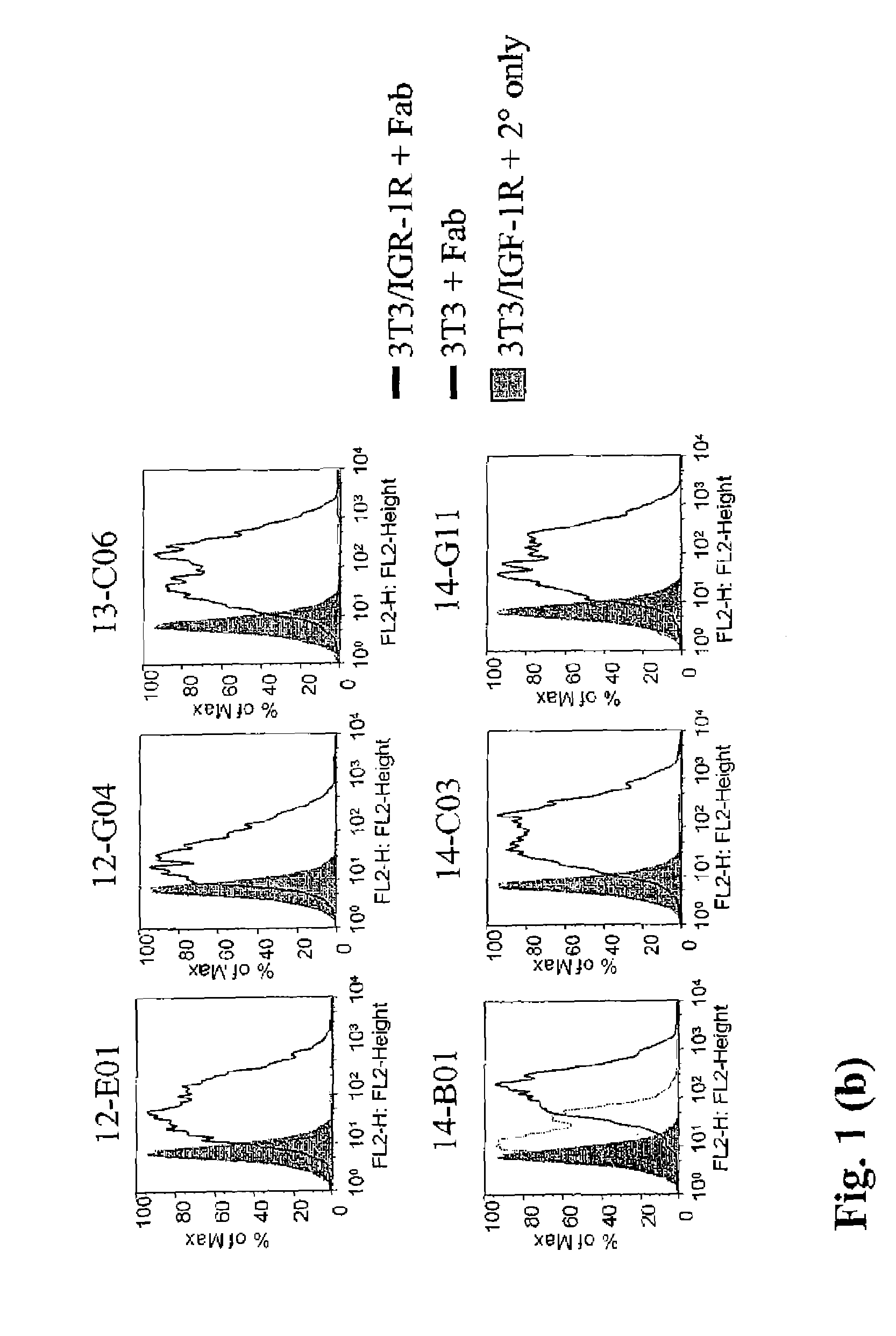
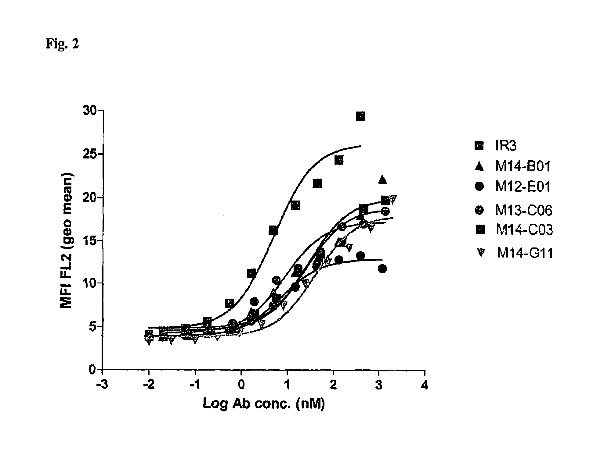
Binding Activity of Fabs to IGF-1R Expressed on Tumor Cells

[0560]The ability of Fabs to bind to the wild type IGF-1R was determined by flowcytometry using MCF-7 tumor cell line.

[0561]MCF-7 cells (Human Breast Adenocarcinoma from NCI) were split 24 hours prior to the setup of the assay to obtain 70% confluent monolayer. Routinely, MCF-7 cell line was maintained within 20 passages. Cells were lifted with cell dissociation buffer (Gibco catalog #13151-014), counted, washed and adjusted to 1×10⁶ cells / ml and one ml of cells were then added to each tube (12×75 mm tube Falcon catalog#352054). Cells were pelleted and supernatant removed by centrifugation at 1200 rpm for 5 min and 100 μl of diluted antibodies were then added to the cell pellet. Purified Fabs were tested at a starting concentration of either 210 or 60 μg / ml with 1:3 dilutions in FACS buffer, down to 0.001 μg / ml. FACS buffer used throughout the assay was PBS (without Ca++ / Mg++) containing 1% BSA (Sigma catalog#A-7906) and 0.1...

example 3

Inhibition of Ligand Binding to IGF-1R by Fabs

[0565]The ability of Fabs to block the binding of IGF-1 and IGF-2 ligands to IGF-1R was determined using a radioimmunoassay (RIA).

[0566]Ligand blocking assay (RIA). Recombinant human IGF-1 (Cat #291-G1), IGF-2 (Cat #292-G2), insulin (Cat #Custom02) human Insulin Receptor (Cat #1544-1R) were purchased from R&D Systems, Inc., Minneapolis, Minn. Insulin (Arg-Insulin, Cat #01-207) was purchased from Upstate Cell Signaling Solutions (Lake Placid, N.Y. (now part of Millipore, Concord, Mass. (USA)). 125I-rhIGF-1 (Cat #IM172), 125I-rhIGF-2 (Cat#IM238) and 125I-rhInsulin (Cat#IM166) were purchased from Amersham Biosciences (Piscataway, N.J.). AffiPure goat anti-human IgG, Fcγ fragment specific antibodies (Cat #109-005-098, Jackson ImmunoResearch, West Grove, Pa.) was used for IGF-1R-Fc capture. As detection antibody, goat anti-mouse IgG HRP (Cat #1030-05, Southern Biotech Birmingham, Ala.) was used.

[0567]As positive controls for IGF-1 and IGF-2 b...


PUM


Abstract

The invention relates to antibodies which bind to insulin like growth factor receptor-1 (IGF-1R) and uses thereof, in particular in the diagnosis and treatment of cancer. Specific human and murine monoclonal antibodies which inhibit IGF-1R-mediated pro-survival and tumor proliferation pathways, and variants, fragments, and derivatives thereof are provided. Also provided are specific human and murine monoclonal antibodies which block the ability of the ligands, insulin like growth factor 1 (IGF-1) and insulin like growth factor 2 (IGF-2) to bind to IGF-1R, as well as fragments, variants and derivatives of such antibodies. The invention also includes polynucleotides encoding the above antibodies or fragments, variants or derivatives thereof, as well as vectors and host cells comprising such polynucleotides. The invention further includes methods of diagnosing and treating cancer using antibodies of the invention.

Description

CROSS-REFERENCES TO RELATED APPLICATIONS[0001]This is application claims benefit under 35 U.S.C. § 119(e) of U.S. provisional application No. 60 / 786,347, filed on Mar. 28, 2006 and of U.S. provisional application No. 60 / 876,554 filed on Dec. 22, 2006. Each of the above-referenced patent applications is hereby incorporated by reference in its entirety.BACKGROUND OF THE INVENTION[0002]A number of epidemiological studies have shown that higher than normal circulating levels of IGF-1 are associated with increased risk for several common cancers, including breast (Hankinson et al, Lancet 1998. 351:1393-6), prostate (Chan et al, Science. 1998. 279:563-6), lung (Yu et al, J. Natl. Cancer Inst. 1999. 91:151-6) and colorectal cancers (Ma et al, J. Natl. Cancer Inst. 1999. 91:620-5). Elevated circulating levels of IGF-2 also have been shown to be associated with increases risk for endometrial cancer (Jonathan et al, Cancer Biomarker & Prevention. 2004. 13:748-52). On the contrary, inverse cor...

Claims


Application Information

Patent Timeline
Patent Type & Authority Patents(United States)
IPC IPC(8): C07K16/00C12P21/08
CPCC07K16/2863A61K2039/505C07K2317/52C07K2316/96C07K2317/21C07K2317/24C07K2317/33C07K2317/41C07K2317/524C07K2317/53C07K2317/55C07K2317/76C07K2317/77C07K2317/92C07K2319/00C07K2317/34C07K2317/73A61P35/00A61P35/02A61P35/04A61P43/00C07K16/00A61K39/395
Inventor HARIHARAN, KANDASAMYGLASER, SCOTTGARBER, ELLENGRAFF, CHRISTILYNREYES, CHRISTOPHER L.DEMAREST, STEPHEN
Owner BIOGEN MA INC
Why Eureka
Try Eureka