Registration of three dimensional image data with X-ray imaging system

a three-dimensional image and coordinate system technology, applied in the field of three-dimensional image data registration with an x-ray imaging coordinate system, can solve the problem of difficult for a physician to become oriented in a three-dimensional setting using a single-plane x-ray image projection display, and achieve the effect of minimizing a cost function

Active Publication Date: 2010-02-02
STEREOTAXIS
View PDF5 Cites 49 Cited by
  • Summary
  • Abstract
  • Description
  • Claims
  • Application Information

AI Technical Summary

Benefits of technology

[0005]The present invention relates to a method for determining a transformation of a three-dimensional pre-operative image data set to obtain a registration of the three-dimensional image data with an X-ray image of a subject body. In one aspect of the present invention, the method comprises the steps of the user identifying at least two extreme opposing points on the object in the subject from at least one X-ray image plane, obtaining from the image data a set of contour points for each of a plurality of section-planes, performing a projection of every contour point in a sampling of the plurality of section-planes, and selecting from the sampling the section-plane with the contour point projecting nearest to the user-identified extreme point. The method further comprises the steps of the user identifying a three-dimensional center point of the object relative to the X-ray imaging coordinate system, defining a first grid having a predetermined number of intervals at a predetermined interval spacing with the grid center at a user-identified center of the pre-operative image, performing a projection of the center of the three-dimensional image data set onto each grid point for evaluation, and selecting the grid point that when projected minimizes a cost function, where the selected grid point is used to define a translation matrix for registering the three-dimensional contour data with a three-dimensional X-ray coordinate system. This method enables registration of a three-dimensional image object such as an organ to an X-ray imaging system, for suitable overlay onto the X-ray image projection.
[0006]In another aspect of the present invention, the method may further refine the search for the closest fit section-plane by defining a narrower sampling of a predetermined number of neighboring section planes on each side of the previous section-plane selected from the prior sampling. The method performs a projection of each contour point on a predetermined number of sequential section-planes that neighbor the previously selected section-plane to create a narrower data set of section-planes and contour points for evaluation, and selecting the section-plane with the contour points projecting nearest to the user-identified extreme points based on minimizing a cost function.
[0007]In another aspect of the present invention, the method may further refine the search for the closest fit grid point by iteratively defining a succeeding grid having a predetermined number of intervals at an interval spacing substantially smaller than the preceding grid spacing, with the grid center at the point selected in the preceding grid, performing a projection of the center of the three-dimensional image data set onto each point in the successive grid for evaluation, selecting a grid point that when projected minimizes a cost function.

Problems solved by technology

In some cases, it may be difficult for a physician to become oriented in a three dimensional setting using a display of a single-plane X-ray image projection.

Method used


Image

  • Registration of three dimensional image data with X-ray imaging system
  • Registration of three dimensional image data with X-ray imaging system
  • Registration of three dimensional image data with X-ray imaging system

Examples


Embodiment Construction

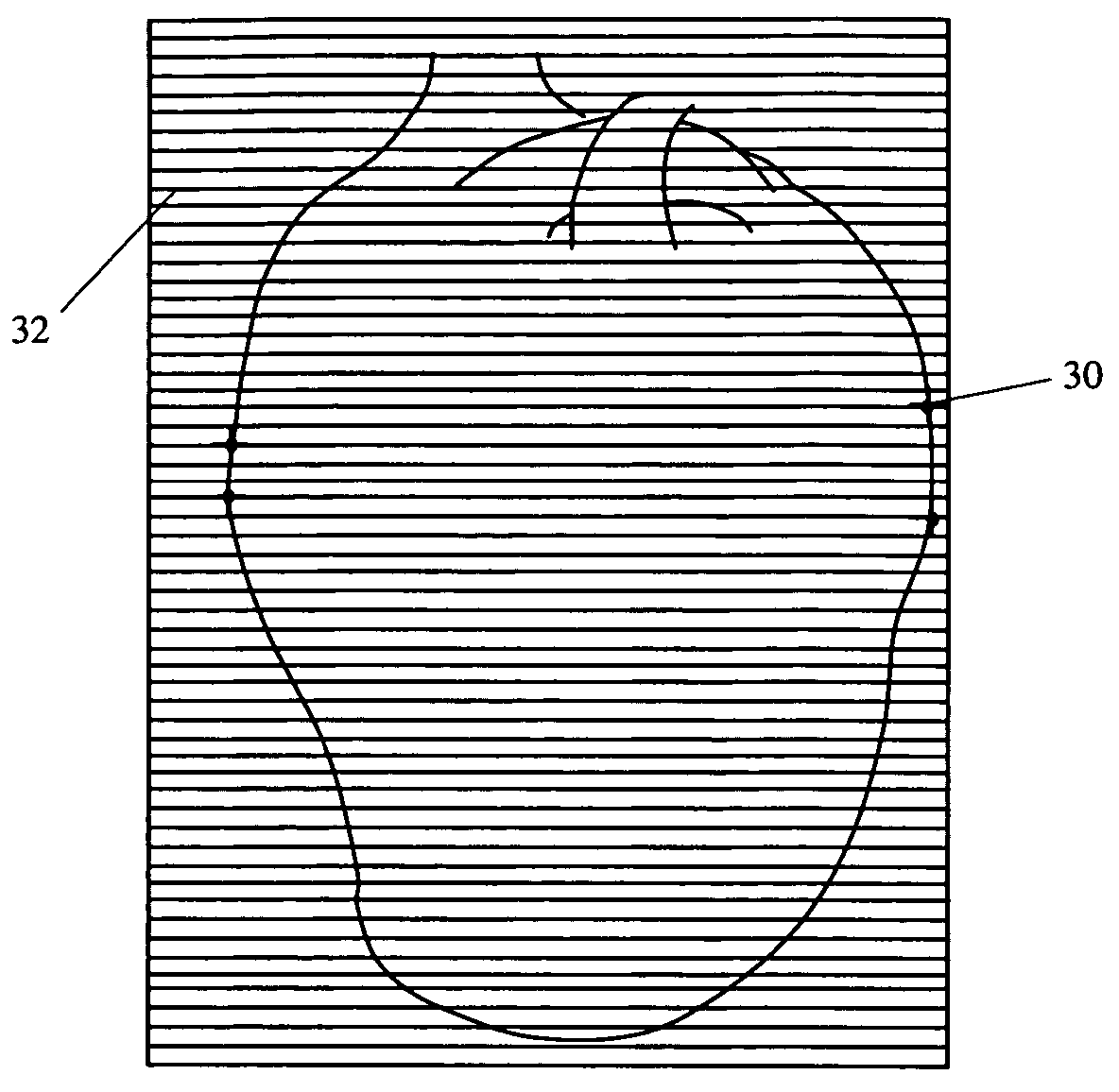
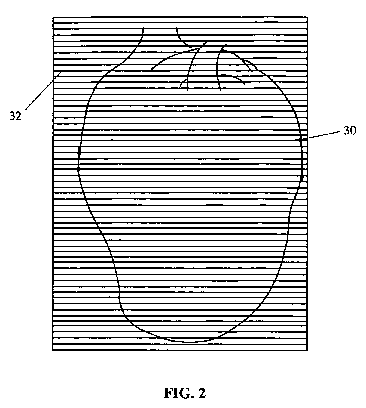
[0013]In one embodiment of the present invention, a method is provided for determining a transformation of a three-dimensional pre-operative image data set to obtain a registration of three-dimensional image data with an X-ray imaging system. The method comprises the steps of identifying at least two extreme opposing points on the object in the subject from at least one X-ray image plane, obtaining from the image data a set of contour points for each of a plurality of section-planes, performing a projection of every contour point in a sampling of the plurality of section-planes, and selecting from the sampling the section-plane with the contour point projecting nearest to the user-identified extreme point. The method further comprises the steps of identifying a three-dimensional center point of the object relative to the X-ray imaging coordinate system, defining a first grid having a predetermined number of intervals at a predetermined interval spacing with the grid center at the us...


PUM


Abstract

A method for determining a transformation of a three-dimensional pre-operative image data set to obtain a registration of the three-dimensional image data with an X-ray imaging system. In one aspect of the present invention, the method comprises the steps of the user identifying a center point and extreme contour points of the object from an X-ray image, obtaining a set of contour points for the image object in each of a plurality of section-planes, and selecting from a sampling of section-planes the points projecting nearest to the user-identified extreme points. The method then defines a grid having a predetermined number of intervals at a predetermined interval spacing with the grid center at the user-identified center of the pre-operative image, and performs a projection of the center point of the three-dimensional image data set onto each grid point for evaluation and selection of the grid point projecting nearest to the user-identified center. Registration of a three dimensional image data will allow, among other things, overlay of a visual representation of a pre-operative image object onto an X-ray image plane that can serve as a visual tool and a surgical navigation aid.

Description

CROSS-REFERENCE TO RELATED APPLICATIONS[0001]This application claims the benefit of U.S. Provisional Patent Application Ser. No. 60 / 678,322, filed May 6, 2005, the entire disclosure of which is incorporated herein.FIELD OF THE INVENTION[0002]This invention relates to translation of three-dimensional data to a reference coordinate system, and more particularly to registration of three dimensional image data with an X-ray imaging coordinate system.BACKGROUND OF THE INVENTION[0003]Interventional medicine is the collection of medical procedures in which access to the site of treatment is made by navigation through one of the subject's blood vessels, body cavities or lumens. Interventional medicine technologies have been applied to manipulation of medical instruments which contact tissues during surgical navigation procedures, making these procedures more precise, repeatable and less dependent of the device manipulation skills of the physician. Some presently available interventional med...

Claims


Application Information

Patent Timeline
Patent Type & Authority Patents(United States)
IPC IPC(8): G06K9/00
CPCG06T5/50G06T7/0028G06T7/0038A61B6/02A61B6/503G06T2207/10072G06T2207/10116G06T2207/30048G06T7/33G06T7/38
Inventor VISWANATHAN, RAJU R.
Owner STEREOTAXIS
Why Eureka
Try Eureka