
System and method for in-vivo sampling and analysis

a sampling and analysis system, applied in the field of in vivo diagnostics, can solve the problems of inability to easily detect the origin of abnormal substances by endoscopy, limited possibility in the upper or lower gi tract, and inability to localize or identify abnormal substances by in vitro sampling

Inactive Publication Date: 2010-03-23
GIVEN IMAGING LTD
View PDF195 Cites 68 Cited by
  • Summary
  • Abstract
  • Description
  • Claims
  • Application Information

AI Technical Summary

Benefits of technology

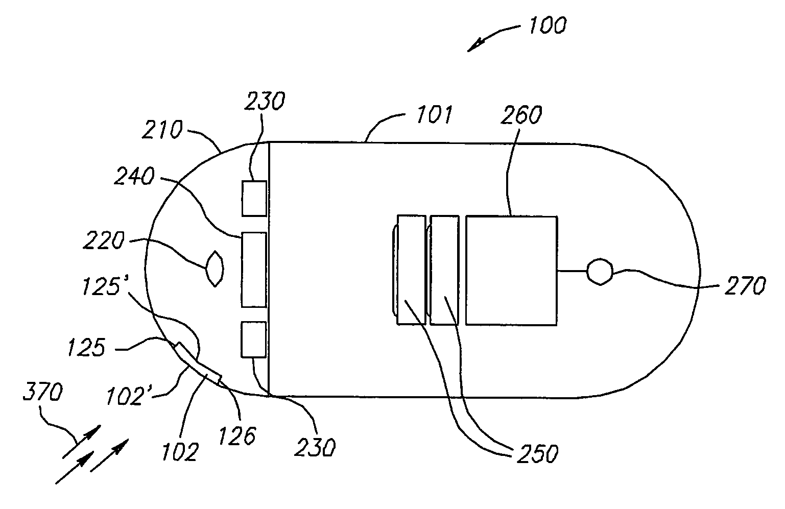
The invention is a system and method for in vivo and in situ sampling and analyzing. It includes an image sensor, an illumination source, and agglutinative particles that can adhere to an analyte in a sample. The system can be inserted into the body lumen and used to collect samples for analysis. The collected samples can be illuminated and imaged to observe agglutination, which indicates the presence of an analyte. The system can also include an autonomous device that can traverse the GI tract and collect samples for analysis. The device includes an optical window for imaging the body lumen and the agglutinative particles can be immobilized to the window for further analysis. The invention provides a non-invasive and efficient way to sample and analyze the body lumen.

Problems solved by technology

In vitro testing of samples does not easily enable the localization or identification of the origin of an abnormally occurring substance.
However, this possibility is limited to the upper or lower GI tract.
Thus, conditions in other parts of the GI tract, such as the small intestine, are not easily detected by endoscopy.

Method used


Image

  • System and method for in-vivo sampling and analysis
  • System and method for in-vivo sampling and analysis
  • System and method for in-vivo sampling and analysis

Examples


Embodiment Construction

[0018]In the following description, various aspects of the present invention will be described. For purposes of explanation, specific configurations and details are set forth in order to provide a thorough understanding of the present invention. However, it will also be apparent to one skilled in the art that the present invention may be practiced without the specific details presented herein. Furthermore, well-known features may be omitted or simplified in order not to obscure the present invention.

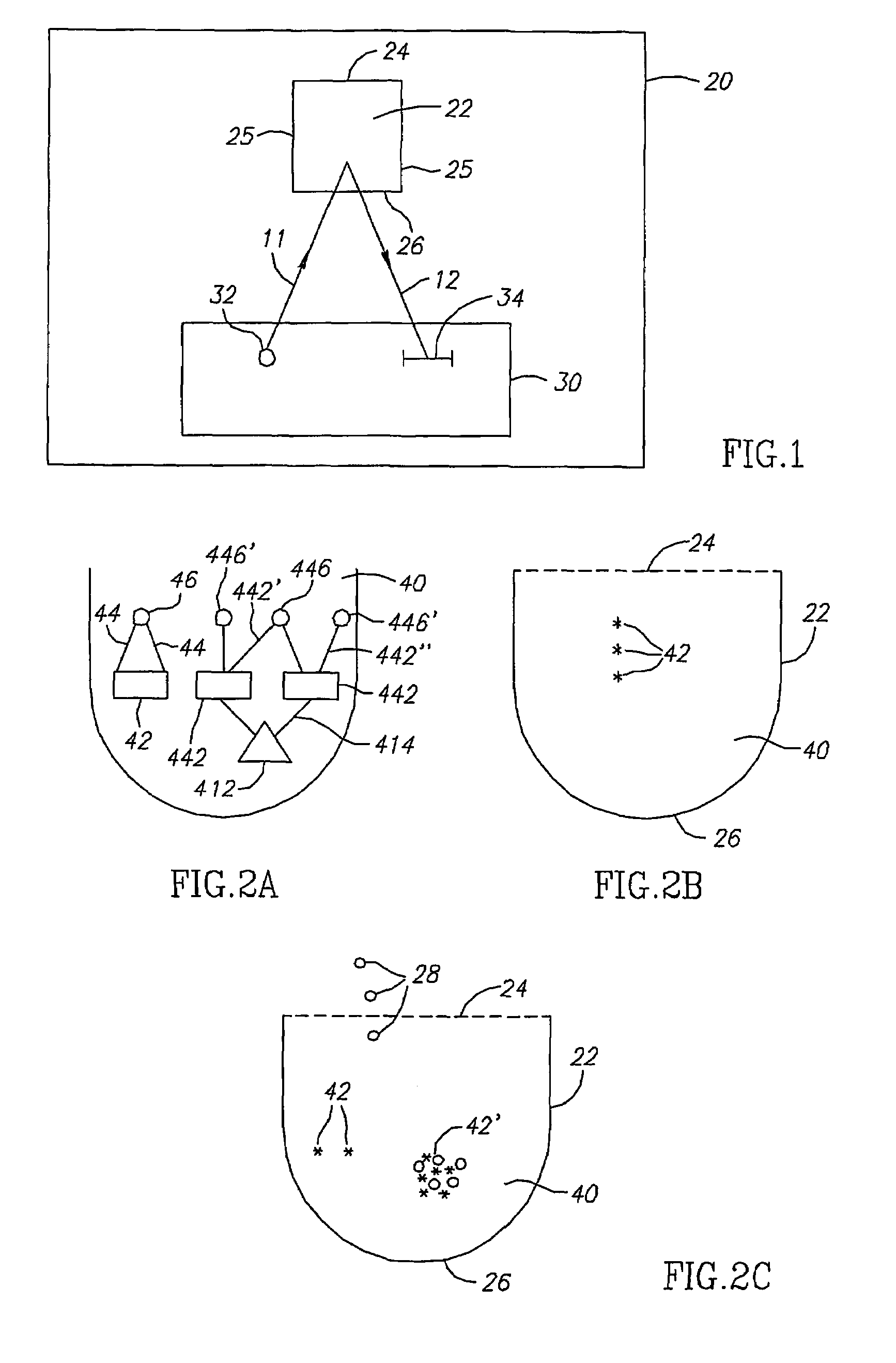
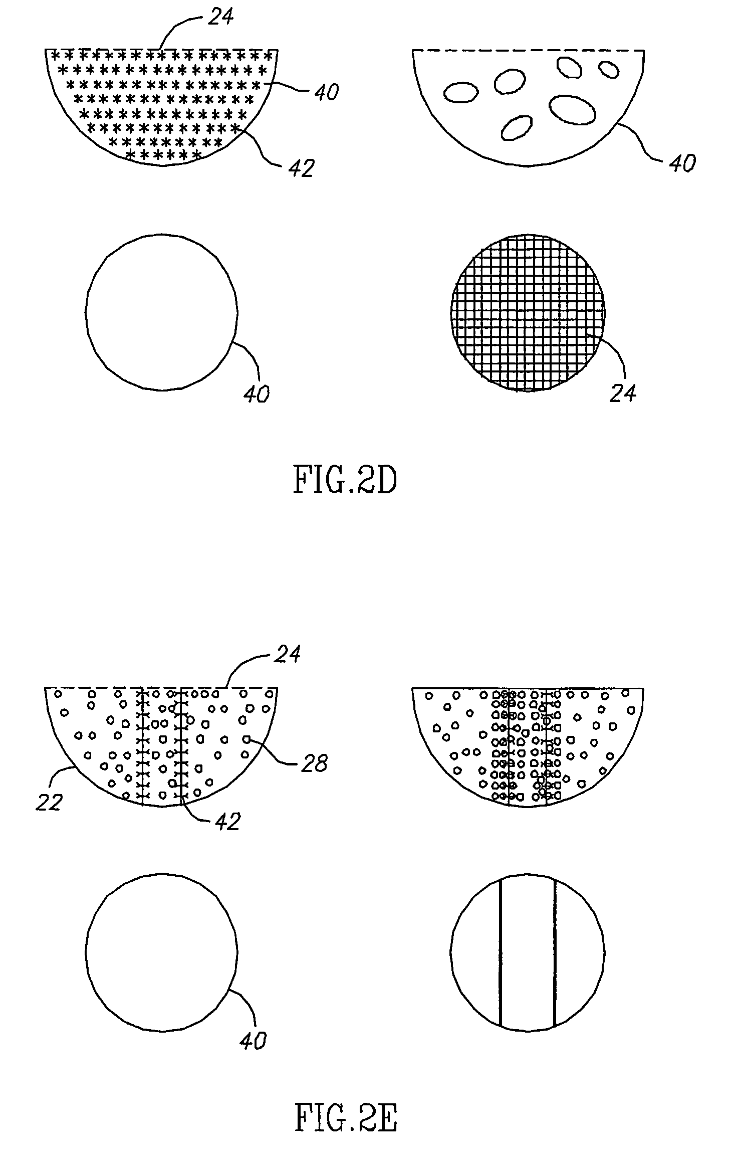
[0019]A system, according to embodiments of the invention, is typically designed to be inserted in and / or passed through a body lumen for sampling contents of the body lumen. A sample or samples may be collected into one or more sample chamber(s). The sample chamber, which typically contains agglutinative particles, may be illuminated and imaged while it is in a body lumen such that optically discernable indication of the presence of a specific analyte may show up in the images.

[0020]An ...


PUM


Abstract

A system for in vivo analysis which includes agglutinative particles capable of interacting with at least one analyte so as to cause an optical change; and at least one in vivo imaging system (220, 230, 240) configured for detecting the optical change in vivo. The system may be incorporated within an ingestible capsule (100).

Description

CROSS-REFERENCE TO RELATED APPLICATIONS[0001]This application is a National Phase Application of PCT International Application No. PCT / IL03 / 00651, International Filing Date Aug. 7, 2003, which claims priority of US Provisional Patent Application, 60 / 402,703, filed Aug. 13, 2002, both of which being incorporated by reference herein in their entirety.FIELD OF THE INVENTION[0002]The present invention relates to the field of in vivo diagnostics. More specifically, the present invention relates to a system and method for in vivo and in-situ sampling and analysis of conditions prevailing in a body lumen.BACKGROUND OF THE INVENTION[0003]An atypical concentration or presence of substances in body fluids or in body lumens may be indicative of the biological condition of the body. For example, the presence of elevated concentrations of red blood cells in the gastrointestinal (GI) tract may indicate different pathologies, depending on the location of the bleeding along the GI tract. Likewise, ...

Claims


Application Information

Patent Timeline
Patent Type & Authority Patents(United States)
IPC IPC(8): A61B5/1455A61B1/05A61B1/31A61B10/00
CPCA61B1/041A61B5/0084A61B5/073A61B5/14546G03B17/02G03B29/00A61B10/0038
Inventor PALTI, YORAM
Owner GIVEN IMAGING LTD
Learn More