Color unmixing and region of interest detection in tissue samples

a color unmixing and tissue sample technology, applied in the field of color unmixing and region of interest detection in tissue samples, can solve the problems of not revealing the spectrum of immunohistochemical dye analysis, and the prior art further does not disclose the color concentration of the second dye normalization

Inactive Publication Date: 2010-03-30
RABINOVICH ANDREW M
View PDF3 Cites 27 Cited by
  • Summary
  • Abstract
  • Description
  • Claims
  • Application Information

AI Technical Summary

Problems solved by technology

The prior art further does not disclose analyzing the image in the spectrum of the immunohistochemical dye for areas of interest, e.g. organs as opposed to stroma (for example fatty or other intermediate tissue).
Further, the prior art does not disclose normalizing the color concentration of the second dye with respect to an area of interest to generate a color index having diagnostic significance.

Method used


Image

  • Color unmixing and region of interest detection in tissue samples
  • Color unmixing and region of interest detection in tissue samples
  • Color unmixing and region of interest detection in tissue samples

Examples


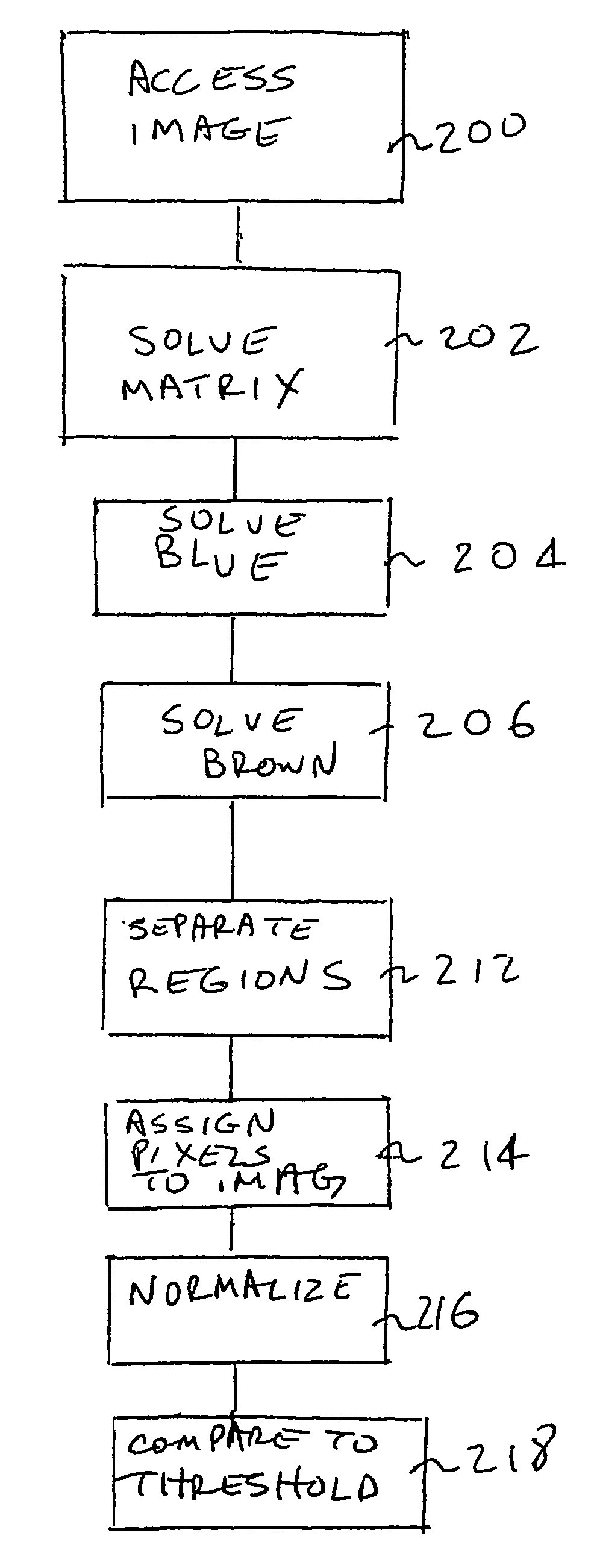
Embodiment Construction

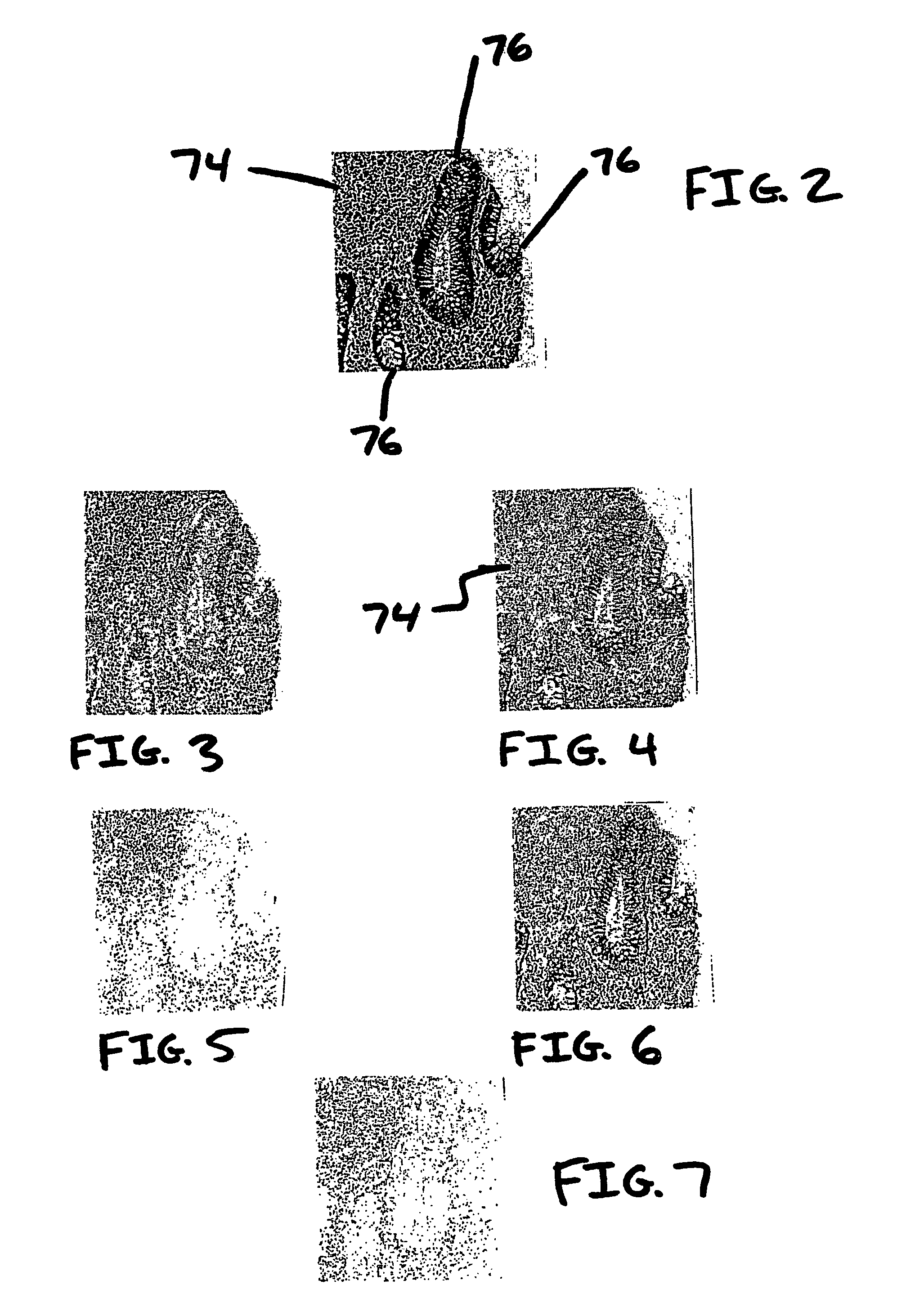
[0032]FIG. 1 is a block diagram illustrating a system for producing images of a slide 10 having mixed colors and analyzing the images to provide deconvoluted images in which colors are unmixed. One color may be used simply for improved visibility of all tissue in the slide, while the other color may bind to protein in tissue affected by a particular disease. The slide 10, which holds a tissue sample, is further described with respect to FIG. 2 below. The system of FIG. 1 includes a microscope 20, a camera 25 and a computer 30 which may have a monitor 32.

[0033]In the microscope 20, the slide 10 is supported on an X-Y stage 40. The X-Y stage 40 includes a platform that is movable in two degrees of freedom so that an operator, e.g. a pathologist, can view selected areas of the slide 10. Commonly, an illumination source 42 is located beneath the X-Y stage 40. The illumination source 42 is a white source that will have a broad spectrum of component wavelengths. The slide 10 is viewed by ...


PUM


Abstract

In a method and system and machine-readable medium, colors in an image (200) are unmixed (202) using a matrix X=AS, where A is a ns. ×nd matrix of spectral definitions, where ns is the number of spectral components and nd is the number of dyes into which the image is decomposed, and where S is a nd×l matrix of amounts of each dye at every pixel, where l is the number of pixels; the matrix X is constrained for solution by an unsupervised matrix decomposition method having constraints consistent with properties of an additive color model; and nd is determined. A texture is determined to identify areas of interest. Further processing may automatically measure cell dye concentration in the determined regions of interest.

Description

CROSS REFERENCE TO RELATED APPLICATIONS[0001]The present application claims priority from U.S. Provisional Patent Application 60 / 475,071 filed May 30, 2003, the disclosure of which is incorporated by reference herein.FIELD OF THE INVENTION[0002]The present subject matter relates to processes, systems, and apparatuses for performing deconvolution of colors in an image and automated analysis of areas of interest in a non-uniform sample.BACKGROUND OF THE INVENTION[0003]One form of tissue analysis comprises staining tissue with a dye and placing this stained tissue on a microscope slide. The tissue may be stained with more than one dye for a number of different reasons. For example, two dyes may be used to identify the presence of two different proteins. In another application, one dye may be specific to bind with a particular cell component such as cytoplasm, while the other dye may stain nuclei. The microscope slide is then illuminated and an electronic image is produced. A digital vi...

Claims


Application Information

Patent Timeline
Patent Type & Authority Patents(United States)
IPC IPC(8): G06K9/00G06K9/46G06K9/62G06T
CPCG06K9/00127G06K9/0014G06K9/4619G06K9/624G06V20/69G06V20/695G06V10/449G06V10/7715G06F18/2134
Inventor RABINOVICH, ANDREW M.
Owner RABINOVICH ANDREW M
Why Eureka
Try Eureka