Method and apparatus for diagnosing conditions of the eye with infrared light

a technology of infrared light and eye conditions, applied in the field of eye conditions or diseases, can solve the problems of reduced ability, severe limitation of the value of this technique, and elevation of intraocular pressure and glaucoma

US7712901B2Inactive Publication Date: 2010-05-11PREDICTEK
2 Cites 9 Cited by

Patent Information

Authority / Receiving Office
US · United States
Patent Type
Patents(United States)
Current Assignee / Owner
Publication Date
2010-05-11
Estimated Expiration
Not applicable · inactive patent

Smart Images

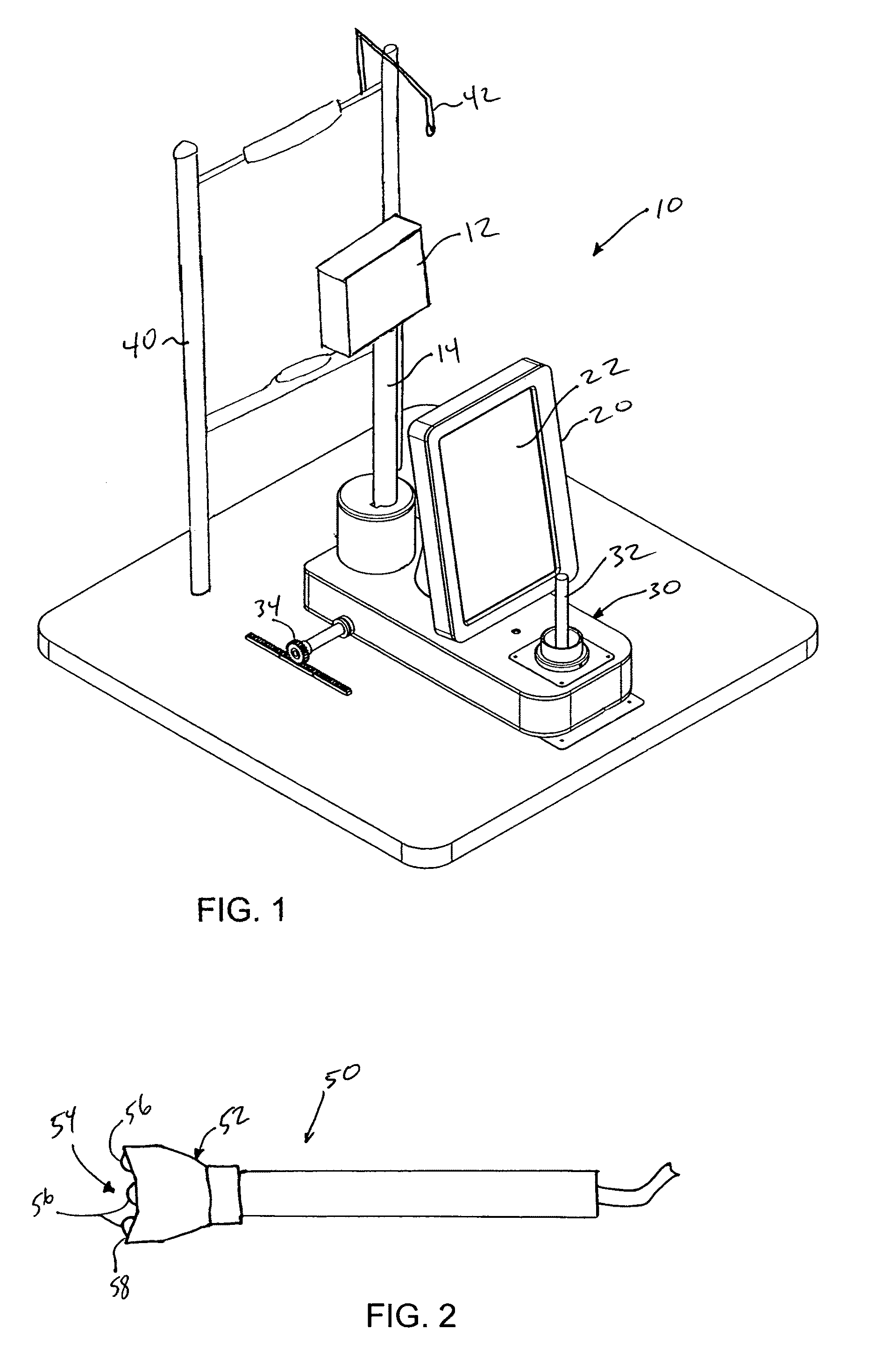
  • Figure 1
    Figure 1
  • Figure 2
    Figure 2
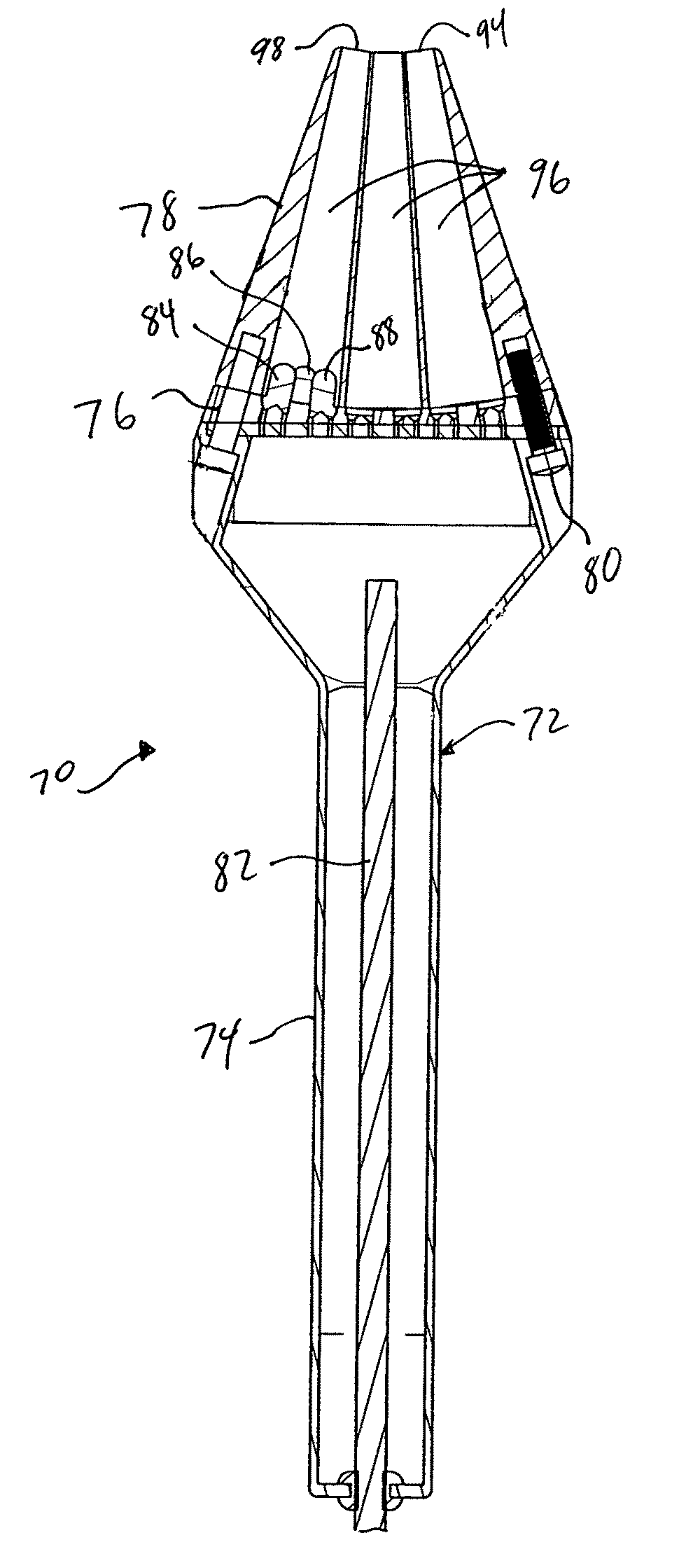
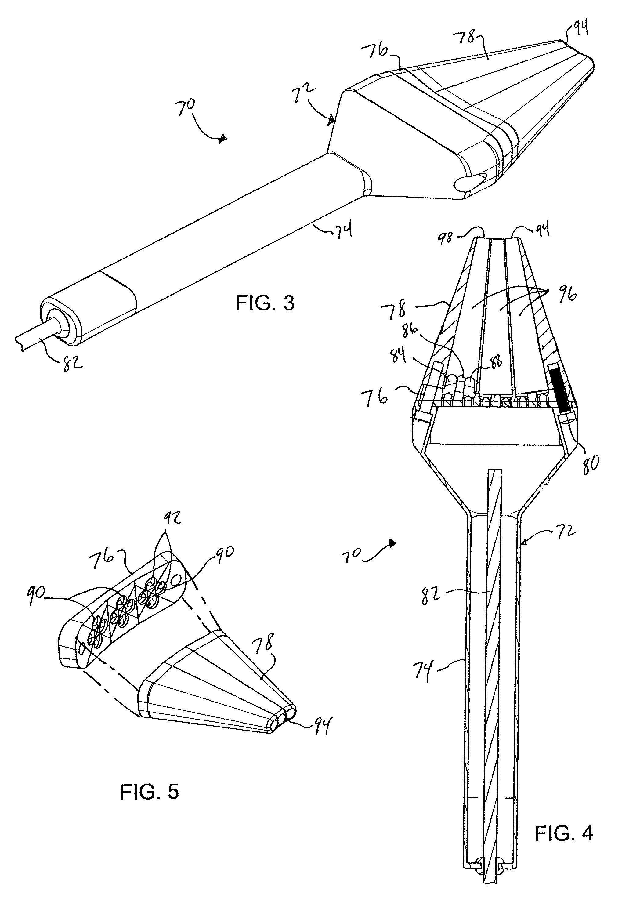
  • Figure 3
    Figure 3
Patent Text Reader

Abstract

A method and apparatus for imaging an eye of a patient and detecting any condition or disease in the eye. A near infrared light beam is emitted into the eye through the skin adjacent to the eye. A digital image is acquired from the light reflected from the eye. The image shows details that are not perceivable using current examination techniques. Images are acquired using more than one infrared wavelength using a multispectral illumination device for a more thorough analysis.
Need to check novelty before this filing date? Find Prior Art

Description

CROSS REFERENCE TO RELATED APPLICATION

[0001] This application claims the benefit of U.S. provisional patent application Ser. No. 60 / 873,669, filed on 8 Dec. 2006. The provisional patent application is hereby incorporated by reference herein in its entirety and is made a part hereof, including but not limited to those portions which specifically appear hereinafter.BACKGROUND OF THE INVENTION

[0002] This invention relates generally to a method and apparatus for diagnosing eye conditions or diseases.

[0003] There are many disorders that affect the iris, which is the pigmented structure at the front of the eye. One such disorder is pigment dispersion syndrome (PDS), a disorder that is characterized by a mechanical debridement of pigment from the back surface of the iris. As a result of PDS, pigment granules are deposited on structures within the anterior segment of the eye, most importantly the trabecular meshwork (the structure within the eye that permits drainage). Prolonged deposition of ...

Examples

examples

[0048]The method of this invention was tested in the clinics of the Illinois Eye Institute (IEI) for acquisition of iris transillumination images of patients. Two groups of subjects were recruited from an existing patient pool at IEI, without preference to age or sex. The first group consisted of 12 Caucasian subjects, of which six were normal and the other six had PDS. The second group consisted of 12 African-American subjects, of which six were normal and the other six had PDS.

[0049]All 12 African-American subjects had dark-colored irides (dark brown). Among the Caucasian group, of the six normal subjects, four had blue eyes, one light hazel, and one light brown; of the six PDS subjects, three had blue eyes, one light hazel, two dark brown. These two groups were chosen to determine whether large variations in iris transmission spectra can be accounted for in image acquisition and processing.

[0050]These subjects were imaged using an apparatus as described above, in a total of 24 se...