
Method and apparatus for diagnosing conditions of the eye with infrared light

a technology of infrared light and eye conditions, applied in the field of eye conditions or diseases, can solve the problems of reduced ability, severe limitation of the value of this technique, and elevation of intraocular pressure and glaucoma

Inactive Publication Date: 2008-06-12
PREDICTEK
View PDF2 Cites 16 Cited by
  • Summary
  • Abstract
  • Description
  • Claims
  • Application Information

AI Technical Summary

Benefits of technology

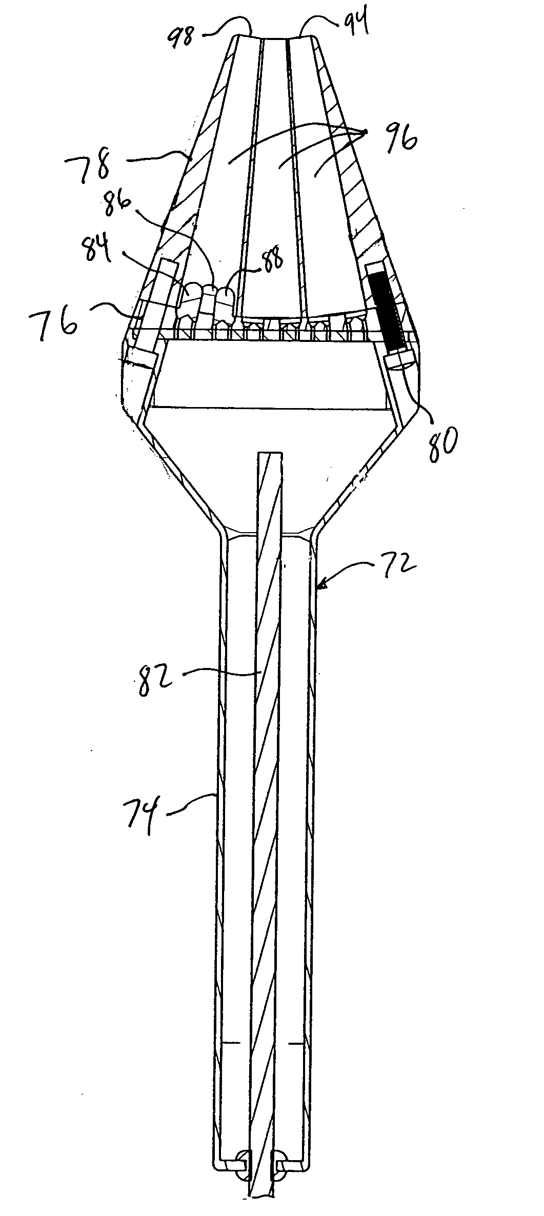
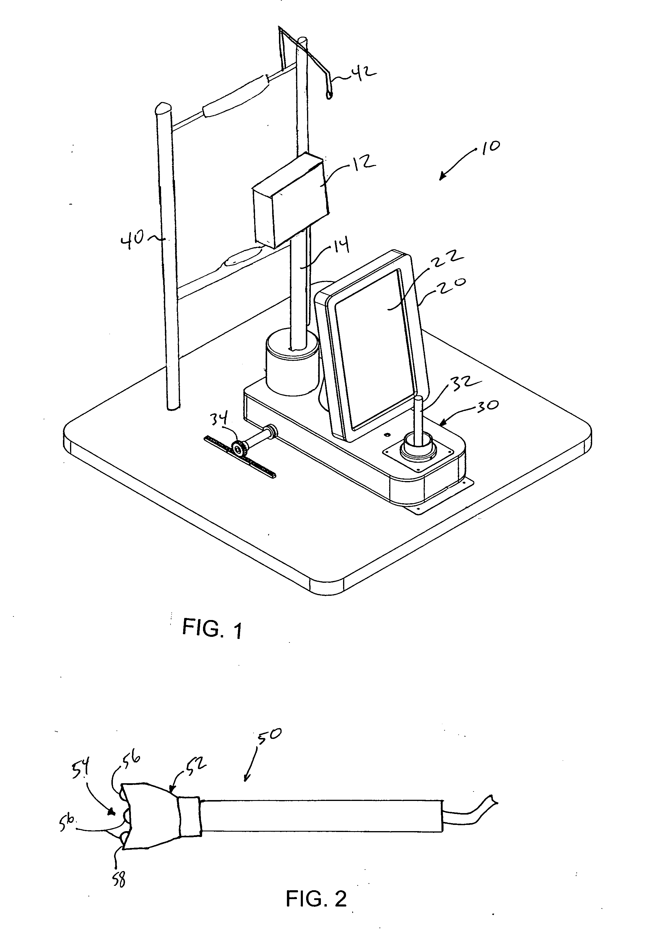
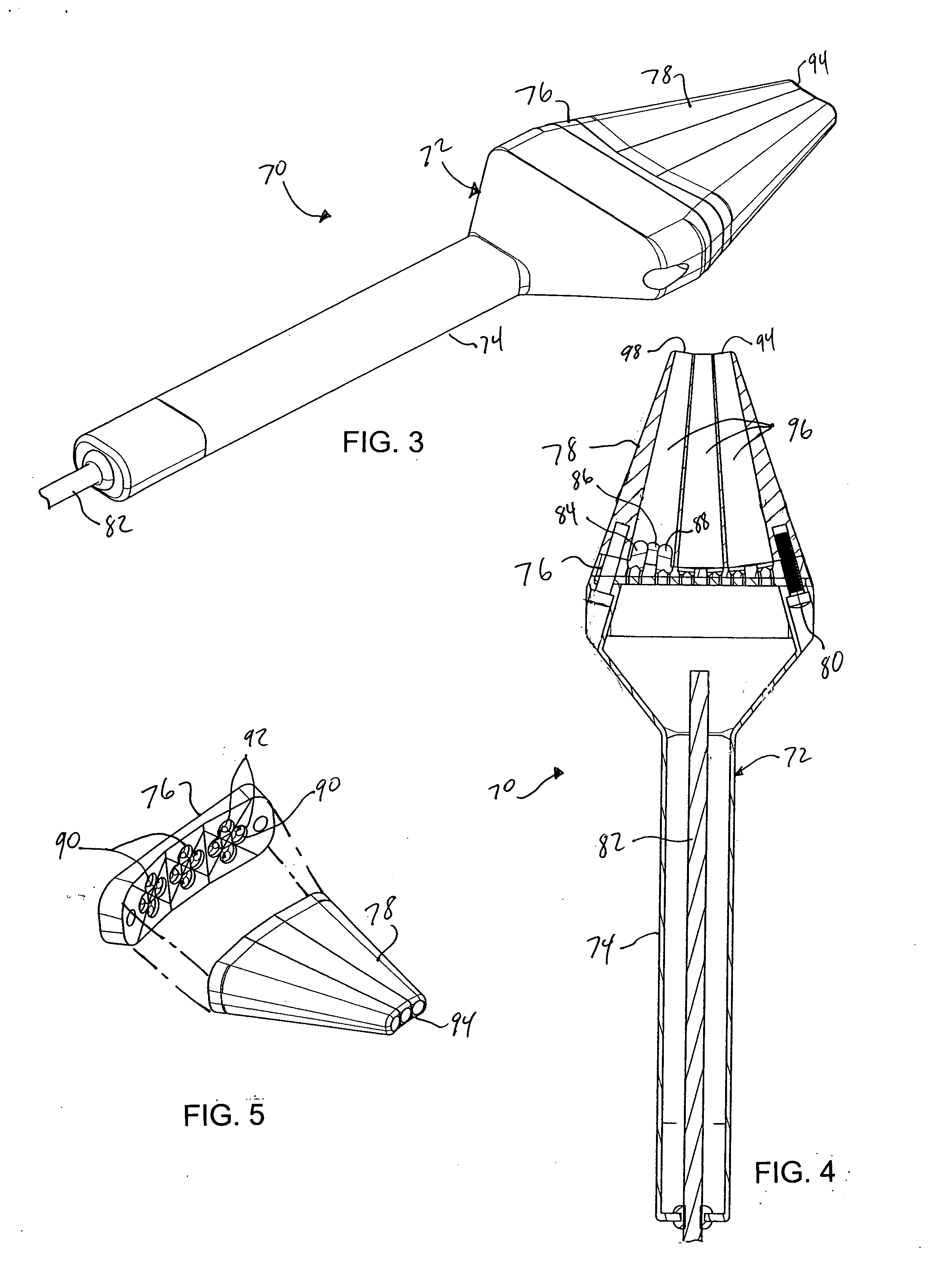
[0015]The apparatus of this invention is a new diagnostic imaging device for use by eye care practitioners. The apparatus collects and, desirably, automatically analyzes multispectral infrared images (e.g., multiple images, each taken using a different wavelength of light) of the iris (the pigmented structure at the front of the eye), permitting the visualization and automatic detection of iris defects associated with pigment dispersion syndrome (PDS) and other ocular conditions that can lead to glaucoma. The multispectral imaging according to this invention produces useful, quality images in spite of complicating variations in eye and skin pigmentations of patients. Multispectral imaging also permits abnormalities to be detected by virtue of different appearances at different wavelength spectra.
[0017]Whereas iris defects show very limited contrast when transilluminating the iris with visible light, these defects are clearly visualized when using NIR light. The imaging technique of this invention can readily detect iris defects that are difficult or impossible to observe by visual examination, even in patients with darkly pigmented eyes and / or skin for whom traditional examination techniques perform very poorly. The multispectral capability of the apparatus allows adaptive optimization of imaging performance to each subject's pigmentation type, and the spectral signatures within the images can be used to help discriminate defects from normal iris tissue.

Problems solved by technology

Prolonged deposition of pigment granules in this structure may cause a reduction in the ability of the trabecular meshwork to transport fluid from the eye, which can result in an elevation of the intraocular pressure and glaucoma.
However, the value of this technique is severely limited by the low transmission coefficient of the iris and skin for light in the visible regime.
However, the true prevalence is probably much greater than this because mild presentations go undetected by traditional examination methods.
It has been widely assumed that PDS is very rare in black patients; however, recent research has shown that this observation is probably due to the poor sensitivity of standard diagnostic methods, especially for patients having dark eyes and / or skin, which can severely limit visible-light transillumination.
This second method suffers from the same limitations as the first method.
However, their success is limited by the low contrast of the features that are observed, which is especially problematic for patients with brown eyes and / or dark skin, or in cases when the disease is subtle or in an early stage of progression.
Thus, thinned areas may not be appreciated.
Currently, there are no devices available to the clinician for routine imaging or automated detection of iris defects.

Method used


Image

  • Method and apparatus for diagnosing conditions of the eye with infrared light
  • Method and apparatus for diagnosing conditions of the eye with infrared light
  • Method and apparatus for diagnosing conditions of the eye with infrared light

Examples


examples

[0048]The method of this invention was tested in the clinics of the Illinois Eye Institute (IEI) for acquisition of iris transillumination images of patients. Two groups of subjects were recruited from an existing patient pool at IEI, without preference to age or sex. The first group consisted of 12 Caucasian subjects, of which six were normal and the other six had PDS. The second group consisted of 12 African-American subjects, of which six were normal and the other six had PDS.

[0049]All 12 African-American subjects had dark-colored irides (dark brown). Among the Caucasian group, of the six normal subjects, four had blue eyes, one light hazel, and one light brown; of the six PDS subjects, three had blue eyes, one light hazel, two dark brown. These two groups were chosen to determine whether large variations in iris transmission spectra can be accounted for in image acquisition and processing.

[0050]These subjects were imaged using an apparatus as described above, in a total of 24 se...


PUM


Abstract

A method and apparatus for imaging an eye of a patient and detecting any condition or disease in the eye. A near infrared light beam is emitted into the eye through the skin adjacent to the eye. A digital image is acquired from the light reflected from the eye. The image shows details that are not perceivable using current examination techniques. Images are acquired using more than one infrared wavelength using a multispectral illumination device for a more thorough analysis.

Description

CROSS REFERENCE TO RELATED APPLICATION[0001]This application claims the benefit of U.S. provisional patent application Ser. No. 60 / 873,669, filed on 8 Dec. 2006. The co-pending provisional patent application is hereby incorporated by reference herein in its entirety and is made a part hereof, including but not limited to those portions which specifically appear hereinafter.BACKGROUND OF THE INVENTION[0002]This invention relates generally to a method and apparatus for diagnosing eye conditions or diseases.[0003]There are many disorders that affect the iris, which is the pigmented structure at the front of the eye. One such disorder is pigment dispersion syndrome (PDS), a disorder that is characterized by a mechanical debridement of pigment from the back surface of the iris. As a result of PDS, pigment granules are deposited on structures within the anterior segment of the eye, most importantly the trabecular meshwork (the structure within the eye that permits drainage). Prolonged dep...

Claims


Application Information

Patent Timeline
Patent Type & Authority Applications(United States)
IPC IPC(8): A61B3/10
CPCA61B3/1216
Inventor WERNICK, MILES N.ROBERTS, DANIEL K.LUKIC, ANA S.YANG, YONGYI
Owner PREDICTEK
Learn More