
Heat diffusion based detection of structures of interest in medical images

a technology of heat diffusion and detection of structures, applied in the field of medical imaging, can solve the problems of unavoidably limited detection accuracy and time-consuming conventional examination of source images

Inactive Publication Date: 2010-06-01
THE BOARD OF TRUSTEES OF THE LELAND STANFORD JUNIOR UNIV
View PDF9 Cites 22 Cited by
  • Summary
  • Abstract
  • Description
  • Claims
  • Application Information

AI Technical Summary

Problems solved by technology

Conventional examination of these source images is rather time-consuming and the detection accuracy is unavoidably limited by human factors such as attention span and eye fatigue.

Method used


Image

  • Heat diffusion based detection of structures of interest in medical images
  • Heat diffusion based detection of structures of interest in medical images
  • Heat diffusion based detection of structures of interest in medical images

Examples


Embodiment Construction

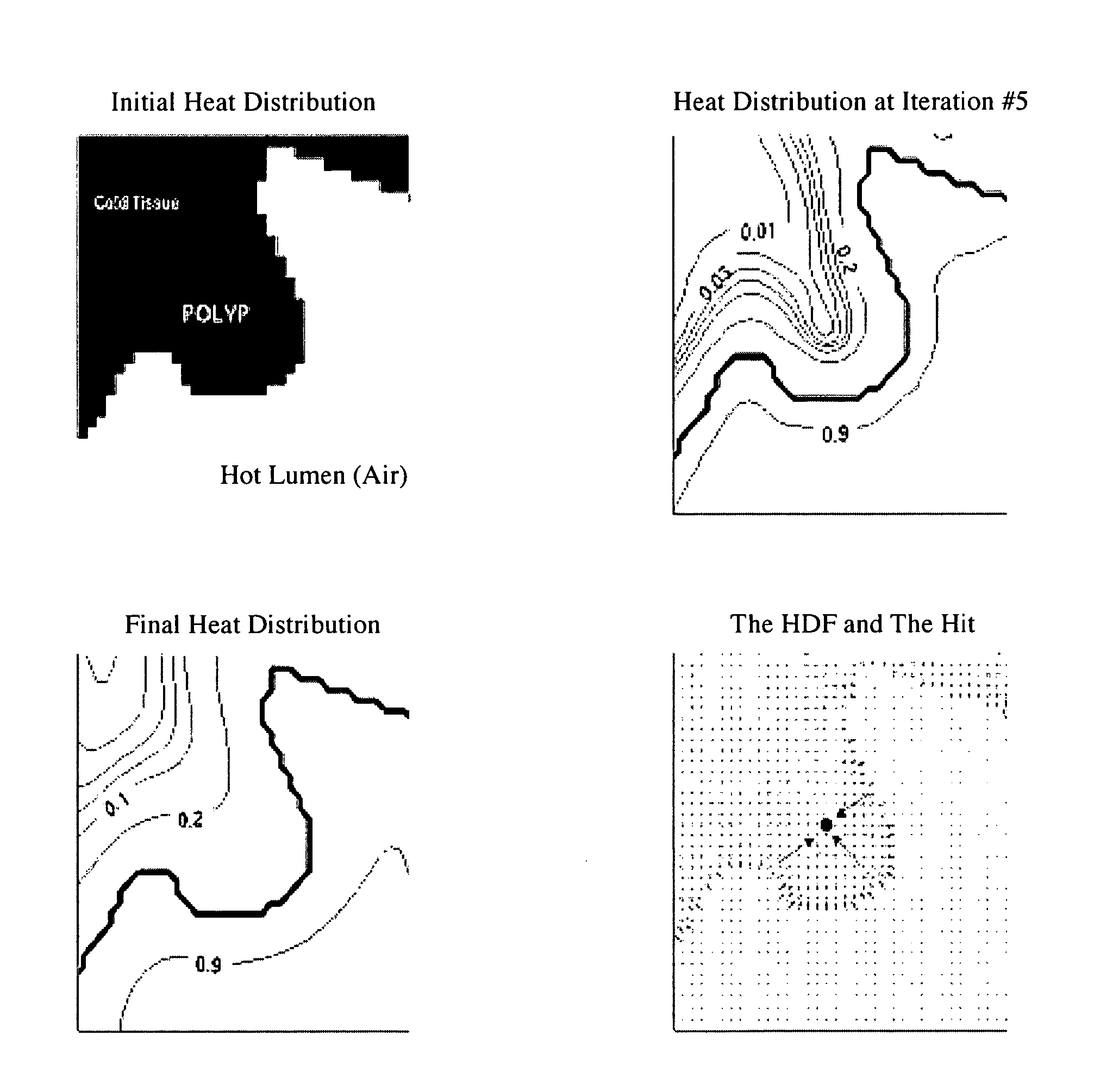
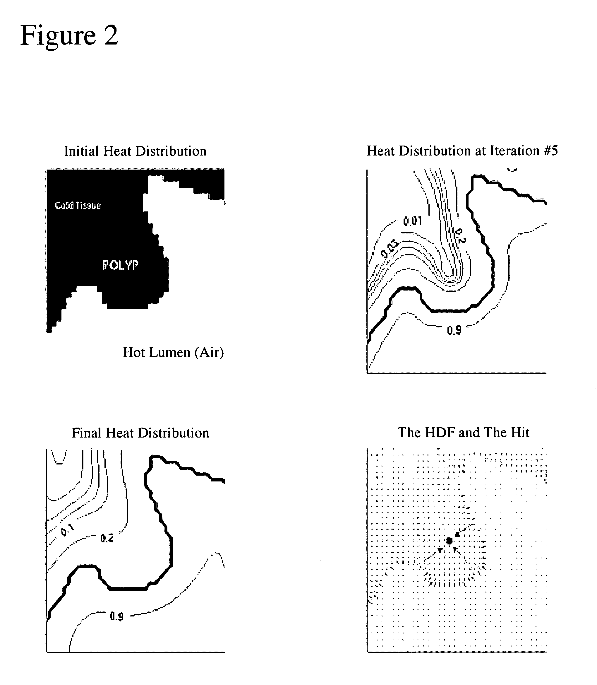
[0014]The present method is aimed at detecting structures of interest from volumetric images and discriminate them from other protruding structures, non-protruding structures and / or stand-alone structures. In an exemplary embodiment, the protruding structures are for instance polyps, haustral folds, etc., whereby the protruding structures of interest are polyps. The non-protruding structures are for instance elongated structures, flat structures or structures in between haustral folds. In this example, both types of structures are on the flexible colon, which is herein considered the volume of interest. In another exemplary embodiment, the structures of interest are stand-alone structures such as lung nodules. In this example, the structures are within a lung, which is then considered the volume of interest. The following description contains details on the first embodiment. However, a person of average skill in the art would readily appreciate that these teachings would also apply ...


PUM


Abstract

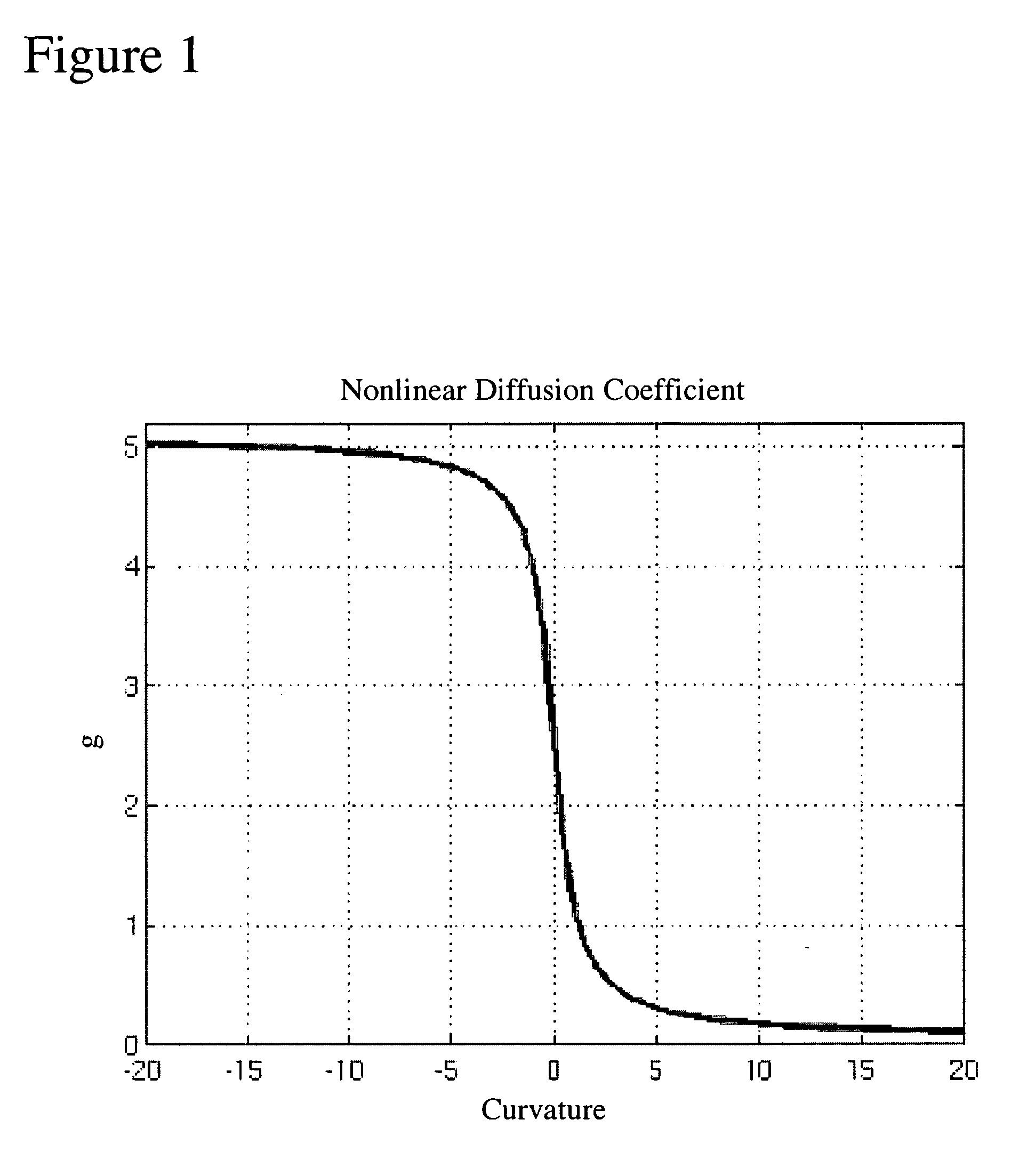
A method for detecting and identifying structures of interest such as colonic polyps or similar structures like lung nodules in volumetric (medical) images data is provided. The method includes obtaining a heat diffusion field (HDF) by applying a heat diffusion scheme to a volume of interest that includes structures. The obtained heat diffusion field is then used for identifying a structure of interest from the structures in the volume of interest using a geometrical analysis of the heat diffusion field. The heat diffusion scheme is, at least partly, governed by non-linear diffusion parameters. The identification includes two parts: (i) the computation of a spherical symmetry parameter, and (ii) the performance of a local analysis of the volume of interest and computation of a triangulization parameter.

Description

CROSS-REFERENCE TO RELATED APPLICATIONS[0001]This application is cross-referenced to and claims priority from U.S. Provisional Application 60 / 526,849 filed Dec. 3, 2003, which is hereby incorporated by reference.STATEMENT REGARDING FEDERALLY SPONSORED RESEARCH OR DEVELOPMENT[0002]This invention was supported in part by grant number R01 CA72023 from the National Institutes of Health (NIH / NCI). The U.S. Government has certain rights in the invention.FIELD OF THE INVENTION[0003]The present invention relates generally to medical imaging. More particularly, the present invention relates to methods of detecting polyps in medical images.BACKGROUND[0004]Computed Tomographic Colonography (CTC) is a minimally invasive technique that employs X-Ray CT imaging of the abdomen and pelvis following cleansing and air insufflation of the colon. Originally proposed in the early 1980's, it became practical in the early 1990's following the introduction of helical CT and advances in computer graphics. C...

Claims


Application Information

Patent Timeline
Patent Type & Authority Patents(United States)
IPC IPC(8): A61B6/00G06F15/00G06T5/00G06T7/60
CPCG06T7/0012G06T7/604G06T7/608G06T2207/30032G06T2207/20148G06T2207/20152G06T7/64G06T7/68G06T7/136
Inventor ACAR, BURAKKONUKOGLU, ENDERBEAULIEU, CHRISTOPHER F.NAPEL, SANDY A.PAIK, DAVID S.
Owner THE BOARD OF TRUSTEES OF THE LELAND STANFORD JUNIOR UNIV
Learn More