Method and device for biochemical detection and analysis of subcellular compartments from a single cell

a biochemical detection and analysis technology, applied in the field of single cell biochemical detection and analysis methods and devices, can solve the problems of insufficient nanometer scale inability to analyze ultrasmall structures at the level of single copies, and inability to achieve nano-scale chemical analysis of subcellular structures. achieve the effect of high spatial resolution

Inactive Publication Date: 2010-08-03
UNIV OF WASHINGTON
View PDF6 Cites 57 Cited by
  • Summary
  • Abstract
  • Description
  • Claims
  • Application Information

AI Technical Summary

Benefits of technology

[0008]Certain aspects of the present invention provide methods, devices, and systems for ultra sensitive biochemical detection and analysis of micro- or nano-scale subcellular structures from a single biological cell. The devices and methods of the present invention provide comprehensive biochemical information on individual subcellular compartments with high spatial resolution. Further aspects of the invention provide methods for isolating subcellular components and molecular components from within a single biological cell using laser-assisted micro- or nano-surgery or using hydrophobic reagents that can permeate a cell membrane of the single biological cell and label a molecular component within a subcellular component of the cell. The device and methods of the present invention can provide spatial information with regard to intracellular localization of molecular components, for example, nucleic acids, proteins, lipids, colloidal entities, small ions, nanoparticles, or other macromolecules, thus providing a powerful complement to many techniques in genomics and in proteomics.
[0015]When encapsulating the subcellular component within a microdroplet, the method further provides contacting the subcellular component in the aqueous microdroplet with an organic phase, adjusting the starting volume of the organic phase and the period of time of contacting the aqueous microdroplet with the organic phase, and controlling the concentration of the molecular component in the aqueous microdroplet.

Problems solved by technology

Although powerful imaging systems exist for studying the morphologies of cells and subcellular architectures, no system currently exists to analyze the biochemical compositions of ultrasmall structures at the level of single copies.
In comparison with these single-cell studies, the chemical analysis of nanometer scale subcellular compartments presents formidable challenges.
Most approaches used in single-cell studies are thus inadequate for nano-scale chemical analysis of subcellular structures.

Method used


Image

  • Method and device for biochemical detection and analysis of subcellular compartments from a single cell
  • Method and device for biochemical detection and analysis of subcellular compartments from a single cell
  • Method and device for biochemical detection and analysis of subcellular compartments from a single cell

Examples


example 1

On-Chip Integration

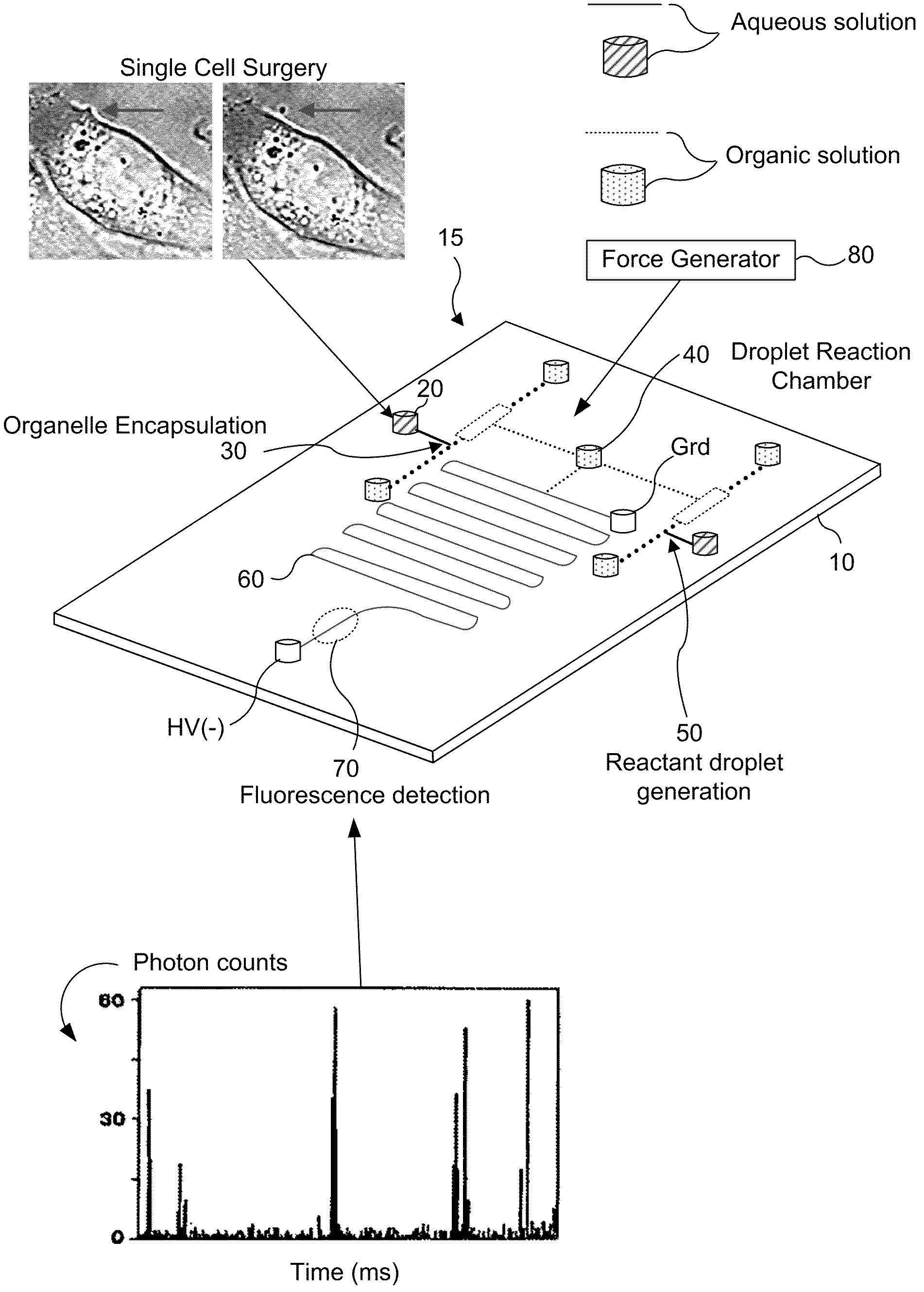
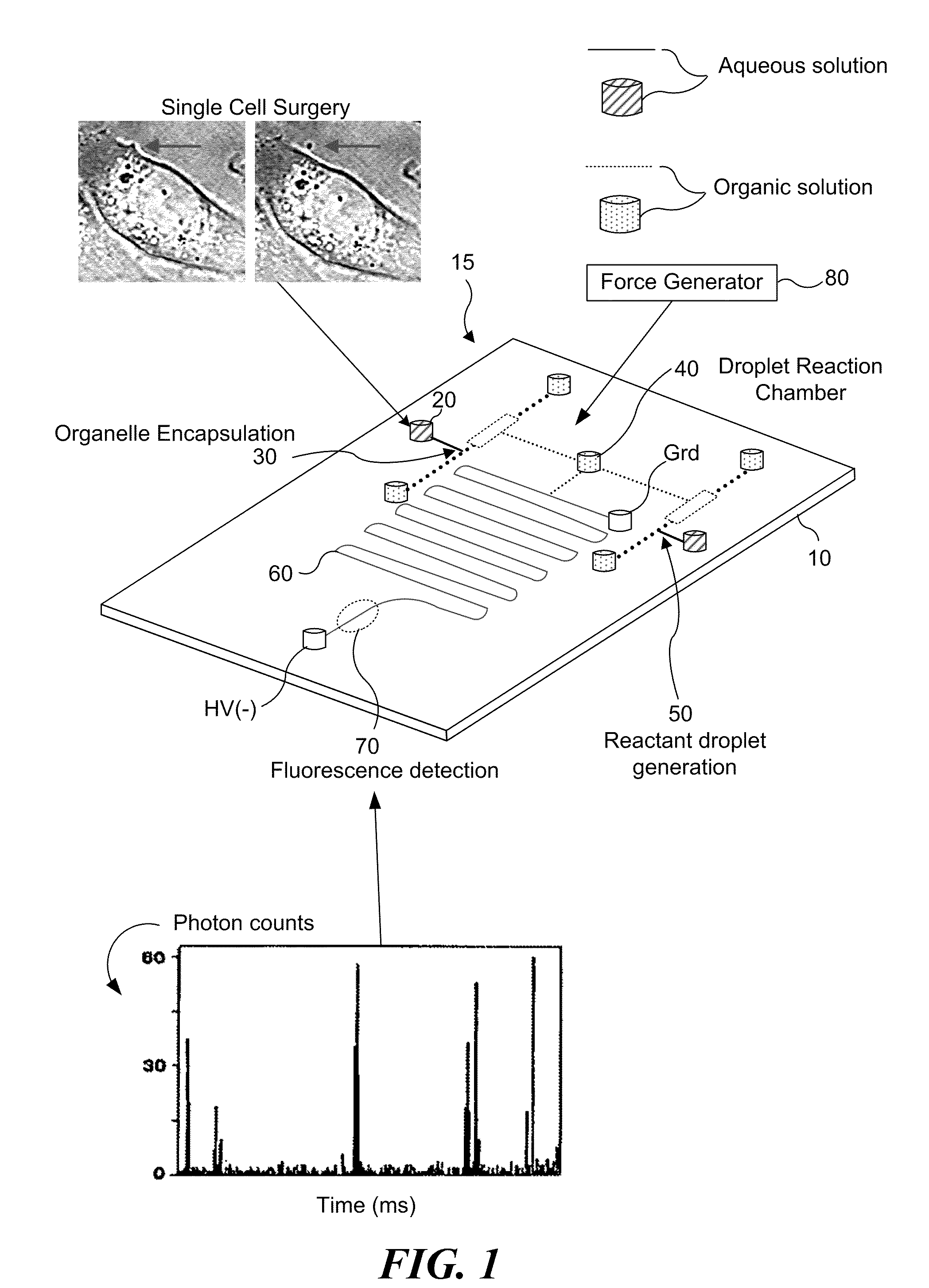
[0084]The interface combination of these techniques was performed on a single chip device 15 as illustrated in FIG. 1 (e.g., fabricated in a substrate 10, of a material such as polydimethylsiloxane, polymethylmethacrylate, glass, silicon, polyester, and / or other materials, and combinations thereof) with micro- and nano-fluidic channels, in which single-cell surgery was performed on the substrate 10 in surgery chamber 20, followed by the in-channel droplet generation at the reactant microdroplet generator 50, along with the micro- and / or nano-scale chemical reaction in droplet reaction chamber 40, with each component of the reacted mixture subsequently separated by CE in separation microchannel 60. In the case of laser-induced fluorescence detection, the separated components are detected within the constricted region of the micro- and / or nano-channel using detector 70. In the case of mass spectrometric detection, the separated components or the intact organelles ar...

example 2

Single-Cell Micro- or Nano-Surgery and Micro- or Nano-Scale Chemical Reaction

[0085]To isolate single subcellular compartments by single cell nano-surgery in surgery chamber 20, optical trapping was used as a force generator 80 to first manipulate and transport the identified organelles close to the cell membrane (FIG. 7A). Optical trapping has been used in a wide range of applications, from the manipulation of micro- and nano-meter sized particles to single cells and subcellular structures. A single nanometer scale organelle was then moved from the cellular interior to a location adjacent to the membrane surface (FIGS. 7B and 7C, arrow). Once parked at the membrane, the organelle is transported across the cell membrane. A focused ultraviolet (UV) or near infrared (IR) laser (FIGS. 7D-7G), was used to “open” a small nanometer-sized patch on the cell membrane to isolate the organelle from the cell for chemical analysis. Because the cell is a heavily compartmentalized structure, this a...

example 3

Sensitive Detection of Single Fluorescent Molecules

[0089]The detection of single fluorescent molecules using detector 70 can be achieved with an excellent signal-to-noise ratio, and has become more facile and routine with improved detectors and optics. The detection volume of a single-molecule confocal fluorescence microscope was defined latitudinally by the laser focus and longitudinally by the pinhole present at the image plane. This far-field method for single-molecule detection shows excellent signal-to-noise ratio at approximately 1-ms photon integration time, which is illustrated in FIG. 9 (which illustrates a photon trace detection of single carboxyrhodamine 6G molecule diffusing in solution, in which each spike signals the presence of a single dye molecule within the probe volume). One criterion employed to achieve such high signal-to-noise ratio is to minimize the laser probe volume by using a diffraction limited laser focus, which is approximately 0.5 μm in diameter and 2 ...


PUM

PropertyMeasurementUnit
volumeaaaaaaaaaa
volumeaaaaaaaaaa
diameteraaaaaaaaaa

Abstract

A method and system for performing biochemical detection or analysis on micro- and nano-scale subcellular component within a single biological cell is provided. An integrated platform device and method to perform the biochemical analysis is also provided.

Description

CROSS REFERENCE TO RELATED APPLICATION[0001]This application claims the benefit of U.S. Provisional Application No. 60 / 497,874, filed Aug. 25, 2003, the entirety of which is incorporated by reference herein.GOVERNMENT SUPPORT[0002]The invention was made with U.S. Government support under grant Nos. RO1 GM65293 and R21 DA16249 awarded by the National Institutes of Health (NIH), and under grant No. 0135109 awarded by the National Science Foundation (NSF). The Government has certain rights in the invention.FIELD OF THE INVENTION[0003]This invention relates to a method and a system to perform biochemical detection or analysis on a micro- and nano-scale subcellular component within a single biological cell. The invention further relates to an integrated platform device and method to perform the biochemical analysis.BACKGROUND OF THE INVENTION[0004]Although powerful imaging systems exist for studying the morphologies of cells and subcellular architectures, no system currently exists to an...

Claims


Application Information

Patent Timeline
Patent Type & Authority Patents(United States)
IPC IPC(8): C12Q1/68C12Q1/00C12M3/00B01L99/00C12M1/36G01N15/06G01N21/00C12M1/34B01L3/02B01L3/00C12N15/10G01N15/14G01N33/53
CPCB01L3/502761B01L3/502784B82Y5/00B82Y10/00B82Y30/00C12N15/1003G01N15/147G01N15/1484G01N33/53B01L2200/0668B01L2200/0673B01L2200/16B01L2300/0816B01L2300/089B01L2400/0415B01L2400/0427B01L2400/0454B01L2400/0487G01N15/1475
Inventor CHIU, DANIEL T.SUN, BINGYUNSHELBY, JAMES PATRICKEDGAR, JOHN SCOTTJEFFRIES, GAVINLORENZ, ROBERT M.KUO, JASON S.HE, MINGYANALLEN, PETER B.MUTCH, SARAHKUYPER, CHRISTOPHER L.FIORINI, GINA S.LIM, DAVID S. W.
Owner UNIV OF WASHINGTON
Why Eureka
Try Eureka