Fiber optic device for sensing analytes and method of making same

a fiber optic device and analyte technology, applied in the field of fiber optic devices for sensing analytes and making same, can solve the problems of slow progress in these approaches, inaccurate measurement of sensors, and difficulty in patient compliance, and achieve the effect of facilitating detachmen

Inactive Publication Date: 2010-08-31
BECTON DICKINSON & CO
View PDF85 Cites 27 Cited by
  • Summary
  • Abstract
  • Description
  • Claims
  • Application Information

AI Technical Summary

Benefits of technology

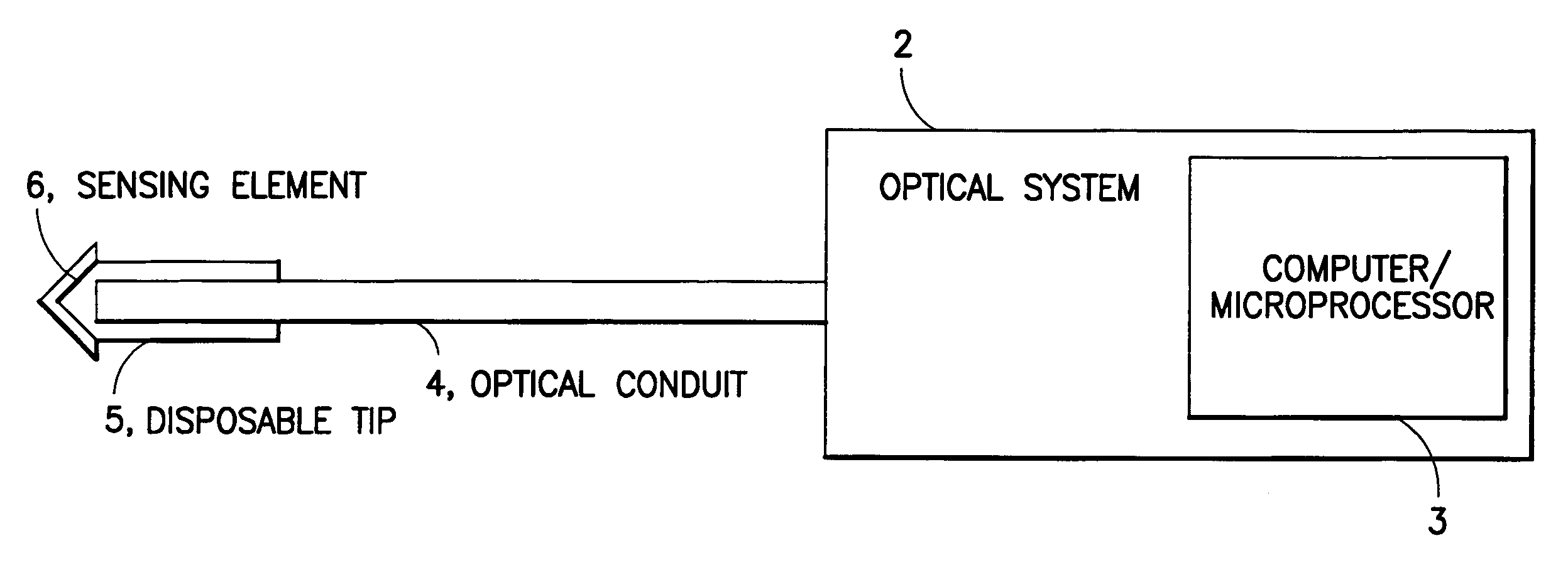
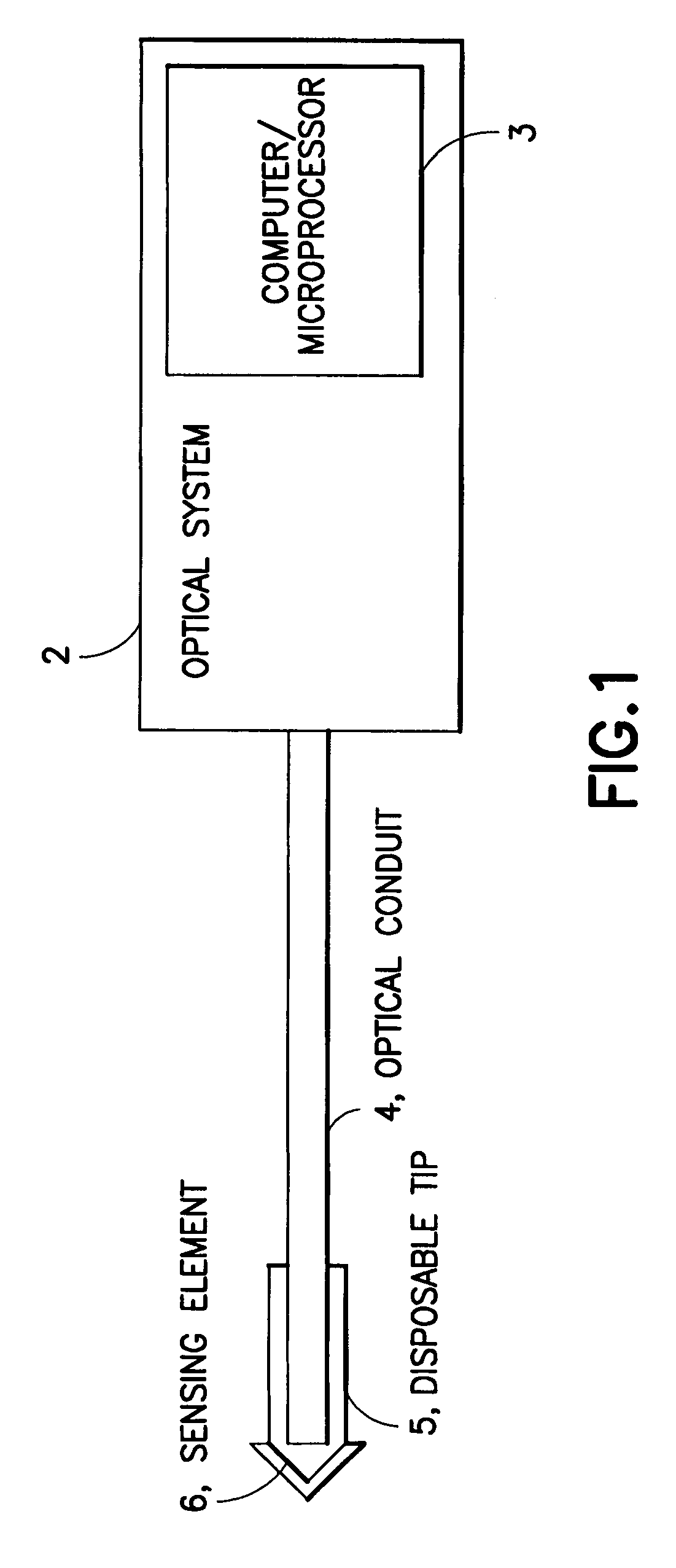
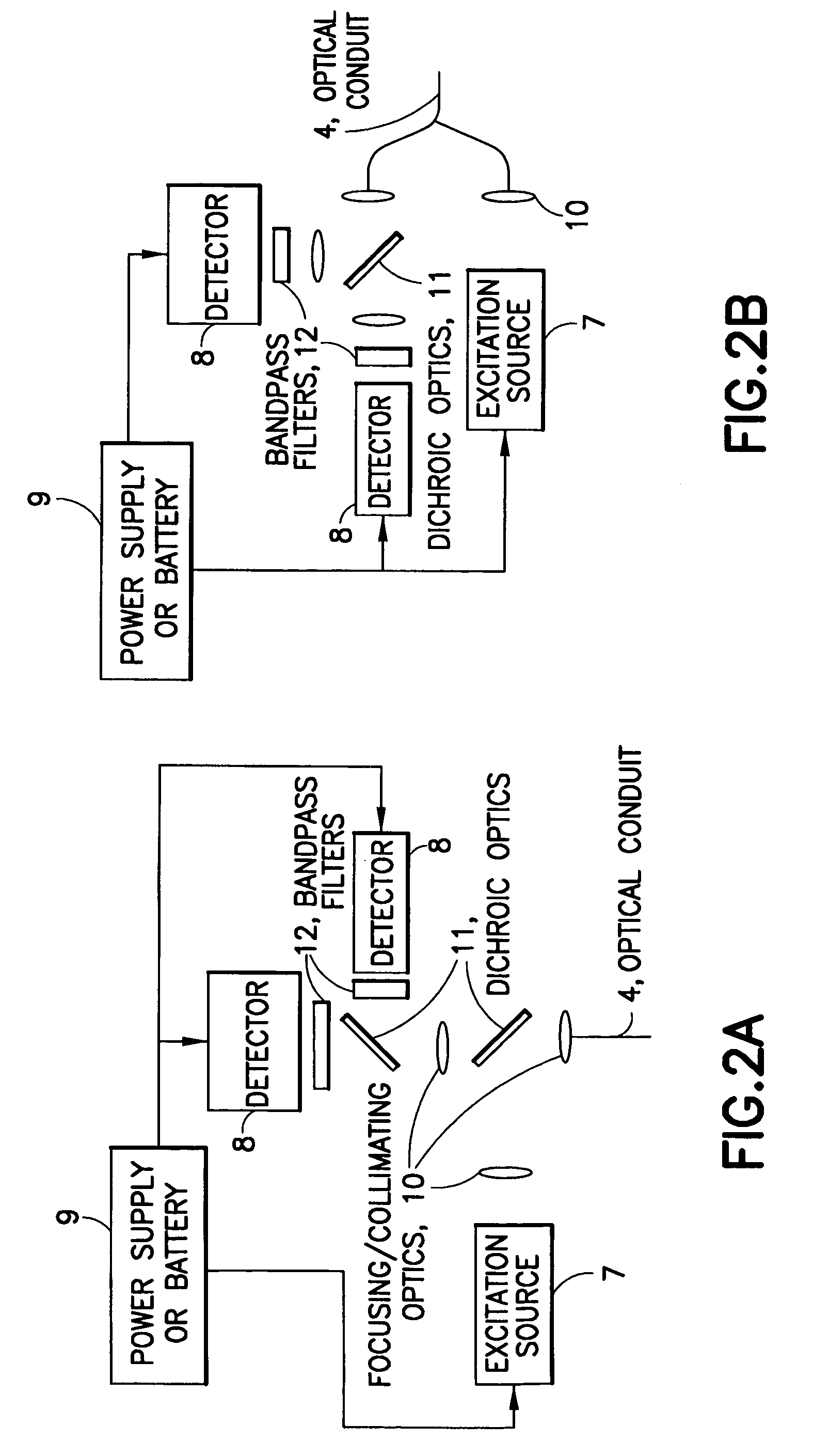
[0015]The optical system interrogates the sample either continuously or intermittently by coupling one or more interrogating wavelengths of light into the optical conduit. The one or more interrogating wavelengths then pass through the optical conduit and illuminate the sensing element. A change in analyte concentration results in a change of the wavelength, intensity, lifetime, energy transfer efficiency, and / or polarization of the luminescence of the reporter group, which is a part of the sensing element. The resulting changed luminescence signal passes back through the optical conduit to the optical system where it is detected, interpreted, and stored and / or displayed. In certain embodiments, the optical system comprises multiple excitation sources. One or more of these sources may be modulated to permit dynamic signal processing of the detected signal, thereby enhancing signal-to-noise and detection sensitivity. Modulation may also be used to reduce power consumption by the device or to increase the lifetime of the sensing element by minimizing undesirable phenomena such as photobleaching. The optical system can also include one or more electromagnetic energy detectors that can be used for detecting the luminescence signal from the reporter and optional reference groups as well as for internal referencing and / or calibration. The overall power consumption of the optical system is kept small to permit the device to be operated using battery power.
[0018]The sensing element is in optical proximity to the optical conduit. “Optical proximity” means that components of the device are close enough to one another such that an optical signal can be transmitted to or received from one object by another. The sensing element may be placed in optical proximity to the optical conduit in a number of ways, for example: attached directly to the optical conduit; attached to a connector that is attached to the optical conduit; attached to a polymer chain or a polymer matrix that is attached to the optical conduit; or attached to a polymer chain or a polymer matrix that is attached to a connector that is attached to the optical conduit. The sensing element may be permanently affixed to the optical conduit or replaceably attached such that the sensing element can be replaced conveniently and economically.
[0020]The sensing element (typically this refers to the binding protein with the associated reporter group and optional reference group) may be attached directly to the distal end of the optical conduit using for example covalent, ionic, or van der Waals interactions, dip coating, spin coating, plasma coating, or vacuum deposition. The sensing element may also be attached to a connector, which allows the sensing element to be readily detachable so that it becomes replaceable.
[0021]In another embodiment, the sensing element is attached to or immobilized in a polymeric matrix. As used herein, the term “matrix” may be any two dimensional or three-dimensional structure that is permeable to an analyte. The matrix may optionally prevent substantial interference from other biomolecules and may be substantially biocompatible. In one embodiment, the matrix allows the binding protein to retain some degree of conformational and / or orientational mobility. The matrix may consist of multiple layers, with an inner layer serving to retain the binding protein, and one or more outer layers to control the permeability and / or achieve biocompatibility. For example, the polymer matrix may be any one of those described in co-pending, commonly owned U.S. application Ser. No. 10 / 428,295, filed May 2, 2003. The entire contents of which are incorporated herein by reference. The immobilization may be accomplished either by covalently linking the sensing element to the polymer matrix or by physically entrapping the sensing element within the matrix. In the instance where the polymer matrix physically entraps the sensing element, the matrix pores are sized to retain the sensing element. In the embodiment where the sensing element is attached to the polymeric matrix, the sensing element is attached to the matrix using, for example, covalent or ionic linkage. The polymer matrix can be attached to the distal end of the optical conduit using adhesives, dip or spin coating, plasma coating, covalent, ionic, or van der Waals interactions, a mechanical connector or combinations thereof.
[0024]The device may further comprise a connector that may be used to attach the components of the device to one another. The connector may be, for example, any mechanical device, such as standard fiber optic connectors, luer locks, plastic, metal, or glass sleeves, or spring-loaded housings. For instance, the connector may be used to attach the sensing element to the optical conduit, or to attach the optical conduit to the optical system. The primary purpose of the connector is to provide a component that allows the other components to be readily detachable so that the component becomes replaceable.

Problems solved by technology

Currently, most diabetics use the “finger stick” method to monitor blood glucose level, and patient compliance is problematic due to pain caused by frequent finger sticks.
Progress with these approaches has been slow due to the requirement for frequent calibration, reproducible sample illumination, and variances in spectroscopic backgrounds between individuals.
These sensors are subject to inaccurate measurement due to oxygen deficiency and buildup of oxidation by-products.
Also, the electrochemical reaction itself generates a buildup of oxidation byproducts that may inhibit and degrade both the enzyme and its protective layer.
However, the potential of optical sensing for in vivo analyte detection has not yet been realized.
Although this is an optical biosensor, with the fluorescence signal level varying with changing oxygen levels, such a sensor is subject to the same problems as amperometric devices based on this same chemistry: oxygen deficiency and enzyme degradation.
Labeled concanavalin A and dextran have been used to create a competitive FRET assay; however, this system requires entrapment of both components, and the dynamic range of the assay is limited.
While a number of groups have developed GGBP mutations capable of responding to glucose in the physiological range, there have been no reports of a functional biosensor device based on binding protein technology that is suitable for in vivo or in vitro analyte monitoring.
Since binding proteins require orientational control and conformational freedom to enable effective use, many physical absorption and random or bulk covalent surface attachment or immobilization strategies as taught in the literature generally are either suboptimal or unsuccessful.
However, coupling of binding proteins to one end of an optical fiber is subject to the above-mentioned challenge of preserving conformational and / or orientational mobility of the protein.
In addition, fiber optic cabling is often impractical from a patient-use point of view since patients may need to remove or replace the sensor periodically.
Replacement of the entire fiber can be costly and inconvenient.
The optical system must also be sufficiently sensitive to detect signal from reporter dyes without relying on high power consumption and / or large-sized elements that would render the system unportable and hence unwearable.

Method used


Image

  • Fiber optic device for sensing analytes and method of making same
  • Fiber optic device for sensing analytes and method of making same
  • Fiber optic device for sensing analytes and method of making same

Examples


example 1

[0054]According to one embodiment of the present invention, glucose galactose binding protein (GGBP) was used with a triple mutation including a cysteine substituted for an glutamic acid at position 149, an arginine substituted for an alanine at position 213 and a serine substituted for leucine at position 238 (E149C / A213R / L238S). The protein was labeled at the 149 position with N,N′-dimethyl-N-(iodoacetyl)-N′-(7-nitrobenz-2-oxa-1,3-diazol-4-yl)ethylenediamine (IANBD amide)oxy. This mutated GGBP (E149C / A213R / L238S) is specific for glucose, and the reporter group undergoes a fluorescence intensity change in response to glucose binding.

[0055]A multicoated or multilayer matrix was prepared as follows. A core matrix was formed by mixing 1 part luminescent or dye-labeled binding protein (15 uM in PBS buffer, pH 7.4, prepared as described in PCT / US03 / 00203) with 2 to 4 parts 3 wt % alginate (v / v) in a scintillation vial and vortexing at slow speed. 3 mL of the resulting protein-alginate m...

example 2

[0058]In another embodiment, the binding protein was glucose galactose binding protein (GGBP), with a cysteine substituted for an glutamic acid at position 149, an arginine substituted for an alanine at position 213 and a serine substituted for leucine at position 238 (E149C / A213R / L238S). The protein was labeled at the 149 position with N,N′-dimethyl-N-(iodoacetyl)-N′-(7-nitrobenz-2-oxa-,3-diazol-4-yl)ethylenediamine (IANBD amide). The biosensor was prepared by inserting the tip of a 400 micron core diameter fiber into a short piece of catheter tubing, and allowing the catheter tubing to overhang the fiber tip by 0.1-1 mm. The fiber comprised a silica core, silica cladding, and polyimide buffer. The fiber diameter was 400 / 440 / 470 microns, where the slashes denote diameters measured from the core cladding buffer exteriors.

[0059]The immobilization matrix was a crosslinked alginate-based hydrogel, prepared by covalently crosslinking Pronova™ UP LVG alginate through the carboxyls with a...

example 3

[0063]In another embodiment of the present invention, a biosensor was formed by covalent attachment of a thin film to the surface of an optical fiber. The binding protein was glucose galactose binding protein (GGBP), with a cysteine substituted for a glutamic acid at position 149, an arginine substituted for an alanine at position 213 and a serine substituted for leucine at position 238 (E149C / A213R / L238S). The protein was labeled at the 149 position with N,N′-dimethyl-N-(iodoacetyl)-N′-(7-nitrobenz-2-oxa-1,3-diazol-3-yl)ethylenediamine (IANBD amide).

[0064]The biosensor was prepared by covalent attachment of an alginate matrix to the amine-functionalized surface of a silica fiber. The fiber comprised a silica core, silica cladding, and polyimide buffer. The fiber diameter was 400 / 440 / 470 microns, where the slashes denote diameters measured from the core / cladding / buffer exteriors.

[0065]The polyimide buffer was removed from the tip of the optical fiber by exposing the last few millime...


PUM

PropertyMeasurementUnit
lengthaaaaaaaaaa
distanceaaaaaaaaaa
heightaaaaaaaaaa

Abstract

A device for sensing analyte concentration, and in particular glucose concentration, in vivo or in vitro is disclosed. A sensing element is attached to the distal end of an optical conduit, and comprises at least one binding protein adapted to bind with at least one target analyte. The sensing element further comprises at least one reporter group that undergoes a luminescence change with changing analyte concentrations. Optionally, the optical conduit and sensing element may be housed within a cannulated bevel.

Description

CROSS-REFERENCE TO RELATED APPLICATIONS[0001]The present application is a continuation-in-part of co-pending U.S. patent application Ser. No. 10 / 721,797, filed Nov. 26, 2003, the content of which is incorporated by reference in its entirety.FIELD OF THE INVENTION[0002]The present invention relates to a device that can be used to monitor concentrations of physiologically relevant compounds.BACKGROUND OF THE INVENTION[0003]Monitoring in vivo concentrations of physiologically relevant compounds to improve diagnosis and treatment of various diseases and disorders is a desirable goal and would enhance the lives of many individuals. Advances in this area show particular promise in the area of facilitating adequate metabolic control in diabetics. Currently, most diabetics use the “finger stick” method to monitor blood glucose level, and patient compliance is problematic due to pain caused by frequent finger sticks. As a consequence, there have been efforts to develop non-invasive or minima...

Claims


Application Information

Patent Timeline
Patent Type & Authority Patents(United States)
IPC IPC(8): A61B5/145G01N21/77G01N33/542G01N33/543
CPCA61B5/14532A61B5/1455A61B5/1459A61B5/4839A61B5/6848G01N21/274G01N21/7703G01N33/542G01N33/54373A61B5/1495G01N2201/1211G01N2021/7786A61B2560/0223
Inventor ALARCON, JAVIERWEIDEMAIER, KRISTINAMIS, TERRY J.DENUZZIO, JOHN D.HERDMAN, CHRISTOPHER C.JACOBSON, ROSS W.PITNER, J. BRUCESHERMAN, DOUGLAS B.KEITH, STEVEN
Owner BECTON DICKINSON & CO
Why Eureka
Try Eureka