Supercharge Your Innovation With Domain-Expert AI Agents!

Endoscope processor, computer program product, endoscope system, and endoscope image playback apparatus

a technology of endoscope and processor, which is applied in the field of image signal processing, can solve the problems of difficult to distinguish and the outer appearance of a lesion that exists deep under the surface of an organ is not clearly defined from that of healthy tissue, and achieves the effect of easy to distinguish

Inactive Publication Date: 2010-10-26
HOYA CORP
View PDF8 Cites 1 Cited by
  • Summary
  • Abstract
  • Description
  • Claims
  • Application Information

AI Technical Summary

Benefits of technology

The processor generates an emphasized image that clearly differentiates lesions from normal tissue, improving the ability to identify lesions like polyps without relying on skilled observation, by magnifying differences in brightness and color within the image.

Problems solved by technology

However, the outer appearance of a lesion that exists deep under the surface of an organ is not clearly defined from that of healthy tissue.
Therefore, it is often difficult to distinguish such a lesion.

Method used


Image

  • Endoscope processor, computer program product, endoscope system, and endoscope image playback apparatus
  • Endoscope processor, computer program product, endoscope system, and endoscope image playback apparatus
  • Endoscope processor, computer program product, endoscope system, and endoscope image playback apparatus

Examples


Embodiment Construction

[0015]The present invention is described below with reference to the embodiments shown in the drawings.

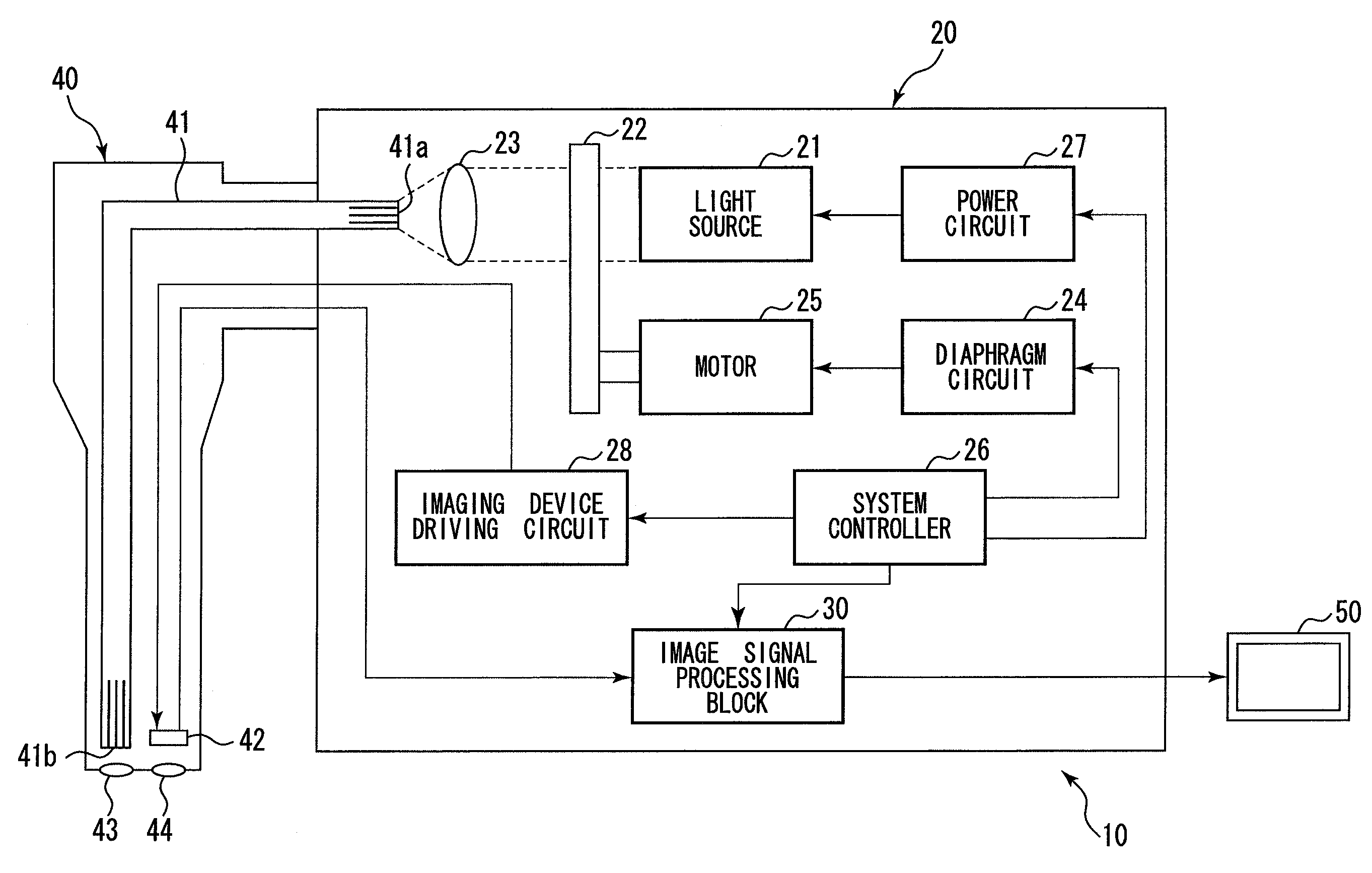
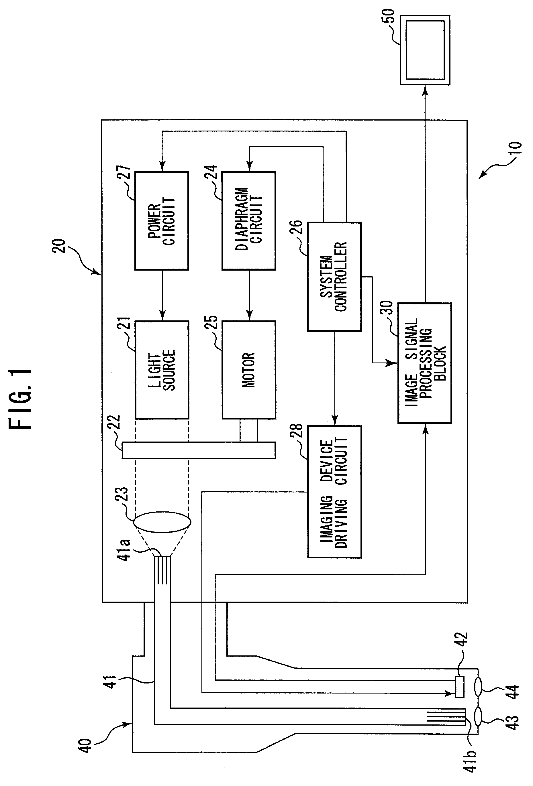
[0016]In FIG. 1, an endoscope system 10 comprises an endoscope processor 20, an electronic endoscope 40, and a monitor 50. The endoscope processor 20 is connected to the electronic endoscope 40 and the monitor 50 via connectors (not depicted).

[0017]The whole structure of the endoscope system 10 is briefly explained. A light source 21 for illuminating an object (not depicted) is housed in the endoscope processor 20. The light emitted from the light source 21 is irradiated onto an object (not depicted) via a light guide 41 housed in the electronic endoscope 40.

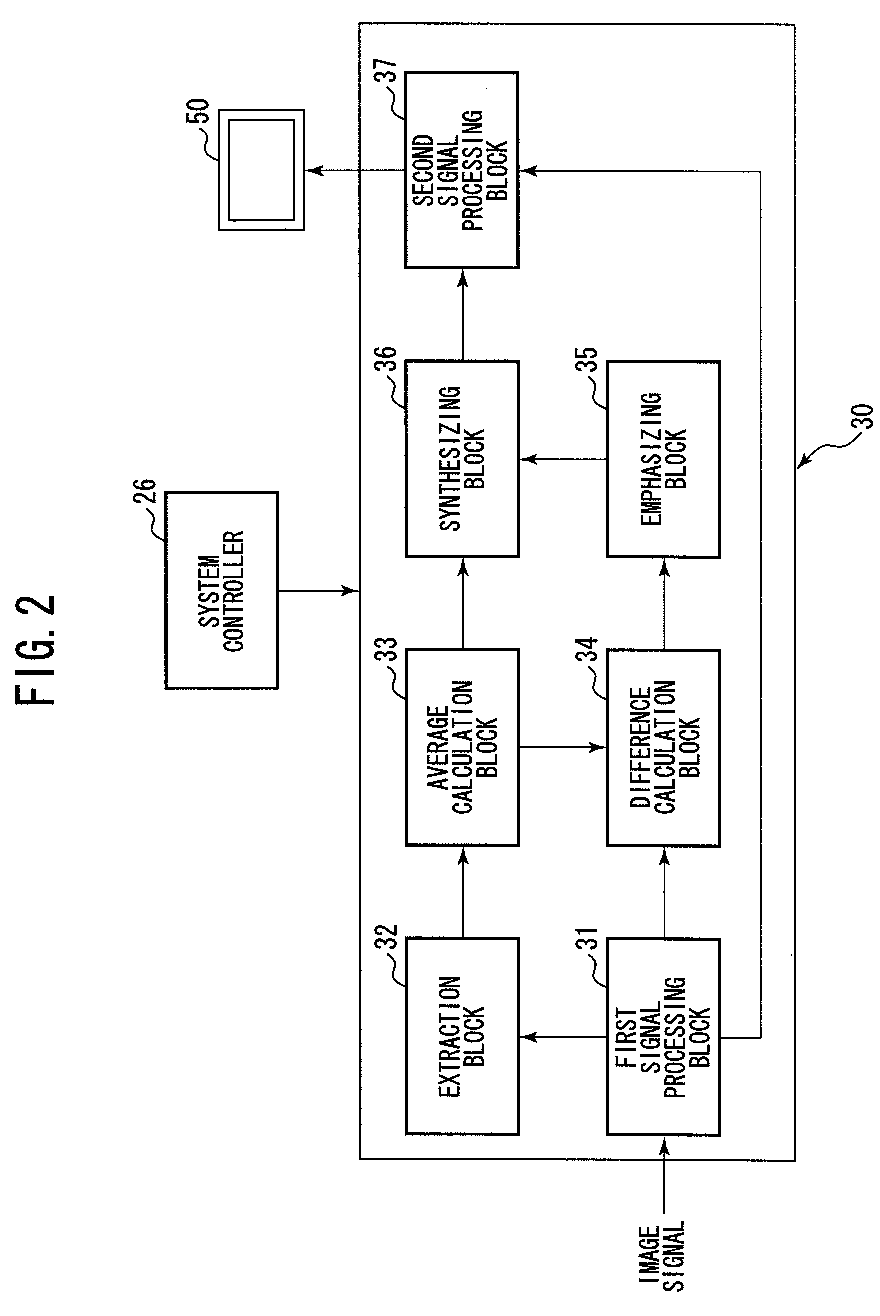
[0018]An imaging device 42, such as a CCD image sensor, is mounted in the electronic endoscope 40. The image of an object which is irradiated by the illumination light is captured by the imaging device 42. Subsequently, an image signal corresponding to the image of the captured object is generated by the imaging device 42. The ...


PUM


Abstract

An endoscope processor comprising a signal receiver, an average calculator, a difference calculator, an emphasizer, a synthesizer, and an output block is provided. The signal receiver receives an image signal generated by an imaging device. The image signal comprises a plurality of pixel signals. The average calculator calculates a signal average value. The difference calculator calculates a signal difference value. The emphasizer calculates an emphasized value by multiplying the signal difference value by a predetermined gain. The synthesizer generates an emphasized image signal, in which the pixel signal for each pixel is replaced with the sum of the emphasized value for each pixel and the signal average value.

Description

BACKGROUND OF THE INVENTION[0001]1. Field of the Invention[0002]The present invention relates to image signal processing that enables a user to discriminate a desired object from an entire image captured with an electronic endoscope.[0003]2. Description of the Related Art[0004]An electronic endoscope, having an imaging device at the end of an insertion tube, is used for medical examinations, industrial examinations, and so on. Light is irradiated from the end of the insertion tube to illuminate an object for observation. An optical image formed by the reflected light is captured by the imaging device, and the captured image is displayed on a monitor.[0005]A medical endoscope is used for identifying abnormal tissue or a lesion of internal organs. The appearance of abnormal tissue or a lesion is different from that of healthy tissue. Based on the user's observation, the abnormal tissue or lesion can be identified.[0006]However, the outer appearance of a lesion that exists deep under t...

Claims


Application Information

Patent Timeline
Patent Type & Authority Patents(United States)
IPC IPC(8): G06K9/00
CPCH04N5/262H04N7/183H04N2005/2255H04N23/555
Inventor IKEMOTO, YOSUKE
Owner HOYA CORP
Features
  • R&D
  • Intellectual Property
  • Life Sciences
  • Materials
  • Tech Scout
  • Unparalleled Data Quality
  • Higher Quality Content
  • 60% Fewer Hallucinations
Learn More