Automatic organ detection using machine learning and classification algorithms

a machine learning and organ detection technology, applied in the field of medical devices scanning methods and apparatuses, can solve the problems of confusing the interpretation of data received, difficult, if not impossible, complex interior of the human body, etc., and achieve the effect of accurate analysis

Active Publication Date: 2011-02-22
SIEMENS MEDICAL SOLUTIONS USA INC
View PDF11 Cites 3 Cited by
  • Summary
  • Abstract
  • Description
  • Claims
  • Application Information

AI Technical Summary

Benefits of technology

[0009]It is therefore an objective of the present invention to provide a method and an apparatus to automatically determine the presence of an organ whereby the similarity of anatomical features of an individual are accurately analyzed.
[0011]It is also an objective of the present invention to provide a method and apparatus for a single scanning system that will be sufficient to accurately scan multiple organs with sufficient clarity without the need for multiple systems for a patient.
[0012]It is a further objective of the present invention to provide a single scanning system that will allow for visual interpretation of results allowing doctors and / or technicians to evaluate scans.

Problems solved by technology

The complexity of the interior of the human body provides significant challenges for physicians and technicians who wish to evaluate the body for medical purposes.
As the human body is complex in its construction, evaluation of the body by medical imaging is difficult, if not impossible, by conventional imaging systems as these systems lack the ability to automatically differentiate between different organs in the body.
Current medical imaging systems, however, cannot automatically detect and represent a user specified organ within a body because some anatomical features of organs have physical similarities between tissues, thus confusing the interpretation of data received.
Such conventional systems, rather, are prone to significant error in their scanning results as organs blend together in scans as the difference in structure cannot be fully ascertained.
As a result, such conventional systems cannot be used for detailed scanning of a patient or in critical medical cases where specific details on anatomy are necessary.
These conventional systems lack sufficient resolution for detailed analysis by physicians and technicians resulting in the physician taking repetitive scans to try to obtain a best possible differentiated result.
Such conventional systems are expensive to manufacture and require specialized training for each individual operating the system.
Conventional systems, as they are specialized, do not have the substantial capability to be programmed for new tasks of automatically scanning individual organs not originally intended to be evaluated in original design and manufacturing of the equipment.
These inflexible systems cannot be updated with the latest information related to scanning technologies pertaining to other organs.
As a result, these systems have a limited service life and capability.

Method used


Image

  • Automatic organ detection using machine learning and classification algorithms
  • Automatic organ detection using machine learning and classification algorithms
  • Automatic organ detection using machine learning and classification algorithms

Examples


Embodiment Construction

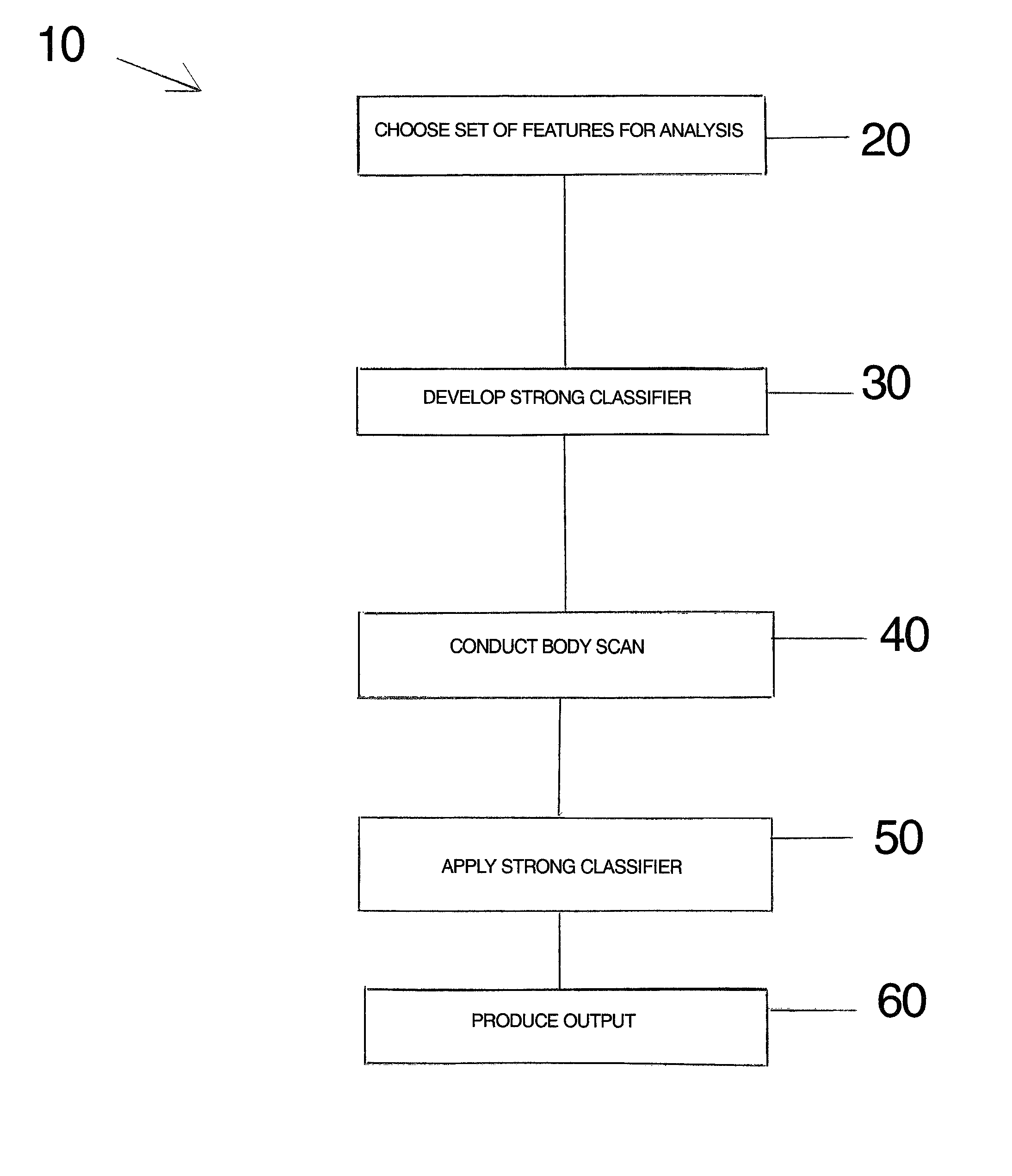
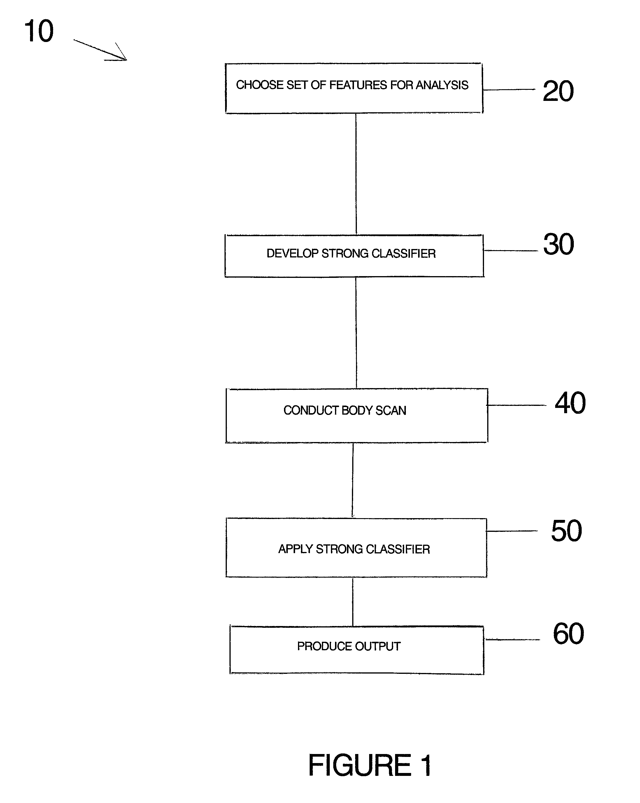
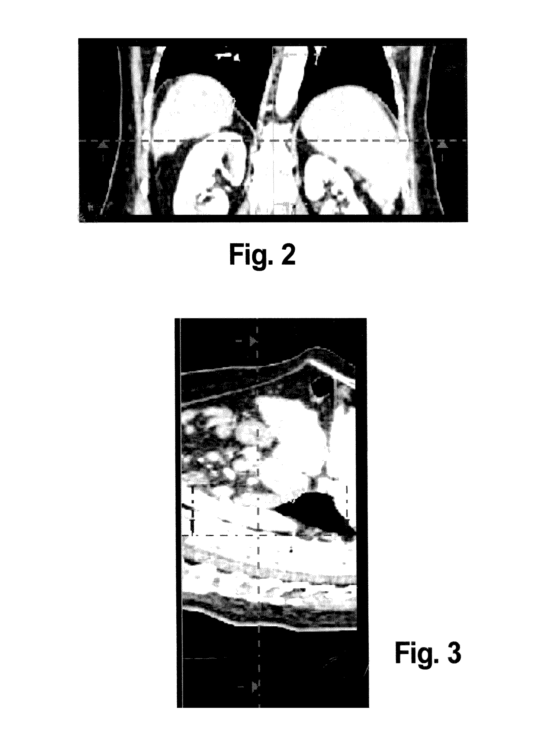
[0029]The present invention relates to automatic organ detection. More specifically, the invention provides for a method and apparatus for processing two dimensional and three dimensional medical images or medical image data sets such that an organ of interest is identified and represented to the physician and / or technician for evaluative purposes. An organ of interest is defined as an organ that is chosen to be evaluated by a medical professional. In the exemplary embodiments provided below, the organs of interest are inside the human body.

[0030]Although described as being applicable to human body organ identification, the system and method can also be used on animals other than humans or on sealed containers to identify objects contained therein. The method and apparatus of the present invention allows the organ of interest to be segregated from other tissues that may have similar construction or tissue features, that are not incorporated, however, into the organ of interest. The ...


PUM


Abstract

A method and apparatus of visually depicting an organ, having the steps of choosing a predefined set features for analysis, the predefined set of features having distinguishing weak learners for an algorithm, wherein the predefined set of features and the weak learners chosen distinguish features of the organ desired to be represented, developing a strong classifier for the algorithm for the organ desired to be represented based upon the weak learners for the organ, one of conducing a body scan to produce a body scan data set and obtaining a body scan data set of information for a patient, applying the strong classifier and the algorithm to the body scan data set to develop a result of a representation of the organ and outputting the result of the step of applying of the strong classifier and the algorithm to the body scan data set to represent the organ.

Description

CROSS-REFERENCE TO RELATED APPLICATIONS[0001]This is a United States non-provisional application of U.S. provisional patent application Ser. No. 60 / 808,011 filed May 23, 2006 by Charles Florin and James P. Williams, the entirety of which application is incorporated by reference herein.FIELD OF THE INVENTION[0002]The present invention relates to scanning methods and apparatus for medical devices. More specifically, the present invention relates to scanning methods and apparatus used to analyze two dimensional and three dimensional medical image scans and data resulting from such image scans to automatically determine and represent user specified organs for medical evaluation.BACKGROUND INFORMATION[0003]The complexity of the interior of the human body provides significant challenges for physicians and technicians who wish to evaluate the body for medical purposes. As the human body is complex in its construction, evaluation of the body by medical imaging is difficult, if not impossibl...

Claims


Application Information

Patent Timeline
Patent Type & Authority Patents(United States)
IPC IPC(8): G06K9/00G06V10/774
CPCG06K9/6256G06T7/70G06K2209/05G06T2207/30092G06T2207/10072G06V2201/03G06V10/774G06F18/214
Inventor FLORIN, CHARLESWILLIAMS, JAMES
Owner SIEMENS MEDICAL SOLUTIONS USA INC
Why Eureka
Try Eureka