Diffusion-based magnetic resonance methods for characterizing bone structure

a magnetic resonance and bone structure technology, applied in the field of improved methods for characterizing materials, can solve the problems of limited clinical value, inability to fully account for the full variation in bone strength, and inability to fully predict fracture risk by methods relying on bone density measurements

Active Publication Date: 2011-02-22
SCHLUMBERGER TECH CORP
View PDF6 Cites 15 Cited by
  • Summary
  • Abstract
  • Description
  • Claims
  • Application Information

AI Technical Summary

Benefits of technology

[0014]According to one embodiment, an NMR technique that is well-established in inorganic porous media may be used to obtain statistical properties of the trabecular structure. This technique, referred to as decay from diffusion in an internal field, or DDIF, can obtain pore structure characteristics at a resolution of about 1 μm. As discussed below, DDIF data on bovine bone samples shows a clear correlation with bone strength. Furthermore, the DDIF data demonstrates a strong correlation with the projected surface-to-volume ratio and exhibits two regimes at the high and low bone strength. These two regimes may be interpreted as two types of bone-weakening behaviors, one by the loss of bone density at the low strength and the other by the change of bone architecture at the high strength. Correlation between the DDIF results and structural properties of bone samples observed in microscopic images suggest that the DDIF data may be used to characterize bone structure. Therefore, in some embodiments, DDIF measurements may provide a reliable indication of bone strength and may thus be used to assess bones, without the need for high resolution imaging.

Problems solved by technology

However, density alone does not account for the full bone strength variation because the internal bone structure, apart from the bone density, contributes significantly to the mechanical strength of the bone.
As a result, methods that rely on bone density measurements do not entirely predict fracture risk, and their clinical value is therefore limited.
Although high resolution magnetic resonance imaging techniques may provide three-dimensional imaging of the trabecular architecture, which may be useful for osteoporosis diagnosis, their clinical application may be difficult.
Specifically, it is difficult to improve the resolution of such imaging techniques far beyond their current levels (about 100 μm) in clinical implementation, primarily due to clinically allowed X-ray radiation dosage and the MRI scan time.
Therefore, these methods also may have limited clinical use.

Method used


Image

  • Diffusion-based magnetic resonance methods for characterizing bone structure
  • Diffusion-based magnetic resonance methods for characterizing bone structure
  • Diffusion-based magnetic resonance methods for characterizing bone structure

Examples


Embodiment Construction

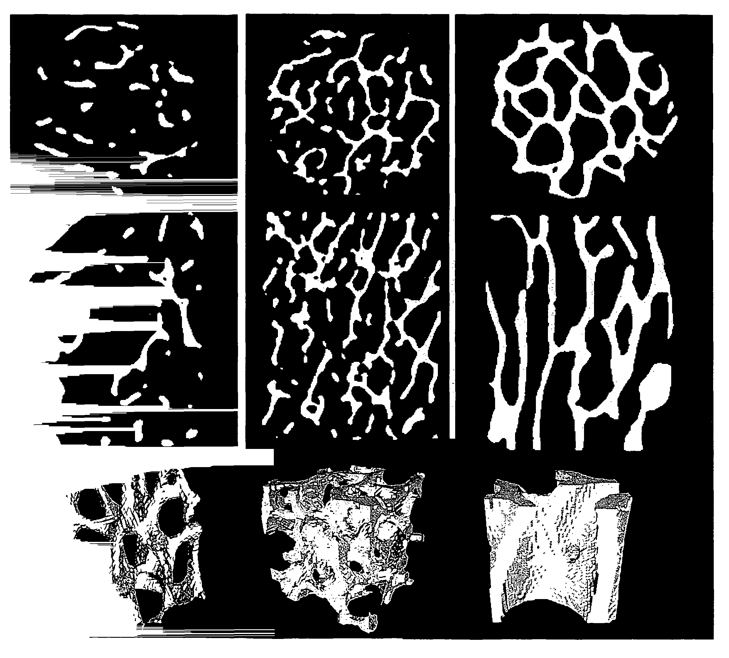
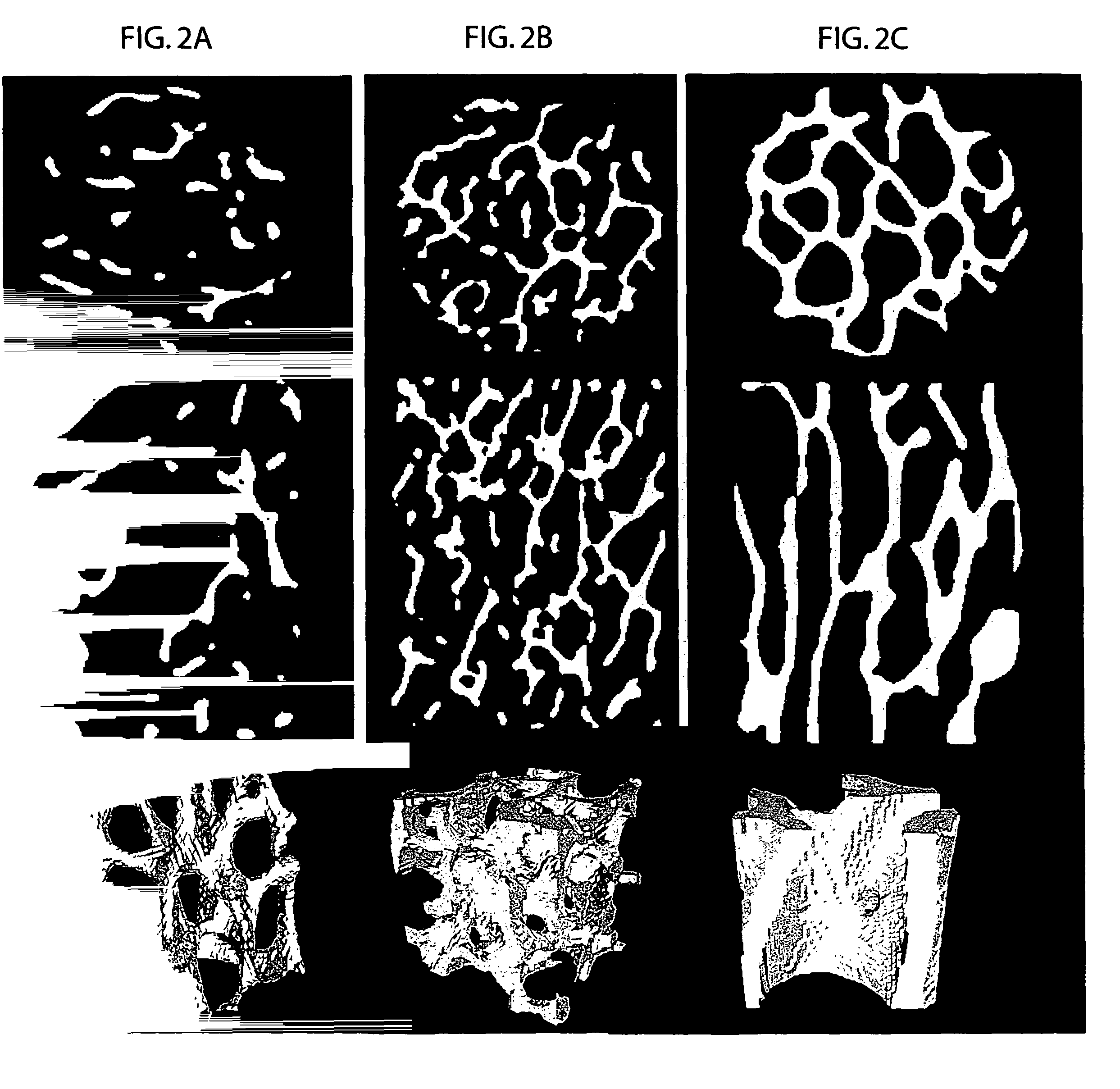
[0046]There are two types of bone in the human body: cortical bone (a dense, compact bone existing in the middle of a long bone) and trabecular or cancellous bone (a more porous type of bone found generally near major joints and in the spine). Trabecular bone is comprised of a complex three-dimensional network of rods and plates. Most of the load-bearing capability of the skeleton is attributed to the trabecular bone region. The development or deterioration of trabecular bone structure is significantly affected by the mechanical forces it experiences.

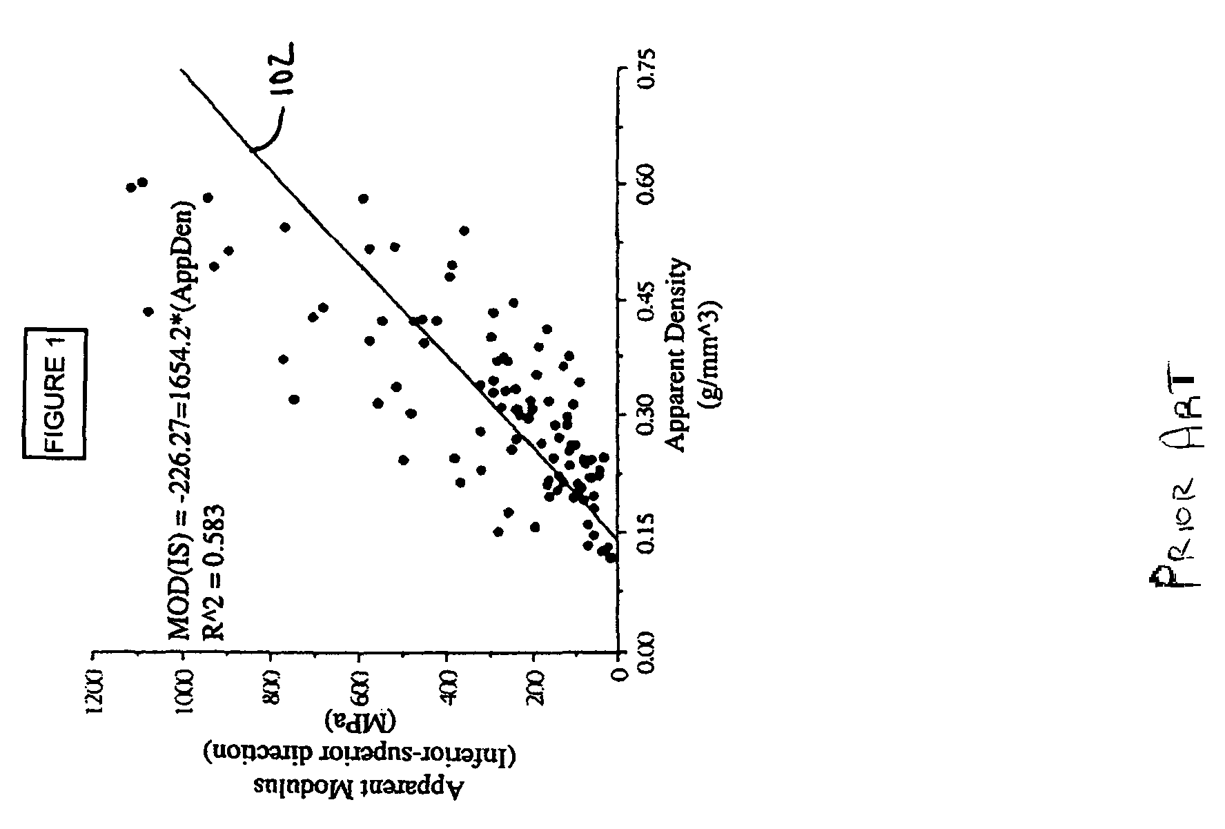
[0047]The mechanical strength of a bone specimen, and thus the risk of its fracturing, depends on several factors. One very important factor in bone strength is the amount of bone material, which is related to various parameters such as bone volume fraction (BVF), porosity, or the most common clinical parameter, bone mineral density (BMD). FIG. 1 illustrates a graph of a relationship between bone strength and BMD. Apparent modulus (in M...


PUM


Abstract

A method of in vitro or in vivo nuclear magnetic resonance and / or magnetic resonance imaging, to determine bone properties by measuring the effects of molecular diffusion inside the bone specimen to derive parameters that are related to the structure of the trabecular bones. The method is a non-invasive probe that provides topological information on trabecular bone without requiring a full high-resolution image of its structure, and is compatible with clinical use.

Description

CROSS-REFERENCE TO RELATED APPLICATIONS[0001]The present application claims the benefit under 35 U.S.C. §119(e) of the earlier filing date of U.S. Provisional Application No. 60 / 761,517, entitled “DIFFUSION-BASED MAGNETIC RESONANCE METHODS FOR BONE STRUCTURE AND EVOLUTION” and filed on Jan. 24, 2006, which is herein incorporated by reference in its entirety.GOVERNMENT INTERESTS[0002]The present invention was developed at least in part with funds from NIH Grant No. 1R21 EB003869-01. The United States Government may have certain rights in the invention.BACKGROUND[0003]1. Field of Invention[0004]The present invention is directed to improved methods for characterizing materials such as trabecular bone.[0005]2. Discussion of Related Art[0006]A major component of the human musculoskeletal system is bone, which supports our body weight, facilitates motion, and plays critical roles in mineral homeostasis and production of blood cells. Osteoporosis is a disorder of the skeleton in which bone...

Claims


Application Information

Patent Timeline
Patent Type & Authority Patents(United States)
IPC IPC(8): A61B5/04
CPCA61B5/055A61B5/4504G01R33/56341G01R33/5617A61B5/4509
Inventor SONG, YI-QIAOSIGMUND, ERIC E.CHO, HYUNGJOON
Owner SCHLUMBERGER TECH CORP
Why Eureka
Try Eureka