Stain-based optimized compression of digital pathology slides

a digital pathology and optimized compression technology, applied in the field of digital imaging, can solve the problems of difficult to compress slide images well, delay in initial diagnosis and subsequent second opinion, and significant visual content in digital pathology slide images

Active Publication Date: 2011-12-13
GENERAL ELECTRIC CO
View PDF7 Cites 16 Cited by
  • Summary
  • Abstract
  • Description
  • Claims
  • Application Information

AI Technical Summary

Problems solved by technology

Due to this manual process, the initial diagnosis and subsequent second opinions may be delayed as the correct slides must be physically delivered to the proper pathologist.
A problem arises in that digital pathology slide images contain significant visual content.
This makes the slide images difficult to compress well while maintaining high visual quality at the same time.

Method used


Image

  • Stain-based optimized compression of digital pathology slides
  • Stain-based optimized compression of digital pathology slides
  • Stain-based optimized compression of digital pathology slides

Examples


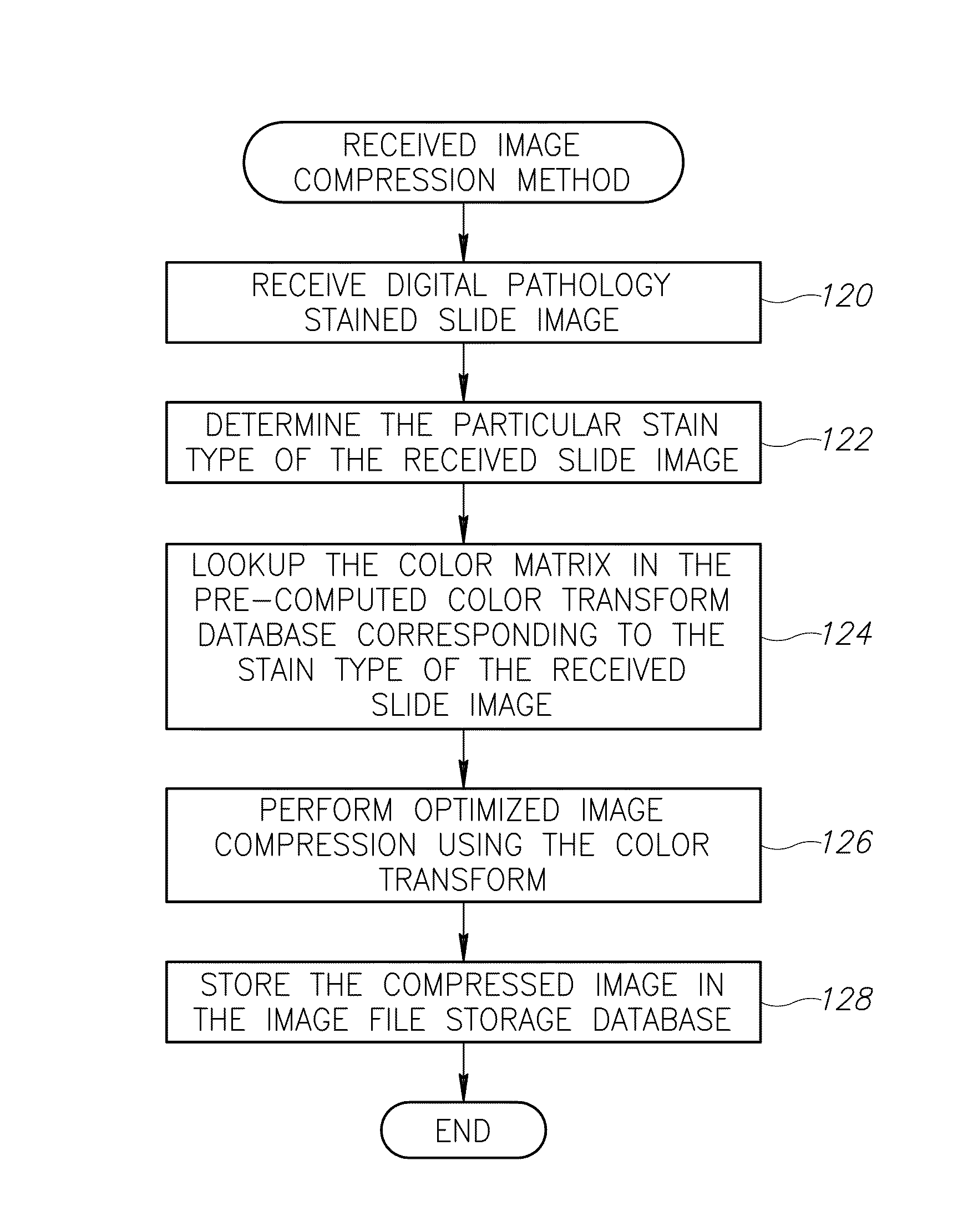
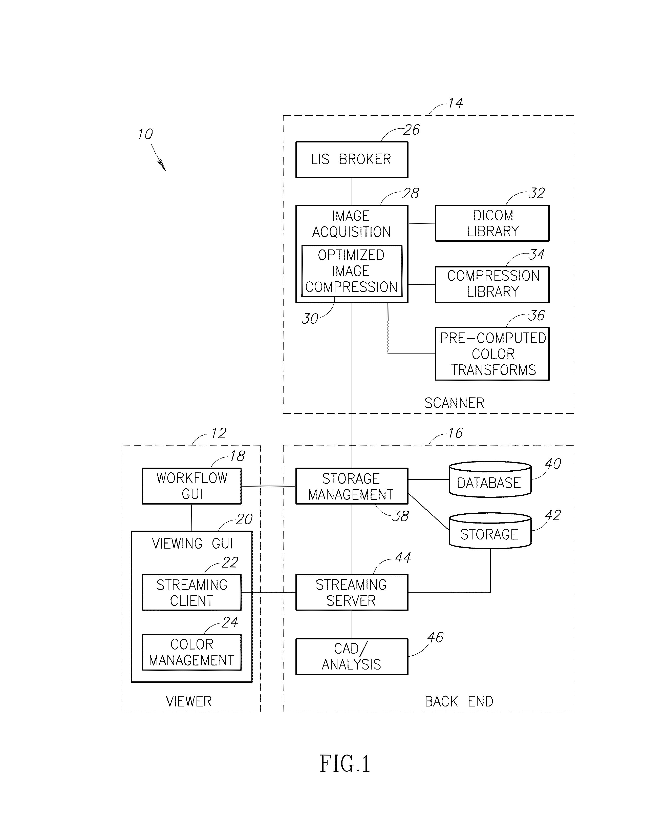
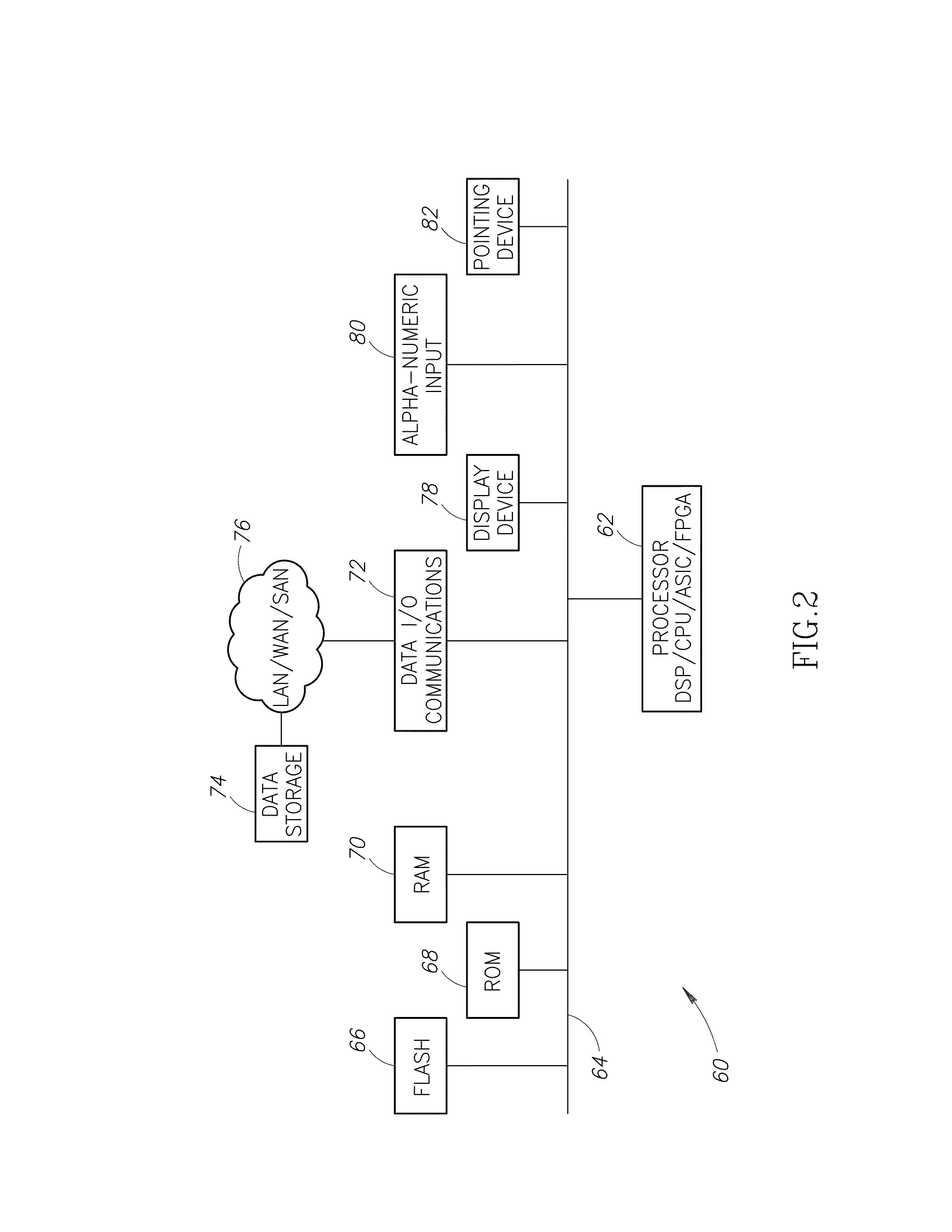
Embodiment Construction

Notation Used Throughout

[0031]The following notation is used throughout this document:

[0032]

TermDefinitionASCIIAmerican Standard Code for Information InterchangeASICApplication Specific Integrated CircuitCADComputer Aided DesignCDROMCompact Disc Read Only MemoryCPUCentral Processing UnitDCTDiscrete Cosine TransformDICOMDigital Imaging and Communications in MedicineDNADeoxyribonucleic AcidDSPDigital Signal ProcessorDVDDigital Versatile DiscDWTDiscrete Wavelet TransformEPROMErasable Programmable Read-Only MemoryFIRFinite Impulse ResponseFPGAField Programmable Gate ArrayFTPFile Transfer ProtocolFWTForward Wavelet TransformGUIGraphical User InterfaceHTTPHyper-Text Transport ProtocolI / FInterfaceI / OInput / OutputIPInternet ProtocolIWTInverse Subband / Wavelet TransformJPEGJoint Photographic Experts GroupKLTKarhunen-Loève TransformLANLocal Area NetworkLISLaboratory Information SystemMACMedia Access ControlNICNetwork Interface CardPCPersonal ComputerPCAPrinciple Component AnalysisPSNRPeak Signa...


PUM


Abstract

A novel and useful method and system of optimized image compression of digital pathology slide images. The optimized image compression mechanism exploits the special color properties of the stained tissue represented by the digital pathology slides and provides an image compression algorithm having improved rate-distortion performance. Optimized color transforms are pre-computed using training sets of pathology slide image scan data for each stain type. The optimized color transforms are used to compress input slide image scans resulting in more efficient image streaming enabling users to review extremely large digital slide scans from any connected location, such as in a hospital, satellite center, home or on a mobile telephone.

Description

FIELD OF THE INVENTION[0001]The subject matter disclosed herein relates to the field of digital imaging, and more particularly relates to a mechanism for stain-based optimized compression of digital pathology slide scans.BACKGROUND OF THE INVENTION[0002]Pathology is the study and diagnosis of diseases by examining body tissues, typically under magnification. Currently, pathologists manually review stained tissue samples on glass slides under an optical microscope to render a diagnosis. Tissue samples are typically prepared with stains by a specialist called a histotechnician. Today, pathologists use an optical microscope to look at slides of tissue samples. This process has not changed much in over 100 years. Due to this manual process, the initial diagnosis and subsequent second opinions may be delayed as the correct slides must be physically delivered to the proper pathologist.[0003]Digitizing the tissue sample images enables easier and faster evaluation without the organization, ...

Claims


Application Information

Patent Timeline
Patent Type & Authority Patents(United States)
IPC IPC(8): G06K9/00G06K9/36
CPCH04N19/63H04N19/60H04N19/85H04N19/186H04N19/136
Inventor DEKEL, SHAIGOOGLE, LEON
Owner GENERAL ELECTRIC CO
Why Eureka
Try Eureka