
System and method for magnetic resonance brain scan planning

a brain scan and magnetic resonance technology, applied in the field of magnetic resonance brain scan planning, can solve problems such as patient discomfort or unnerving, inconsistent scanning execution, and problems with diagnosis

Inactive Publication Date: 2011-12-20
SIEMENS MEDICAL SOLUTIONS USA INC
View PDF12 Cites 1 Cited by
  • Summary
  • Abstract
  • Description
  • Claims
  • Application Information

AI Technical Summary

Problems solved by technology

Even when the same anatomic landmarks are used, the scanning can be executed inconsistently, due to inter- or intra-operator variation.
This can cause variations in the position and orientation of various MR brain scans, which can lead to problems with diagnosis when using MR brain images generated from MR brain scans.
However, such methods require a 3D scout scan for registration.
This may be uncomfortable or unnerving for the patient.
Since an extra non-diagnostic 3D scan is time-consuming, methods requiring a 3D scout image may be undesirable in clinical MR scanning applications.

Method used


Image

  • System and method for magnetic resonance brain scan planning
  • System and method for magnetic resonance brain scan planning
  • System and method for magnetic resonance brain scan planning

Examples


Embodiment Construction

[0023]The present invention is directed to a method for automatic magnetic resonance (MR) brain scan planning. Embodiments of the present invention are described herein to give a visual understanding of the segmentation method. A digital image is often composed of digital representations of one or more objects (or shapes). The digital representation of an object is often described herein in terms of identifying and manipulating the objects. Such manipulations are virtual manipulations accomplished in the memory or other circuitry / hardware of a computer system. Accordingly, is to be understood that embodiments of the present invention may be performed within a computer system using data stored within the computer system. For example, according to various embodiments of the present invention, electronic data representing a target MR image, as well as electronic data representing training images for an active shape model are manipulated within a computer system.

[0024]According to an em...


PUM


Abstract

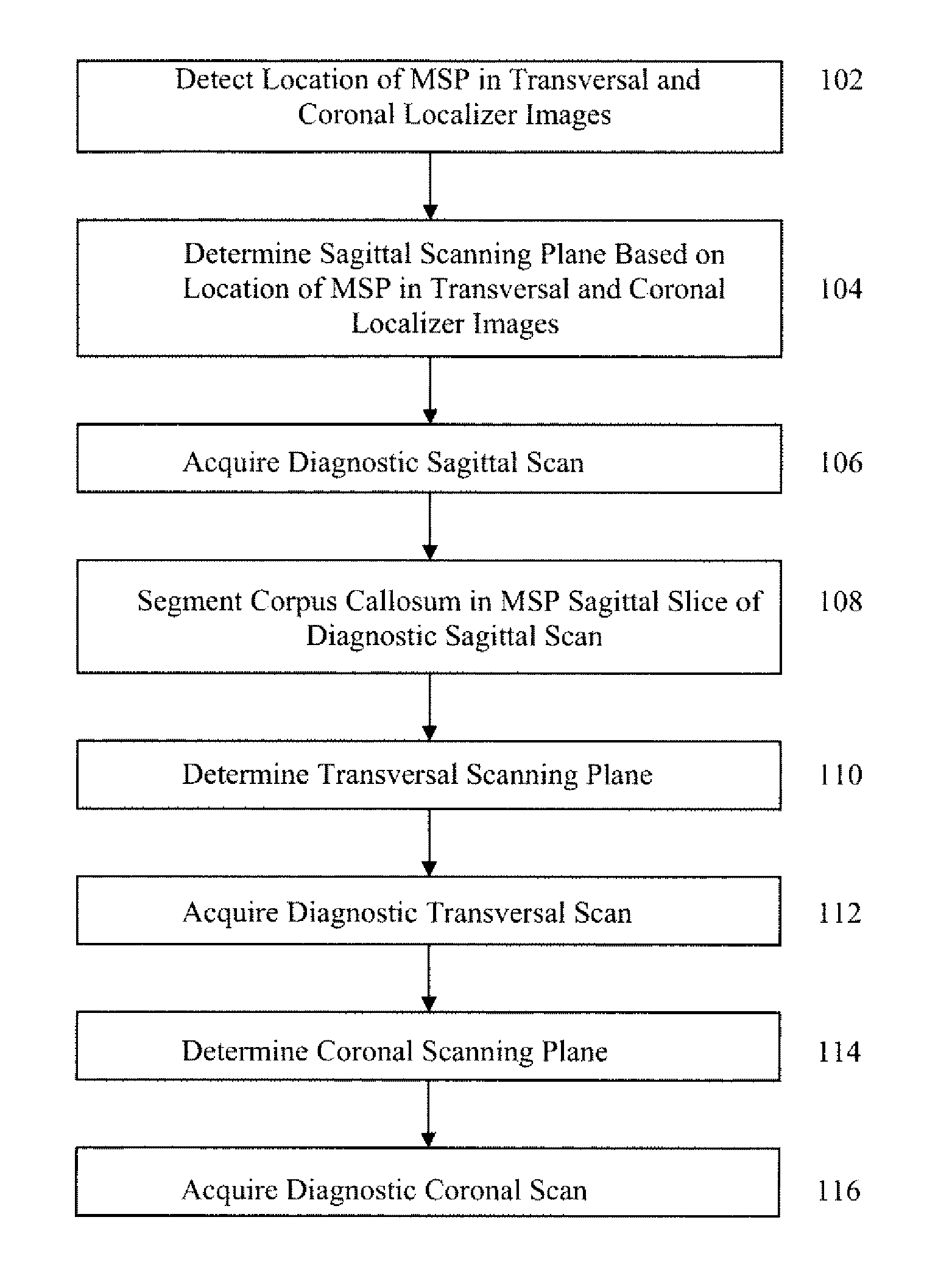
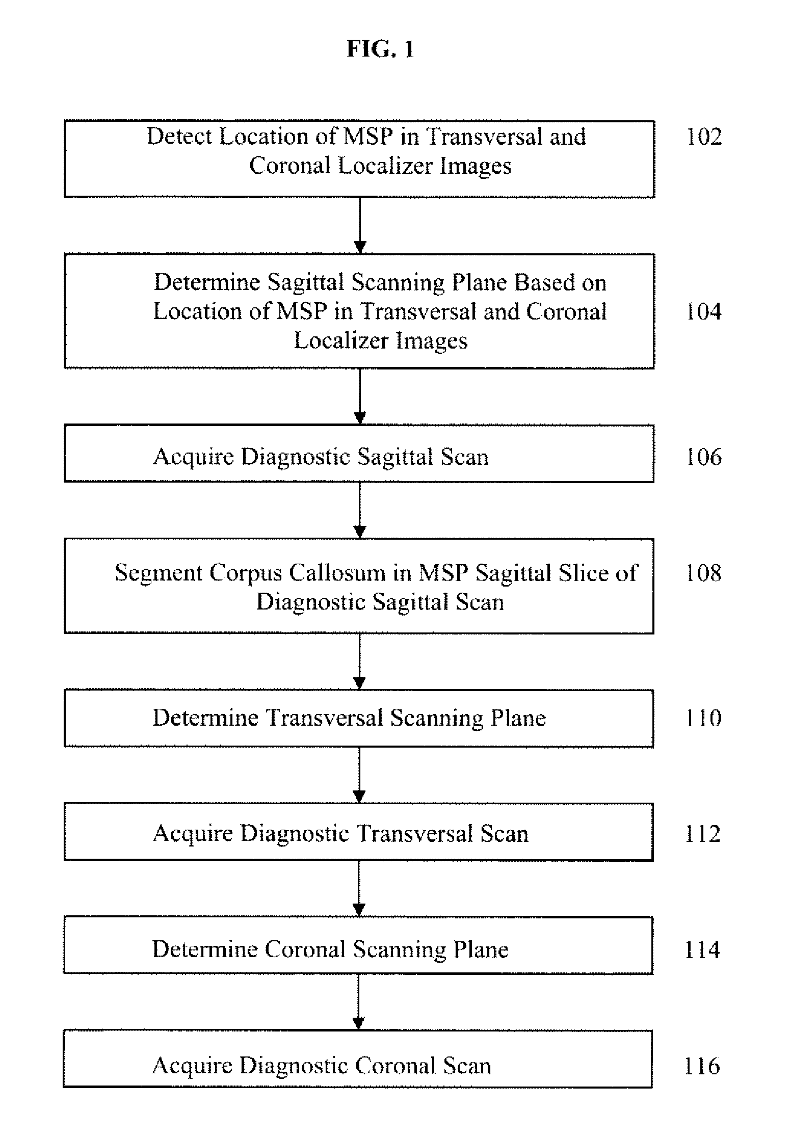
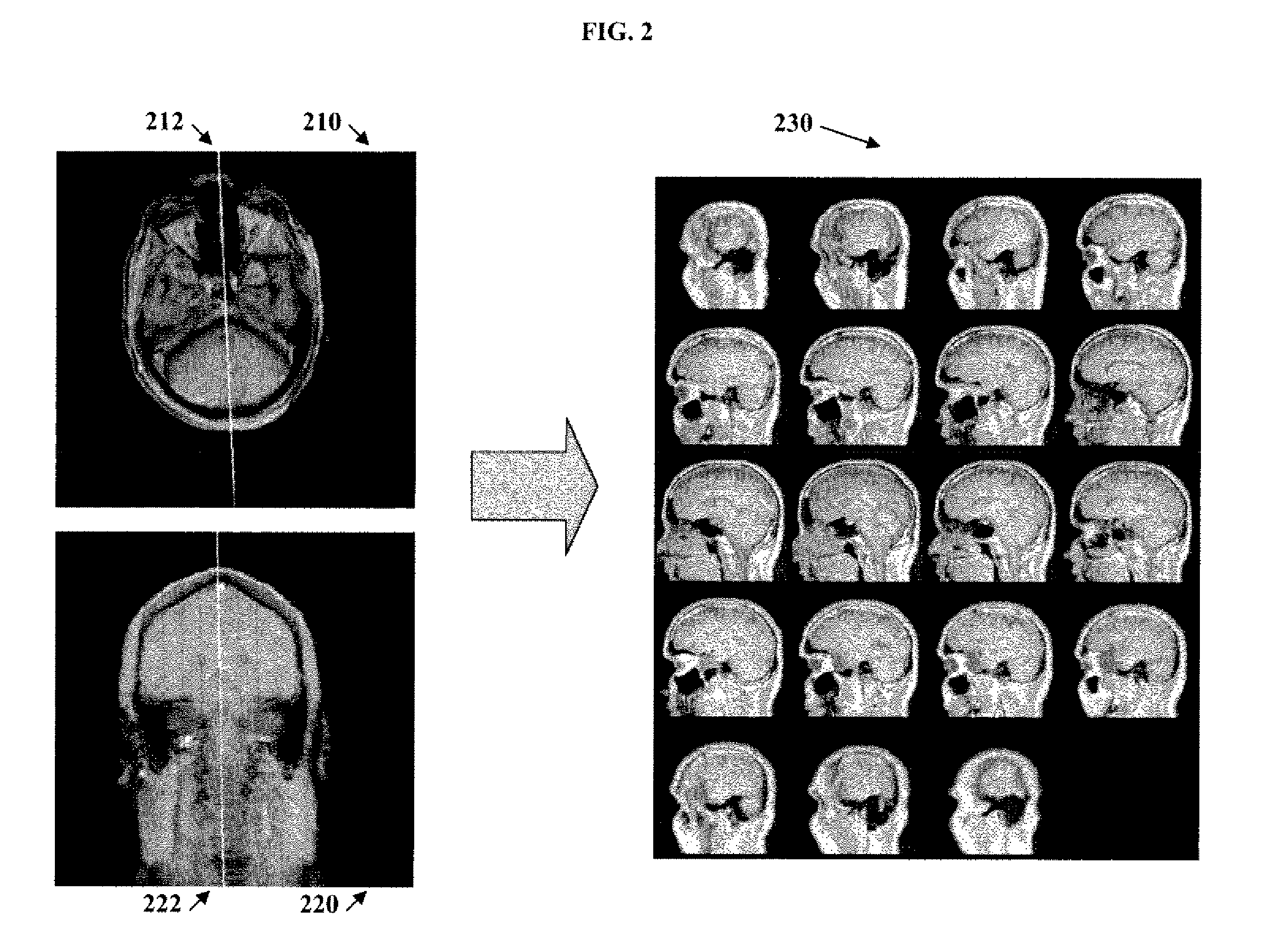
A method and system for automatic MR brain scan planning is disclosed. The method utilizes a set of 2D orthogonal localizer images to determine scanning planes for 3D diagnostic MR scans. A location of the mid-sagittal plane (MSP) is detected in each of a transversal localizer image and a coronal localizer image. A sagittal scanning plane is determined based on the location of the MSP in the transversal and coronal localizer images. A diagnostic sagittal MR scan is then acquired based on the sagittal scanning plane. The corpus callosum CC is segmented in a sagittal MR image slice resulting from the diagnostic sagittal MR scan. A transversal scanning plane can be determined based on a location of the CC in the sagittal MR image slice and the location of the MSP in the coronal localizer image, and a coronal scanning plane can be determined based on the location of the CC in the sagittal MR image slice and the location of the MSP in the transversal localizer image.

Description

[0001]This application claims the benefit of U.S. Provisional Application No. 60 / 826,251, filed Sep. 20, 2006, the disclosure of which is herein incorporated by reference.BACKGROUND OF THE INVENTION[0002]The present invention relates to magnetic resonance brain scanning, and more particularly, to automatic magnetic resonance brain scan planning.[0003]Magnetic Resonance (MR) is a well known technique for imaging internal organs of a patient. MR scanners are typically capable of generating cross-sectional images in any plane of the body, including oblique planes. Accordingly, the MR volume data resulting from MR scans can have many possible orientations. In MR brain scanning, planning is needed in order to determine the orientation and position of the scanning volumes. In a typical MR brain scan process, an MR operator first acquires scout / localizer images by a short time sequence. The operator then manually plans a 3D diagnostic, which typically take a longer time, scan from the loca...

Claims


Application Information

Patent Timeline
Patent Type & Authority Patents(United States)
IPC IPC(8): A61B5/00
CPCA61B5/055A61B5/6814G01R33/4833A61B5/0037
Inventor ZHANG, LINOVAK, CAROLCHEN, HONGXU, QING
Owner SIEMENS MEDICAL SOLUTIONS USA INC
Learn More