Method and device for the separate three-dimensional representation of arteries and veins in an examination object and device

a three-dimensional representation and examination object technology, applied in the field of three-dimensional representation of arteries and veins in an examination object and device, can solve the problems that conventional 3d angiography fails to ensure adequate separation between arterial and venous vascular systems, and achieves the effect of fast and simpl

Active Publication Date: 2012-02-28
SIEMENS HEALTHCARE GMBH
View PDF8 Cites 8 Cited by
  • Summary
  • Abstract
  • Description
  • Claims
  • Application Information

AI Technical Summary

Benefits of technology

[0007]An object underlying the present invention is thus to provide a particularly fast and simple method, which is suitable for generating three-dimensional data records of vessels, which represent either only arteries or only veins, depending on requirements.
[0010]Because it is possible to limit the number of injections of contrast agent to one, the load on the patient due to contrast agent is relatively low. The method can also be coordinated temporally in a simple manner in that the arterial phase and venous phase are each allocated to one filler pass. The patient is also subjected to a relatively low x-ray dose, as the number of x-ray recordings remains relatively small. The short recording time means that patients have to hold their breath and remain motionless for a shorter time and the treatment is therefore less unpleasant. The smaller number of projection recordings per filler pass is then offset by a weighting, which means that the quality of the three-dimensionally reconstructed volumes of the veins and arteries is at least comparable to those of double the number of recordings.
[0038]According to a further embodiment of the invention the tomography x-ray device is formed by a C-arm x-ray device. Such C-arm x-ray devices are able to produce projection images as they circle quickly about a patient, having the advantage that with their relatively small form compared with a CT device, they still ensure easy access to the patient. C-arms supported on industrial robots in particular are extremely flexible.
[0039]Depending on the tomography x-ray device used and its movement pattern, in a preferred embodiment of the invention the mask pass, the first filler pass and the second filler pass are implemented in the same scan direction. In the case of tomography x-ray devices, which can change the scan easily, in other words the C-arm x-ray device for example, the mask pass, the first filler pass and the second filler pass are implemented in an alternating manner in different scan directions. It is possible to save time in this manner, as the x-ray device does not have to be returned to its original position to start the first filler pass and the second filler pass.

Problems solved by technology

Conventional 3D angiography nevertheless fails to ensure adequately clear separation between the arterial and venous vascular systems, by virtue of the recording times and the dynamics of contrast agent propagation.

Method used


Image

  • Method and device for the separate three-dimensional representation of arteries and veins in an examination object and device
  • Method and device for the separate three-dimensional representation of arteries and veins in an examination object and device
  • Method and device for the separate three-dimensional representation of arteries and veins in an examination object and device

Examples


Embodiment Construction

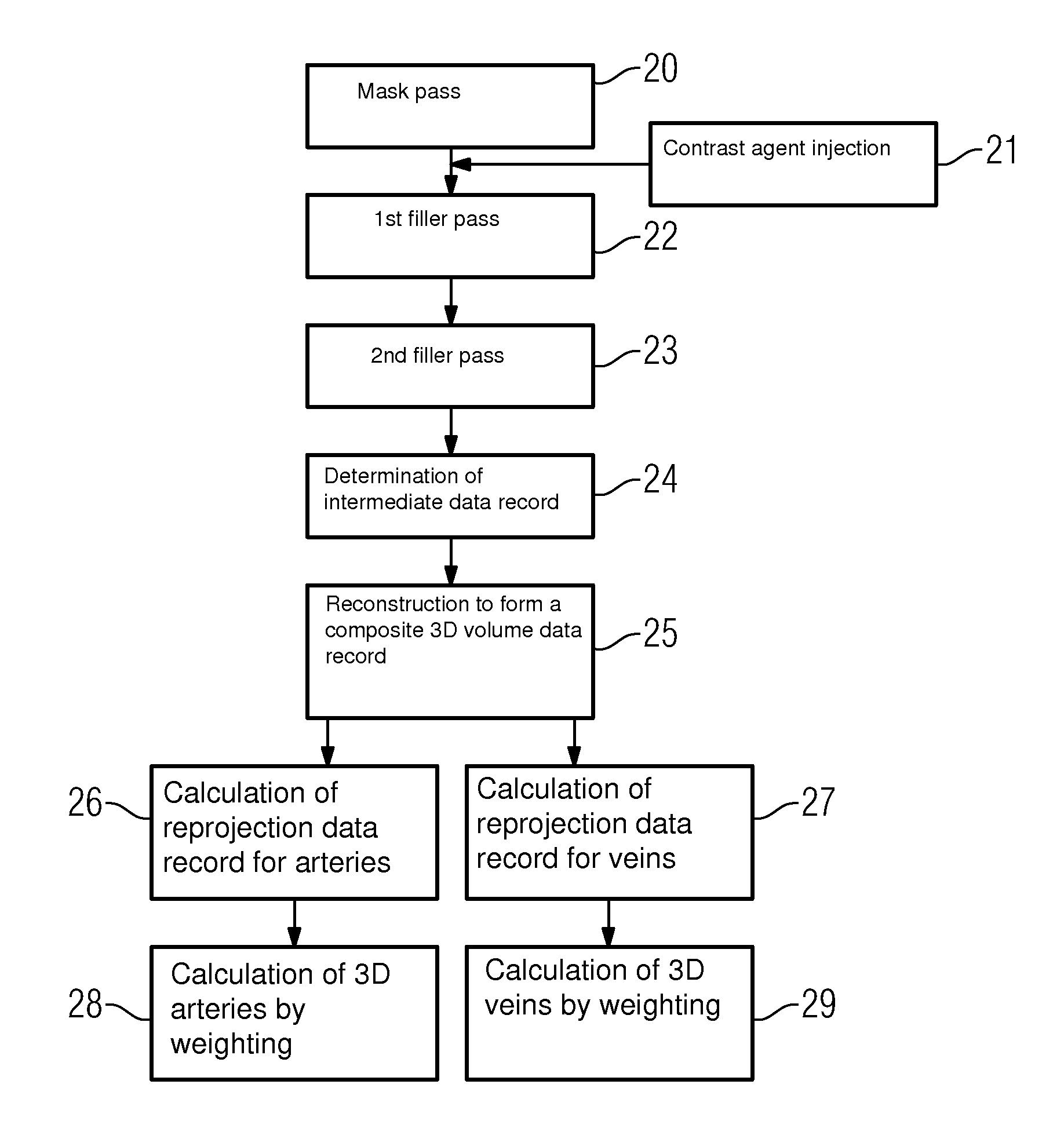
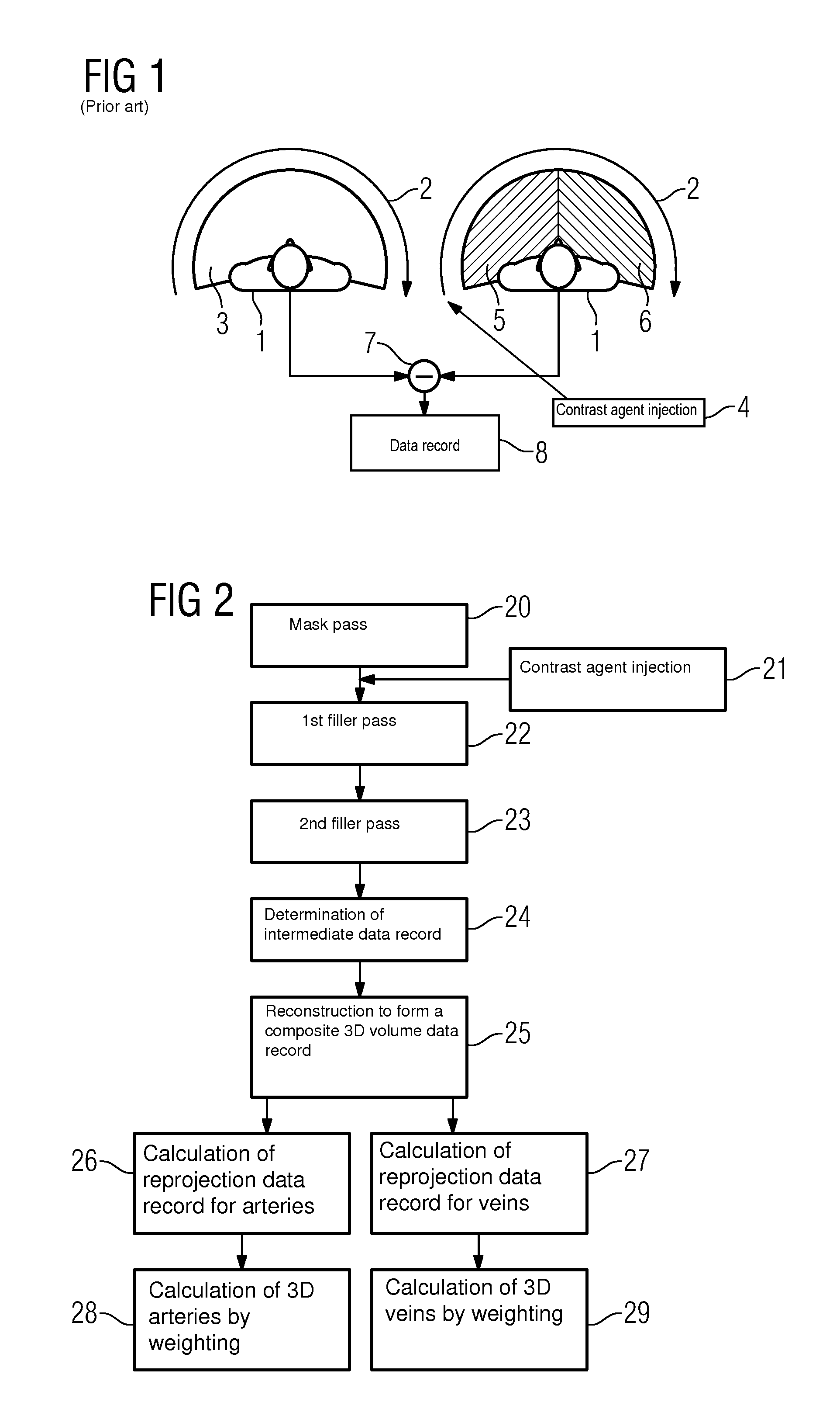
[0046]FIG. 1 shows the principle of the scanning of an examination object 1 according to the prior art. During a mask pass 3 a series of projection images is produced by a recording system circling 2 about an examination object, being recorded from an angular range of approximately 200°. The projection images are in particular recorded by a C-arm x-ray device, on which an x-ray source and an x-ray detector are permanently arranged. A contrast agent is then injected into the vascular system of interest by means of a contrast agent injection 4 and a so-called filler pass is then carried out. This filler pass contains the same number of projection images as the mask pass, which were likewise recorded at the same angular positions as during the mask pass.

[0047]Since in general the rotation time of such a C-arm is around 5 seconds, if a sufficient number of recordings are to be produced for a high-quality reconstruction but the arterial phase 5 of vascular contrasting only lasts around 2...


PUM


Abstract

Method for three-dimensional representation of arteries and / or veins in an object by a tomography x-ray device is proposed. A mask pass with N projections at N angular positions without contrast agent is implemented. A first filler pass with M projections at M angular positions after injecting a contrast agent, where M<N is implemented, followed by a second filler pass with N−M projections at N−M angular positions. A composite three-dimensional volume data record is reconstructed from an intermediate data record determined from the mask pass, the first and the second filler pass. A reprojection for arteries and / or for veins is calculated by reprojection of the first filler pass and / or of the second filler pass from the composite three-dimensional volume data record. A three-dimensional volume data record for arteries and / or for veins is calculated by weighting the composite three-dimensional volume data record taking into account the respective measured and calculated projection images.

Description

CROSS REFERENCE TO RELATED APPLICATIONS[0001]This application claims priority of German application No. 10 2007 045 313.4 DE filed Sep. 21, 2007, which is incorporated by reference herein in its entirety.FIELD OF INVENTION[0002]The present invention relates to a method for the separate three-dimensional representation of arteries and / or veins of a vascular system in an examination object by means of a tomography x-ray device and a device, in particular for executing the method. The invention is preferably applied in C-arm angiographs.BACKGROUND OF INVENTION[0003]There is an increasing demand for the most accurate possible three-dimensional representation of the appearance and pattern of vessels in parts of the body, in particular of arteries and veins, for diagnostic purposes within the field of vascular diseases and the therapy thereof. The examination of cerebral aneurysms represents an important field of application; this also includes an analysis and optimal representation for d...

Claims


Application Information

Patent Timeline
Patent Type & Authority Patents(United States)
IPC IPC(8): A61B6/03
CPCA61B6/466A61B6/481A61B6/504Y10S378/901
Inventor HARER, WOLFGANGZELLERHOFF, MICHAEL
Owner SIEMENS HEALTHCARE GMBH
Why Eureka
Try Eureka