Methods and devices for image-guided manipulation or sensing of anatomic structures

an anatomic structure and image-guided technology, applied in the field of medical devices, can solve the problems of unintentional alteration of neighboring structures, risk of causing harm to patients, transseptal approach, etc., and achieve the effects of minimizing friction, easy image, and improving penetration

Active Publication Date: 2012-05-08
SUNNYBROOK HEALTH SCI CENT
View PDF23 Cites 84 Cited by
  • Summary
  • Abstract
  • Description
  • Claims
  • Application Information

AI Technical Summary

Benefits of technology

[0028]In a particular embodiment of the present invention, a transseptal puncture catheter is adapted to include a forward-looking ultrasound-imaging probe near the tip of the puncture catheter. This embodiment integrates the imaging and interventional components, and enable real-time simultaneous visualization of the needle tip, fossa ovalis and left atrial cavity. The device will facilitate safe and accurate puncture of the inter-atrial septum, positioning of the needle within the left atrium and access to the anatomical targets of the procedure.

Problems solved by technology

Such alterations of structure can facilitate diagnostic or therapeutic procedures, but may be accompanied by risks of causing harm to the patient if the alteration occurs in a manner or location other than intended.
For example, when many structures are adjacent to each other, and the intention of the medical practitioner is to alter a focal region of these structures, harm can come from unintentionally altering neighboring structures.
Although puncture of the fossa ovalis itself is quite safe, the danger of the transseptal approach lies in the possibility that the needle and catheter will puncture an adjacent structure.
After transseptal puncture, the most important problem is to determine whether the tip of the needle is in the left atrium.
The most common complication of the transseptal approach is inadvertent puncture of the wall of the heart or great vessels.
Traditionally, transseptal punctures have been guided by fluoroscopy (see Circulation, 1966 September; 34(3):391-9; Considerations regarding the technique for transseptal left heart catheterization, Ross J Jr) however this form of imaging does not accurately delineate the critical cardiac structures; specifically the fossa ovalis.
Transseptal puncture remains therefore a difficult procedure that is burdened by rare but serious, or even life-threatening complications (see J Invasive Cardiol.
Transesophageal echocardiography (TEE) limits communication with the patient (as it may require the patient's sedation), and creates risks of esophageal bleeding, longer procedure times, and even inadequate location of the fossa ovalis in some cases (see Roelke CCD 1994 32 332; and Pacing Clin Electrophysiol.
Intracardiac echocardiography (ICE) accurately images the fossa ovalis, however it is expensive, invasive and requires placement of an additional device within the heart during the procedure (see Chest.
TEE and ICE are limited by difficulty in imaging the needle tip at the time of the puncture and following penetration, and challenge the operator to coordinate fluoroscopic and ultrasonic images that are acquired from different sources in real-time.
Furthermore, most ICE systems are side-viewing and provide only 2D images.
Therefore, transseptal puncture is a complex procedure limited by difficulty in achieving accurate and simultaneous real-time imaging of the needle tip, fossa ovalis and the surrounding cardiac structures.
These interventions are associated with prolonged procedure times and high doses of x-ray radiation, primarily because of difficulty in visualizing the ablation targets under fluoroscopy (see Curr Probl Cardiol.
), however the above limitations apply.
U.S. Pat. No. 6,755,790 issued Jun. 29, 2004), however this device is limited by an inability to image the needle tip and the fossa ovalis, which may encumber the procedure, and is unable to guide interventions beyond the inter-atrial septum.
However magnetic resonance imaging is expensive, available in few medical centers, contraindicated in patients with implants and incompatible with ferro-magnetic medical devices.
These devices are limited by employing non-imaging modalities.
However this technology is not expected to visualize the advancement of the needle tip beyond the inter-atrial septum, and would therefore not be able to guide the degree of needle advancement into the left atrium so as to avoid puncture of the atrial free wall.
The ring array provides forward-looking 3D imaging, but the imaging quality achieved to date with CMUTs is not adequate for most uses.

Method used


Image

  • Methods and devices for image-guided manipulation or sensing of anatomic structures
  • Methods and devices for image-guided manipulation or sensing of anatomic structures
  • Methods and devices for image-guided manipulation or sensing of anatomic structures

Examples


Embodiment Construction

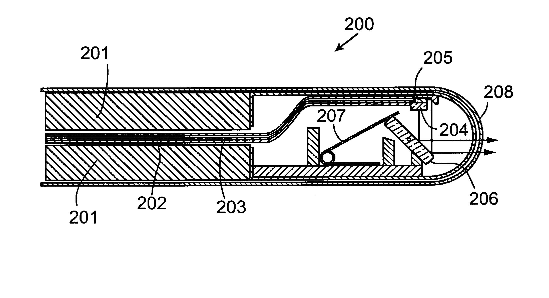
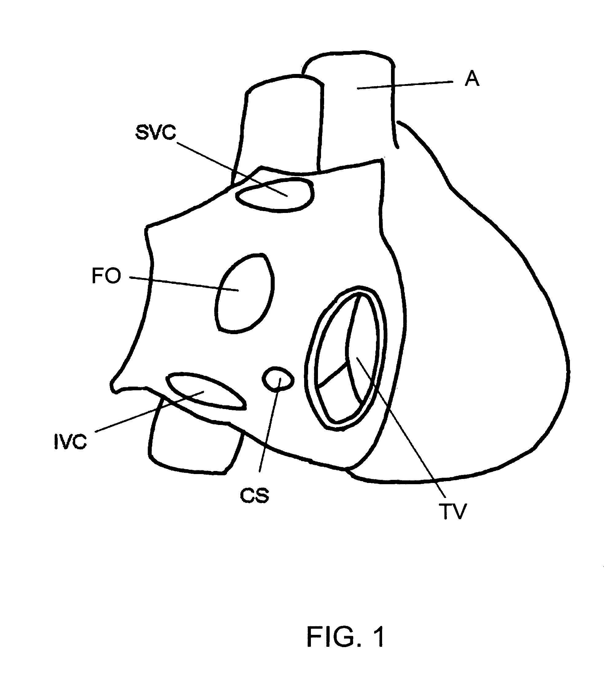
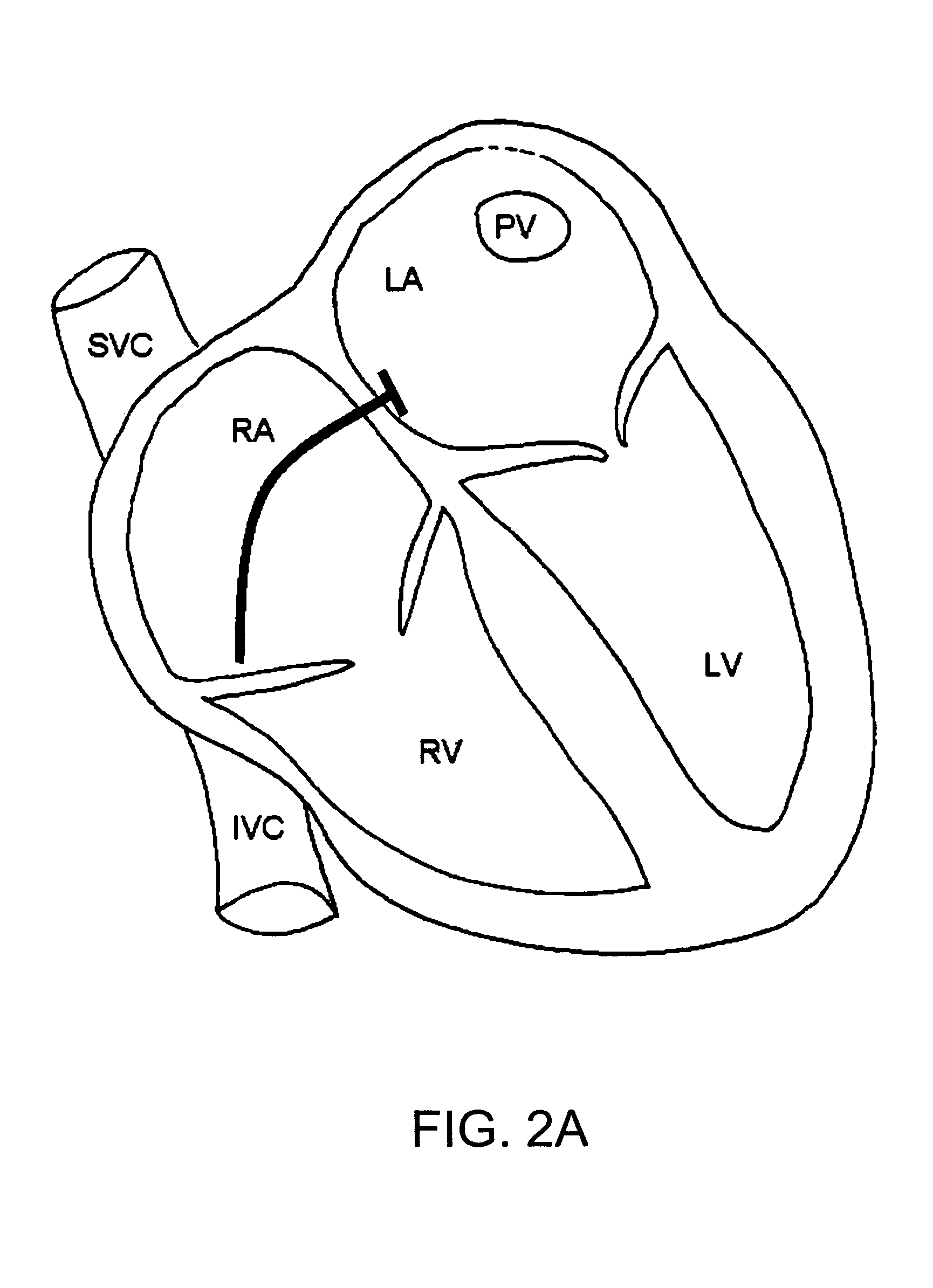
[0060]Without limitation, the majority of the systems described herein are directed to an imaging probe using either optical or ultrasonic (or both) imaging. The imaging probe may include means for estimating a rotational motion near the distal end of a rotating shaft within the probe. As required, embodiments of the present invention are disclosed herein. However, the disclosed embodiments are merely exemplary, and it should be understood that the invention may be embodied in many various and alternative forms.

[0061]The Figures are not to scale and some features may be exaggerated or minimized to show details of particular elements while related elements may have been eliminated to prevent obscuring novel aspects. Therefore, specific structural and functional details disclosed herein are not to be interpreted as limiting but merely as a basis for the claims and as a representative basis for teaching one skilled in the art to variously employ the present invention. For purposes of t...


PUM


Abstract

Forward-looking ultrasound transducers are positioned at the tip of a transseptal catheter to facilitate transseptal puncture and interventions within the left atrium, pulmonary veins and mitral valve. Catheter devices and systems are guided by forward-looking ultrasound or optical coherence tomography imaging for penetrating from one location within a mammalian patient's body to another location, and / or performing diagnostic or therapeutic interventions. A penetrator, diagnostic, or interventional device (eg. probe, biopsy apparatus, electrode, needle) is positioned at the catheter tip and is advanceable from the catheter to a target location outside of the lumen in which the catheter is positioned. The imaging probe uses forward-looking ultrasound or optical coherence tomography that allows one to locate both the target and the medical device. The operator may then adjust the position and / or rotational orientation of the catheter such that when the medical device is subsequently advanced it will either enter or be deployed at the target location.

Description

CROSS REFERENCE TO RELATED U.S. APPLICATIONS[0001]This patent application relates to, and claims the priority benefit from, U.S. Provisional Patent Application Ser. No. 60 / 929,187 filed on Jun. 18, 2007, in English, entitled METHODS AND DEVICES FOR IMAGE-GUIDED MANIPULATION OR SENSING OF ANATOMIC STRUCTURES, which is incorporated herein by reference in its entirety.FIELD OF THE INVENTION[0002]The present invention relates to medical devices in the field of minimally invasive medical interventions. In particular, the present invention provides devices and methods for identifying or observing a precise location in the body through and / or upon which medical procedures may be efficiently and safely performed. By way of example, the present invention can be used to identify or observe cardiac tissue during a medical procedure.BACKGROUND OF THE INVENTION[0003]It may be desired during certain medical procedures to alter anatomic structures, such as by penetrating, or injecting into said st...

Claims


Application Information

Patent Timeline
Patent Type & Authority Patents(United States)
IPC IPC(8): A61B8/12
CPCA61B5/0084A61B8/0833A61B8/0883A61B8/4461A61B8/483A61B8/12A61B5/0066A61B5/0086A61B8/445A61B18/14A61B18/18
Inventor JAFFE, RONENSTRAUSS, BRADLEY H.COURTNEY, BRIAN K.
Owner SUNNYBROOK HEALTH SCI CENT
Why Eureka
Try Eureka