
Method and system of determining the stain quality of slides using scatter plot distribution

a scatter plot and stain quality technology, applied in the field of biological specimen methods and systems, can solve the problems of limitation of traditional pap smear methods, pap smear tests, and other tests, and about one-third of the 15,000 women diagnosed with cervical cancer annually still di

Active Publication Date: 2012-05-22
CYTYC CORP
View PDF9 Cites 0 Cited by
  • Summary
  • Abstract
  • Description
  • Claims
  • Application Information

AI Technical Summary

Benefits of technology

[0012]In accordance with one aspect of the invention, a method of determining the stain quality of a plurality of biological specimens is provided. The method comprises identifying a number of objects of interest (OOIs) within the biological specimen. In a preferred method, this can be accomplished by acquiring images of the biological specimen and processing the images to identify OOIs. The method further comprises measuring a first (e.g., nuclear area) and a second (e.g., nuclear integrated optical density (IOD)) feature of each OOI, and then generating a scatter plot of the measured first features versus the measured second features.

Problems solved by technology

Therefore, about one-third of the 15,000 women diagnosed with cervical cancer annually still die, because the cancer was detected too late.
Another contributing factor that has received much attention is the limitation of the traditional Pap smear method itself.
Thus, Pap smear tests, as well as other tests requiring detailed review of biological material, have suffered from a high false negative rate due to fatigue imposed on the technician.
No automated device, however, has thus far eliminated false negative and false positive results at sufficiently low cost to be practical.
That is, the more slides that a cytotechnologist must review, or the more time it takes for a technician to review each slide slide, the more cost in labor the laboratory incurs.
The occurrence of false positives sometimes results from biological specimens with low-quality stains.
For example, a technician may fail to follow the proper protocol for applying a particular stain to the biological specimens, or if correctly applied, the shelf-life of the stain may have expired.
Oftentimes, specimens are not flagged as having a low-quality stain until after they have been manually reviewed by the cytotechnologist, thereby wasting valuable time.

Method used


Image

  • Method and system of determining the stain quality of slides using scatter plot distribution
  • Method and system of determining the stain quality of slides using scatter plot distribution
  • Method and system of determining the stain quality of slides using scatter plot distribution

Examples


Embodiment Construction

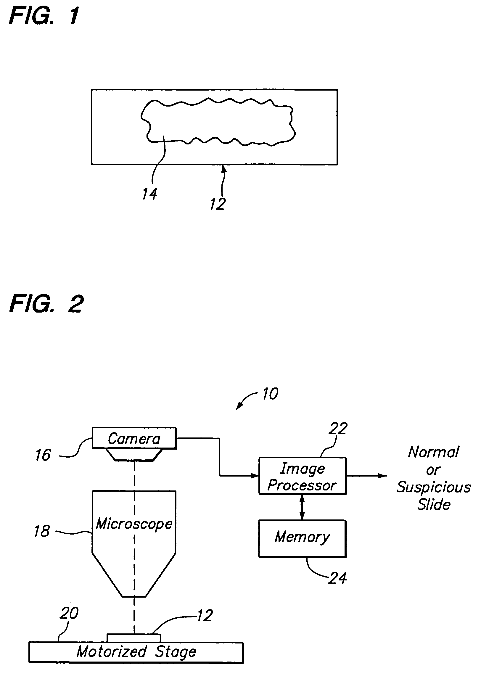
[0025]Referring to FIG. 2, a biological screening system 10 constructed in accordance with one preferred embodiment of the present invention is described. The system 10 is configured to process a series of microscope slides 12 in order to classify a biological specimen 14 (best shown in FIG. 1) as either “normal,” which would not require review by a cytotechnologist, or “suspicious,” which would require review by the cytotechnologist.

[0026]Although the system 10 can be used to classify any biological specimen as normal or suspicious, the system 10 lends itself particularly well to the presentation of cytological cervical or vaginal material, such as that typically found on a Pap smear slide. In this case, the cells may reflect abnormalities (e.g., cytolysis, atrophy, infection, damage), malignancy or premalignancy, such as Low Grade Squamous Intraepithelial Lesions (LGSIL) or High Grade Squamous Intraepithelial Lesions (HGSIL), as well as all other cytologic categories as defined by...


PUM

PropertyMeasurementUnit
diameteraaaaaaaaaa
diameteraaaaaaaaaa
imaging stationaaaaaaaaaa

Abstract

Methods, systems, and computer software for determining the stain quality of a plurality of biological specimens. A number of objects of interest are identified in a biological specimen. A first feature of each object of interest (e.g., nuclear area) and a second feature of each object of interest (e.g., nuclear integrated optical density) are measured, and a scatter plot of the first and second features is generated. The stain quality of the specimens are determined based on the distribution of points within the scatter plot.

Description

RELATED APPLICATIONS[0001]This application claims priority from provisional U.S. Patent Application Ser. No. 60 / 478,434, filed Jun. 12, 2003.FIELD OF THE INVENTION[0002]The field of the present invention generally relates to methods and systems for analyzing biological specimens, and more particularly to methods and systems for classifying biological specimens as normal, abnormal or suspicious, and determining the stain quality of such specimens.BACKGROUND OF THE INVENTION[0003]In the medical industry, there is often a need for a laboratory technician, e.g., a cytotechnologist, to review a cytological specimen for the presence of specified cell types. For example, there is presently a need to review a cervico-vaginal Papanicolaou (Pap) smear slides for the presence of malignant or pre-malignant cells. Since its introduction over fifty years ago, Pap smears have been a powerful tool for detecting cancerous and precancerous cervical lesions. During that time, the Pap smear has been cr...

Claims


Application Information

Patent Timeline
Patent Type & Authority Patents(United States)
IPC IPC(8): G01N33/48G01N1/00G01N1/30G01N15/14G06T7/00
CPCG01N15/1475G06K9/00147G06T7/0012G06T7/401G01N1/30G06T2207/10016G06T2207/10056G06T7/41G06V20/698G01N15/1433
Inventor WONG, KAM LINISENSTEIN, LOUISEZAHNISER, DAVID
Owner CYTYC CORP
Features
  • Generate Ideas
  • Intellectual Property
  • Life Sciences
  • Materials
  • Tech Scout
  • Unparalleled Data Quality
  • Higher Quality Content
  • 60% Fewer Hallucinations
Learn More