Hemolysin and its protein fragments in sero-detection of Anaplasma phagocytophilum
a technology of hemolysin and anaplasma phagocytophilum, which is applied in the field of diagnostic assays for the detection of infectious agents, can solve the problems of prone to sampling errors, tedious microscopic examination, and often false positive results of tests
- Summary
- Abstract
- Description
- Claims
- Application Information
AI Technical Summary
Benefits of technology
Problems solved by technology
Method used
Image

Examples
example 1
Cloning and Expression of Hemolysin
PCR Amplification and Ligation into Plasmid Vector
[0104]In order to determine if hemolysin possesses antibody recognition sites, we cloned and recombinantly expressed the full-length hemolysin protein in Anaplasma phagocytophilum.
[0105]Our cloning strategy involved the design and preparation of synthetic oligonucleotides (˜30 bp in length) and use of them in amplifying the hemolysin gene. As controls, we also cloned two (2) non-TIVSS proteins (i.e., succinate dehydrogenase iron-sulfur subunit and p44 outer membrane protein) and used them for comparison. Table 1 shows the nucleotide sequence of the various oligonucleotides (i.e., SEQ ID NOs: 1-6) used in the PCR amplification reaction.
[0106]Genomic DNA of Anaplasma phagocytophilum (a generous gift from Dr. S. Dumler at Johns Hopkins University) was used as the template for each of the PCR reactions. Synthetic oligonucleotides corresponding to the hemolysin gene were used for the PCR amplification r...
example 2
IgG / IgM ELISA for Recombinantly Expressed Hemolysin Protein
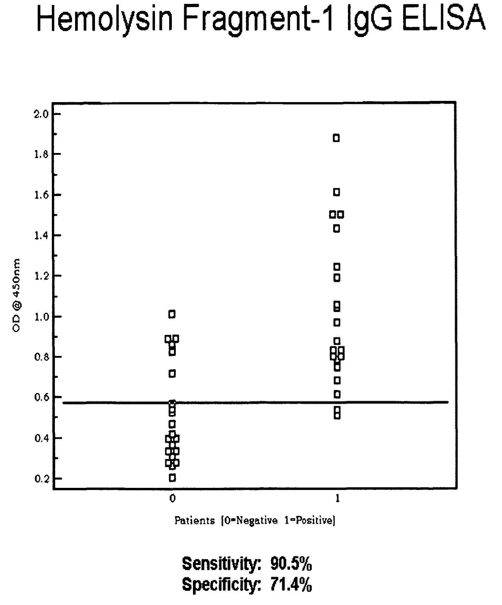
[0151]We adopted IgG and IgM ELISA assays and evaluated the binding activity of the recombinant protein towards IgG and IgM. The ELISA procedure involves: (i) coating 96-well micro-titer plates with the recombinant protein at varying concentrations at 4° C. overnight; (ii) adding 5% non-fat milk to block non-specific binding; (iii) adding patients' sera to allow formation of antibody-antigen complex; (iv) detecting the antibody-antigen complex. IFA sero-positive sera served as positive controls, and IFA sero-negative sera served as negative controls. Detection of antibody-antigen complex was performed with the use of horseradish peroxidase.
[0152]Patient Study: Hemolysin
[0153]IgG ELISA
[0154]Recombinant hemolysin protein, when tested in an IgG ELISA, exhibited a dose-dependent increase in binding towards IgG sero-positive serum as measured by OD450nm. IgG ELISA for recombinant hemolysin attained a sensitivity of 81.0% and a sp...
example 3
Amplification and Cloning of Hemolysin Protein Fragments
[0162]I) PCR Amplification and Ligation into Plasmid Vector
[0163]We cloned and recombinantly expressed hemolysin fragments 1-3 in E. coli. Our cloning strategy involved the design and preparation of synthetic oligonucleotides (˜30 bp in length) and use of them in amplifying the hemolysin fragments.
[0164]Table 4 shows the nucleotide sequence of the various oligonucleotides (i.e., SEQ ID NOs. 10-15) used in the PCR amplification reaction.
[0165]Genomic DNA of Anaplasma phagocytophilum (a generous gift from Dr. S. Dumler at Johns Hopkins University) was used as the template for each of the PCR reactions. Synthetic oligonucleotides corresponding to the hemolysin gene fragments were used for the PCR amplification reactions. Using the synthetic oligonucleotides (sequence listed in Table 4) and genomic DNA from Anaplasma phagocytophilum, we successfully amplified the hemolysin gene fragments; as well as two (2) non-TIVSS genes (i.e., s...
PUM
| Property | Measurement | Unit |
|---|---|---|
| temperatures | aaaaa | aaaaa |
| temperatures | aaaaa | aaaaa |
| temperature | aaaaa | aaaaa |
Abstract
Description
Claims
Application Information
 - R&D
- Intellectual Property
- Life Sciences
- Materials
- Tech Scout
- Unparalleled Data Quality
- Higher Quality Content
- 60% Fewer Hallucinations
Browse by: Latest US Patents, China's latest patents, Technical Efficacy Thesaurus, Application Domain, Technology Topic, Popular Technical Reports.
© 2025 PatSnap. All rights reserved.Legal|Privacy policy|Modern Slavery Act Transparency Statement|Sitemap|About US| Contact US: help@patsnap.com