Single-insertion, multiple sample biopsy device with integrated markers

a biopsy device and multiple sample technology, applied in the field of tissue biopsy sampling device, can solve the problems of inability to obtain more than, inaccurate post-biopsy diagnosis, pain and scarring of the body site, etc., and achieve the effect of preventing a vacuum

Active Publication Date: 2012-09-18
CR BARD INC
View PDF394 Cites 49 Cited by
  • Summary
  • Abstract
  • Description
  • Claims
  • Application Information

AI Technical Summary

Benefits of technology

[0045]In addition, the rigidity of the lumens can be derived from a secondary element that houses the lumen to give it rigidity, meaning a rigid portion of a lumen does not need to be a monolithic structure and the uses of terms such as “rigid lumen” or “rigid portion of a lumen” are not intended to limit the identified lumen structures to single-element structures. For example, a flexible lumen can be guided by a rigid member (for example it can slide within a tube) giving it all the effective rigidity needed to enable the lumen to move a transport member distally within a needle. Or a flexible tube can have a moving rigid guide (tube or other structure) to which it is fixedly attached, to give it all the effective rigidity needed to enable the lumen to move a transport member distally within a needle.
[0048]Although in most of the disclosed embodiments, fluid is provided to the distal end of the needle and permitted to flow proximally as the tissue sample is transported proximally, the fluid itself need not, and in embodiments, preferably is not, sufficient in quantity or velocity to move the tissue sample. That is, preferably, the fluid rate does not produce enough drag on the sample, given the seal between the sample and the stylet, the fluid flow rate, and the hydrodynamic properties of the sample, to transport the sample along the stylet. The fluid is preferably provided to flood the sample chamber and lubricate the passageway for transport. In addition the fluid may be only sufficient to fill in the space behind the bulkhead or bulkheads so that they, and the tissue, move more freely without creating any vacuum, even momentarily, in their wake. Preferably, the bulkheads described in the disclosed embodiments to not form a seal with the stylet or cannula. In this way fluid can flow around them easily. In fact, the fluid used to lubricate movement of the bulkhead(s) and sample may be provided at the middle of the sample chamber or proximal of the sample chamber and allowed to flow around the bulkheads to aid in transporting and preventing a vacuum.

Problems solved by technology

However, there are circumstances where there may be a need to obtain more than one samples.
While the known biopsy needle can be re-inserted multiple times, such technique can cause pain and scarring of the body site.
The additional step and device may not allow the marker to be deposited at the actual biopsied site, which can lead to inaccurate post-biopsy diagnosis.

Method used


Image

  • Single-insertion, multiple sample biopsy device with integrated markers
  • Single-insertion, multiple sample biopsy device with integrated markers
  • Single-insertion, multiple sample biopsy device with integrated markers

Examples


Embodiment Construction

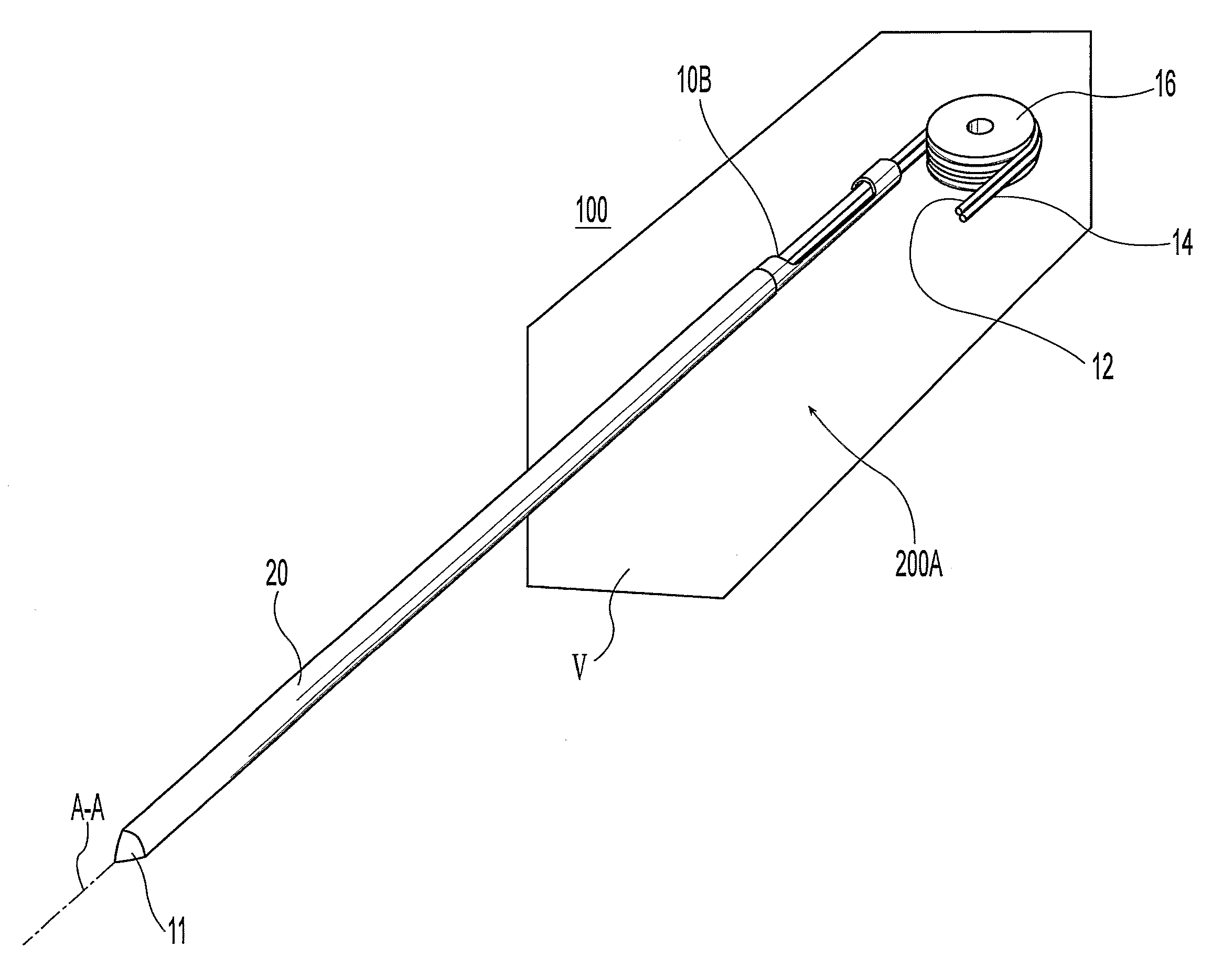
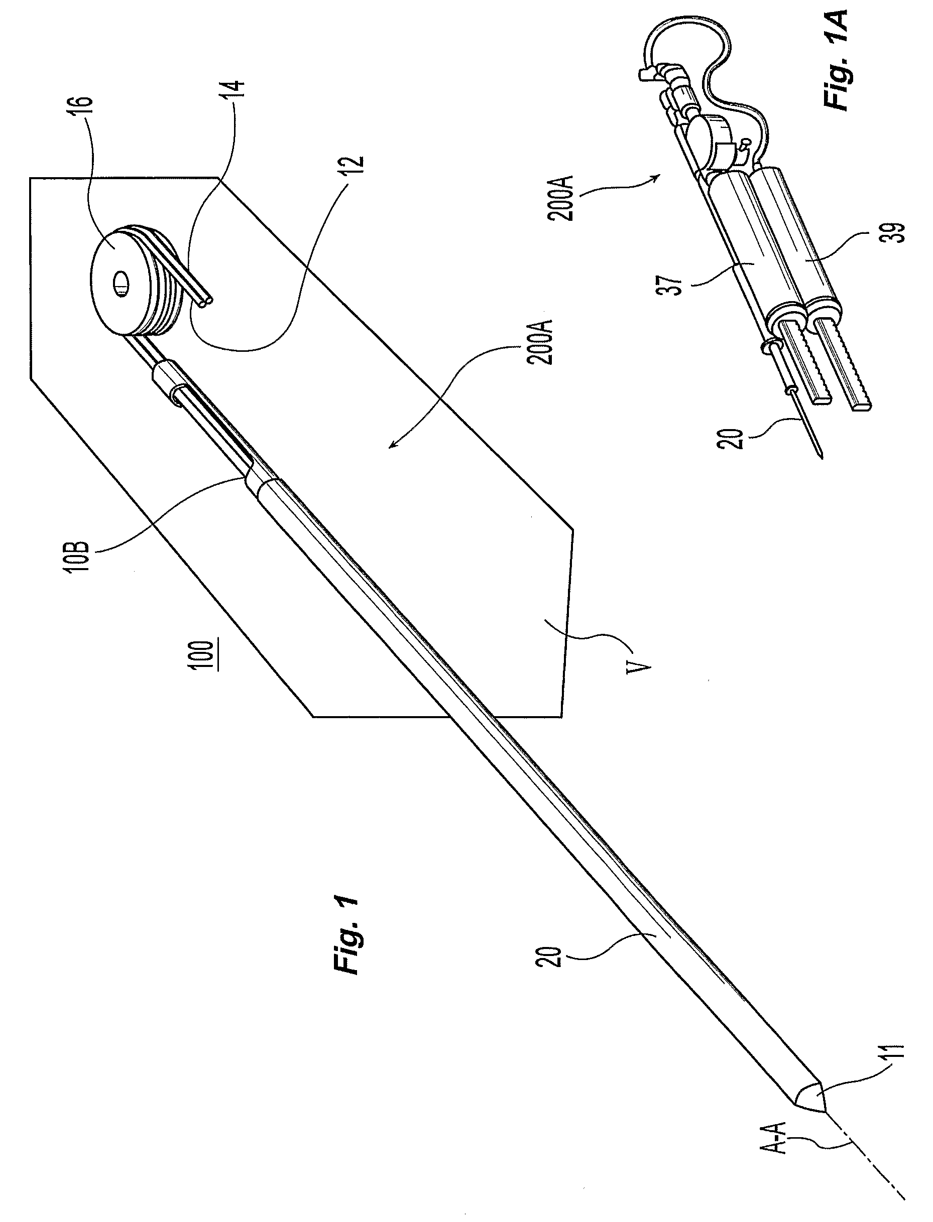
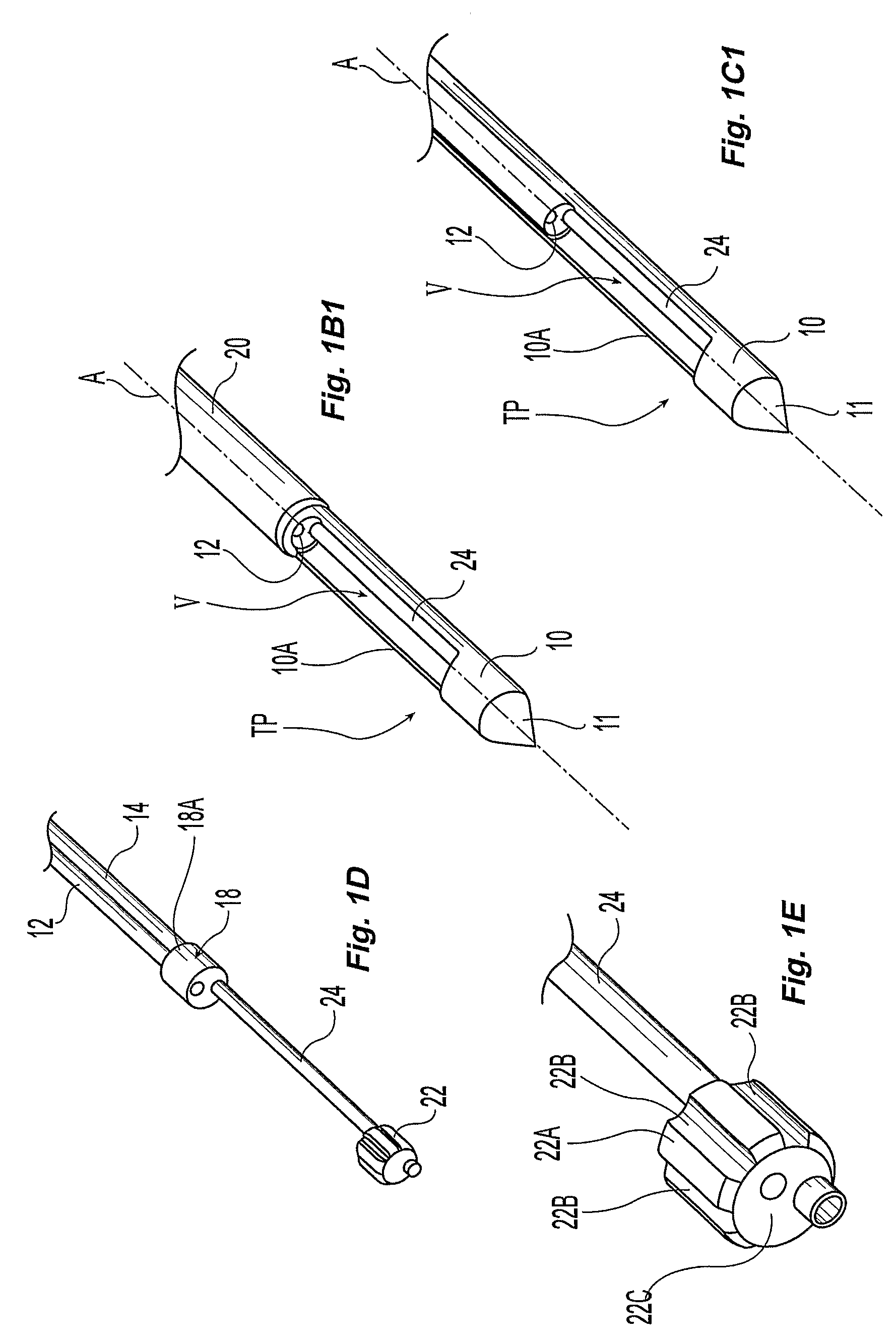
[0073]FIGS. 1-6 illustrate the preferred exemplary embodiments. In particular, FIG. 1 shows a perspective view of a stylet 10 coupled to the single-insertion, multiple samples biopsy device 100 provided with a transport subassembly 200A. The transport subassembly 200A includes the stylet, which has a tip 11 at the distal end and an outer cutting cannula 20 covering a substantial portion of the stylet 10 and a first port 10A. Extending through a hollow portion of the stylet 10 are two flexible lumens 12 and 14 coupled to a common pulley 16 proximate a second port 10B. The transport subassembly 200A can be coupled to ancillary components of the device 100 such as respective saline 37 reservoir and pump and vacuum and air pressure pump 39, a motor drive 200A, and switches and sensors as shown in FIG. 1A.

[0074]Referring to FIG. 1D, the flexible lumens 12 and 14 are coupled to a first bulkhead 18. A second bulkhead 22 is coupled to the first bulkhead via a rigid lumen 24. One of the flex...


PUM


Abstract

The present invention provides for exemplary embodiments of a single-insertion, multiple sample biopsy device. Exemplary embodiments of a single-insertion, multiple sampling device with integrated marker release.

Description

PRIORITY DATA AND INCORPORATION BY REFERENCE[0001]This application is a 35 U.S.C. 371 application of International Application No. PCT / US2006 / 031327, filed Aug. 10, 2006, which claims priority to U.S. Provisional Patent Application No. 60 / 707,229, filed Aug. 10, 2005, the entireties of which are hereby incorporated by reference.FIELD OF THE INVENTION[0002]This invention relates to a tissue biopsy sampling device.BACKGROUND OF THE INVENTION[0003]Often, it is either desirable or necessary to obtain specimens of tissue from humans and other animals, particularly in the diagnosis and treatment of patients with cancerous tumors, premalignant conditions, and other diseases or disorders. For example, when it is discovered that suspicious conditions exist, either by means of x-ray or ultrasound imaging in various tissues of the body, a physician typically performs a biopsy to determine if the cells at the suspected site are cancerous.[0004]A biopsy can be done either by an open or percutane...

Claims


Application Information

Patent Timeline
Patent Type & Authority Patents(United States)
IPC IPC(8): A61B10/00
CPCA61B10/0275A61B10/0283A61B2019/5487A61B2010/0225A61B2010/0208A61B2090/3987
Inventor TAYLOR, JONTHOMPSON, STANLEY O.COONAHAN, TIMOTHY J.GRAY, GREGORY A.
Owner CR BARD INC
Why Eureka
Try Eureka