
Method for tracking of contrast enhancement pattern for pharmacokinetic and parametric analysis in fast-enhancing tissues using high-resolution MRI

a tissue and high-resolution technology, applied in image enhancement, image analysis, instruments, etc., can solve the problems of not being able to distinguish and characterize the cancerous and healthy prostate tissues, the inability of methods to take fast images, and the limitations of high-spatial resolution on temporal resolution

Active Publication Date: 2012-09-25
KONINKLJIJKE PHILIPS NV
View PDF0 Cites 12 Cited by
  • Summary
  • Abstract
  • Description
  • Claims
  • Application Information

AI Technical Summary

Problems solved by technology

Unfortunately, these methods are not always able to differentiate and characterize the cancerous and healthy prostate tissues.
However, the high-spatial resolution poses some limitations on temporal resolution (i.e., how fast images can be taken).
However, dynamic imaging in general, and pharmacokinetic methods in particular, may not provide sufficient temporal resolution to monitor the rapid dynamics of the contrast agent exhibited in fast-enhancing tissues such as prostate tissue.
This is particularly true for the majority of currently deployed MRI scanners, which have limited gradient strength and cannot perform sequences fast enough.

Method used


Image

  • Method for tracking of contrast enhancement pattern for pharmacokinetic and parametric analysis in fast-enhancing tissues using high-resolution MRI
  • Method for tracking of contrast enhancement pattern for pharmacokinetic and parametric analysis in fast-enhancing tissues using high-resolution MRI
  • Method for tracking of contrast enhancement pattern for pharmacokinetic and parametric analysis in fast-enhancing tissues using high-resolution MRI

Examples


Embodiment Construction

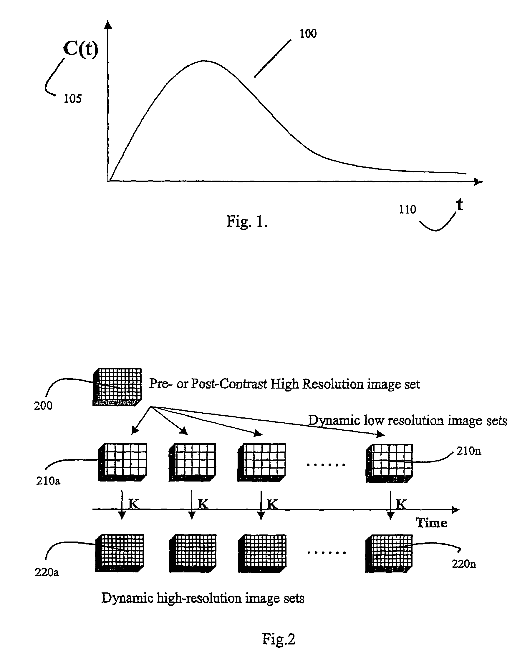
[0027]Enhancement patterns in tissues are mainly determined by the blood flow to the tissue and by the vascular permeability of the tissue vessels. For pharmacokinetic analysis and calculation of physiologic parameters, it is necessary to separate the flow and permeability contributions. Separating the flow and permeability contributions is only possible if the enhancement kinetics can be monitored with sufficient temporal resolution. As previously mentioned, dynamic imaging in general may not provide sufficient temporal resolution to monitor such rapid enhancement behavior. Low-resolution magnetic resonance (MR) images (with a matrix of 128×128 voxels or lower) have been previously employed to examine such fast enhancement behavior. However, this method causes the image voxels (i.e., volume elements) to be large, and volume-averages the enhancement patterns in the image voxels. This result is not acceptable because cancer is known to be very heterogeneous in its enhancement pattern...


PUM


Abstract

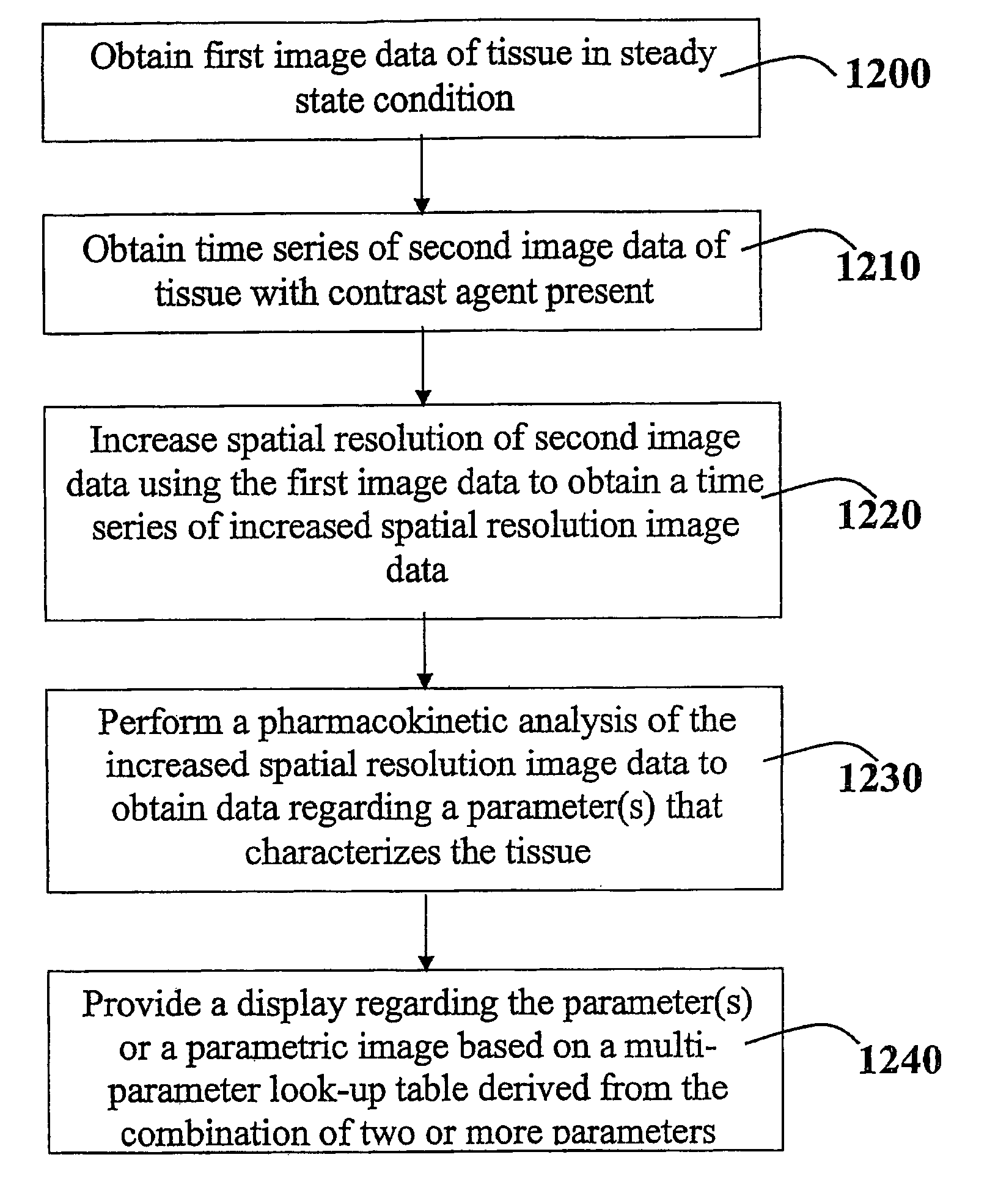
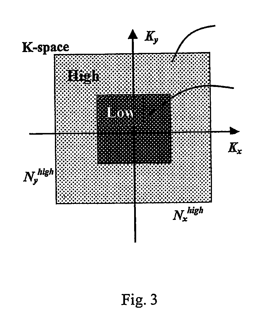
A method for performing a high-resolution pharmacokinetic analysis for calculation of tissue parameters for a fast-enhancing tissue enables medical personnel to accurately determine pharmacokinetic parameters in fast-enhancing tissues. The method includes obtaining mask image data of the tissue when it is in a steady state condition, obtaining a time series of image data of the tissue when the contrast agent is flowing in the tissue, and increasing a spatial resolution of the time series of image data using the mask image data to obtain a time series of increased spatial resolution image data. The method further includes performing a pharmacokinetic analysis to obtain data including at least one parameter that characterizes the tissue, providing a multi-parameter look-up table derived from a combination of two or more parameters, and providing a display including one parameter or a parametric image, where the parametric image is derived from the look-up table.

Description

TECHNICAL FIELD[0001]The present invention relates to a system and method for use with medical imaging and, more particularly to a system and method for obtaining and analyzing images from tissues which exhibit fast uptake of a contrast agent.BACKGROUND INFORMATION[0002]There is a growing trend in the development of non-invasive methods for detection and characterization of tumors. As a second most common cause of cancer-related death in men, prostate cancer is a usual object for many research studies. The appropriate tumor localization and staging are very important for determining the best choice of treatment. The most common diagnostic methods for prostate cancer are transrectal ultrasound and conventional magnetic resonance imaging (MRI). Unfortunately, these methods are not always able to differentiate and characterize the cancerous and healthy prostate tissues.[0003]A comparatively new method for characterization of tumor microvasculature is the dynamic contrast enhanced (DCE)...

Claims


Application Information

Patent Timeline
Patent Type & Authority Patents(United States)
IPC IPC(8): G06K9/00
CPCG06T7/0012G06T2207/30004
Inventor MURADYAN, NAIRAMUEHLER, ANDREAS
Owner KONINKLJIJKE PHILIPS NV
Learn More