Preservation of cell-free RNA in blood samples

a cell-free nucleic acid and blood sample technology, applied in the field of identification and isolation of cell-free nucleic acids in blood samples, can solve the problems of affecting the detection ability affecting the detection efficiency of cell-free nucleic acids, so as to prevent the leakage of cellular rna

Active Publication Date: 2012-11-06
STRECK LLC
View PDF156 Cites 127 Cited by
  • Summary
  • Abstract
  • Description
  • Claims
  • Application Information

AI Technical Summary

Benefits of technology

[0009]The present invention contemplates a screening method for the identification of a disease state, comprising the steps of: contacting a drawn blood sample that includes a plurality of blood cells with a plasma RNA (e.g., mRNA) protective agent; isolating cell-free RNA from the blood sample; and analyzing (e.g., by quantity, quality, or both) the isolated RNA for the presence, absence, or severity of a disease state. The protective agent may be present in an amount and for a time sufficient so that the RNA synthesis is inhibited for at least two hours. The protective agent may be present in an amount so that blood cells of the drawn blood sample are fixed to substantially prevent leaking of cellular RNA into the plasma. The protective agent may be present in an amount so that any cellular RNA that is within the blood cells at the time of the blood draw is substantially preserved to immobilize the protein expression pattern of the blood cells so that the protein expression pattern of the cells remains substantially the same as at the time of the blood draw. The protective agent may be present in an amount so that the RNA that is in the plasma is substantially stabilized against degradation mediated by the combined action of nucleases and proteases.

Problems solved by technology

However, after a blood sample is acquired from a patient, cell lysis begins and the nucleic acids from within the blood cells are mixed with the cell-free nucleic acids, making it difficult if not impossible to isolate and distinguish the cell-free mRNA.
Consequently, the disease indication capability of the cell-free nucleic acids may be diminished as their presence is no longer accurately ascertainable.
However, there is concern that after blood draw the nucleic acids somehow become disassociated from the apoptotic bodies and become vulnerable to nucleases.

Method used


Image

  • Preservation of cell-free RNA in blood samples
  • Preservation of cell-free RNA in blood samples

Examples


example 1

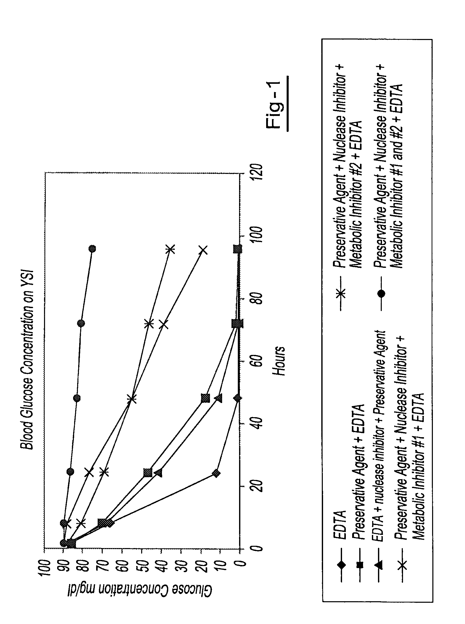
[0042]Blood samples from the same donor are drawn into six separate blood collection tubes (tube 1 through tube 6). Tube 1 contains only EDTA. Tube 2 contains DU and EDTA. Tube 3 contains DU, EDTA and ATA. Tube 4 contains DU, EDTA, ATA and glyceraldehyde. Tube 5 contains DU, EDTA, ATA and sodium fluoride. Tube 6 contains DU, EDTA, ATA, glyceraldehyde and sodium fluoride. All tubes are stored at room temperature and 1 ml aliquots of blood are removed from each tube at hours 1.5, 8, 24, 48, 72 and 96. The blood glucose levels of each sample are measured using a YSI blood glucose meter available from YSI Life Sciences (Yellow Springs, Ohio). The blood glucose concentration of those samples from tube 6 maintained relatively consistent glucose levels over the test period, indicating that the combination of EDTA, DU, ATA, glyceraldehyde and sodium fluoride provided reduced levels of cell metabolism. The results of this example are shown in a graphic format at FIG. 1.

example 2

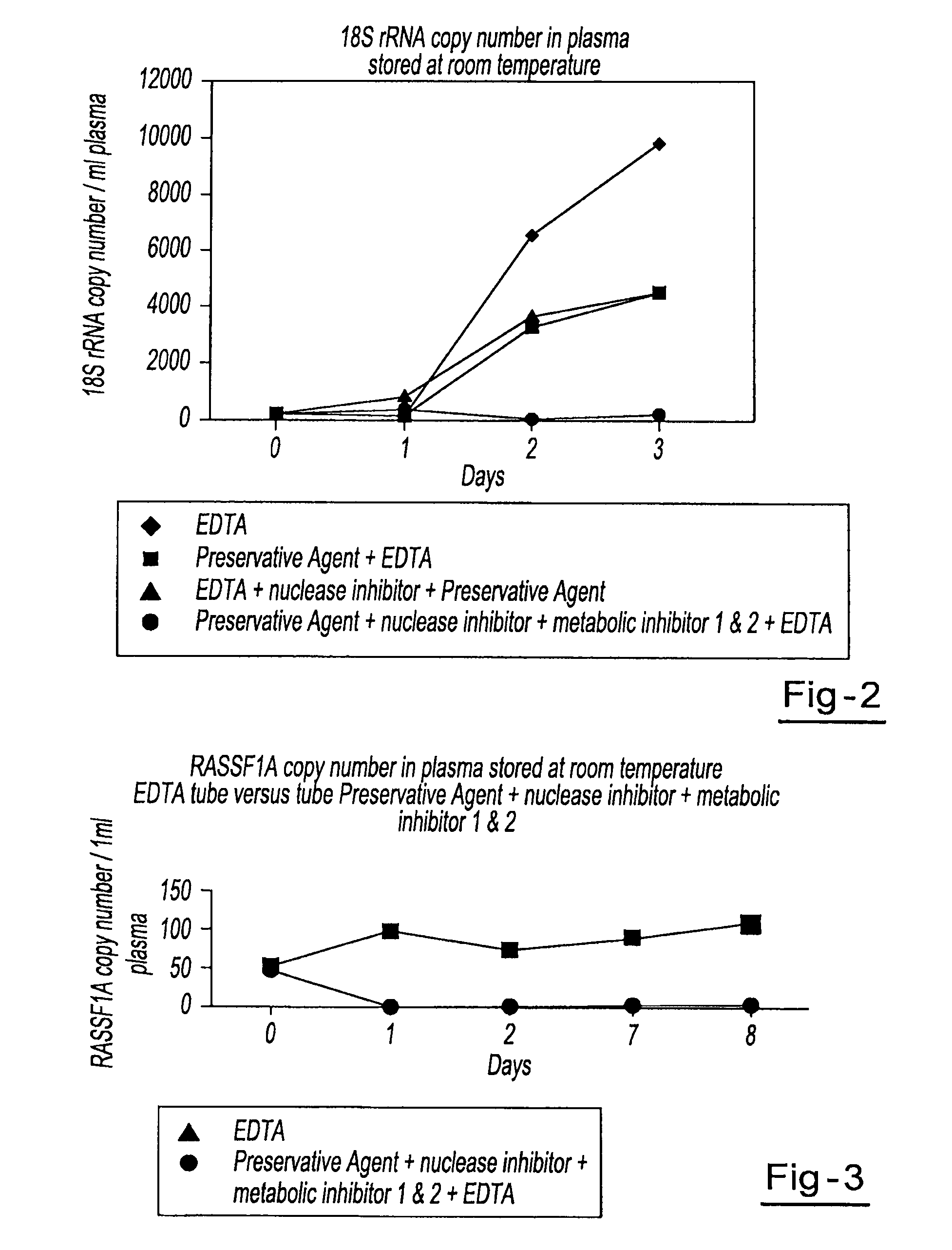
[0043]Four blood samples from the same donor are drawn into four separate blood collection tubes, tube A through tube D. Tube A contains DU, EDTA, ATA, glyceraldehyde and sodium fluoride. Tube B contains DU, EDTA and ATA. Tube C contains DU and EDTA. Tube D contains only EDTA. All tubes are stored at room temperature and 1 ml aliquots of blood are removed from each tube on day 0, day 1, day 2, and day 3 and plasma is separated. All samples are centrifuged at 800 g for 10 minutes at room temperature to separate the plasma. The plasma is then transferred into new tubes and centrifuged at 1500 g for 10 minutes at room temperature. Free circulating RNA is purified using the QIAamp MinElute Virus Spin kit available from Qiagen, Inc. (Valencia, Calif.). RNA is extracted from each plasma sample. The samples are then amplified by Real Time PCR (using TaqMan® RT PCR reagents available from Applied Biosystems, Foster City, Calif.) to identify the 18S rRNA copy number per ml of plasma. Results...

example 3

[0044]Blood samples from the same donor are drawn into two separate blood collection tubes. The first tube contains DU, EDTA, ATA, glyceraldehyde and sodium fluoride. The second tube contains only EDTA. Both tubes are stored at room temperature and 1 ml aliquots of blood are removed from each tube on day 0, day 1, day 2, day 7, and day 8 and plasma is separated. All samples are centrifuged at 800 g for 10 minutes at room temperature to separate the plasma. The plasma is then transferred into new tubes and centrifuged at 1500 g for 10 minutes at room temperature. Free circulating RNA is purified using the QIAamp MinElute Virus Spin kit available from Qiagen Inc. (Valencia, Calif.). RNA is extracted from each plasma sample. The samples are then amplified by Real Time PCR (using TaqMan® RT PCR reagents available from Applied Biosystems, Foster City, Calif.) to identify fragments of β-globin and RASSF1A genes within the plasma. Results showed a consistent relative percentage of RASSF1A ...


PUM

PropertyMeasurementUnit
concentrationaaaaaaaaaa

Abstract

A method for preserving and processing cell-free nucleic acids located within a blood sample is disclosed, wherein a blood sample containing cell-free nucleic acids is treated to reduce both blood cell lysis and nuclease activity within the blood sample. The treatment of the sample aids in increasing the amount of cell-free nucleic acids that can be identified and tested while maintaining the structure and integrity of the nucleic acids.

Description

CLAIM OF PRIORITY[0001]This application claims the benefit of the filing date of U.S. Provisional Application Ser. No. 61 / 153,472, filed on Feb. 18, 2009, the entirety of the contents of that application being hereby incorporated by reference for all purposes.FIELD OF THE INVENTION[0002]This invention relates to the identification and isolation of cell-free nucleic acids in blood samples and more particularly to the preservation of cell-free RNA within a blood sample.BACKGROUND OF THE INVENTION[0003]It has been recognized that mRNA obtained from plasma of a blood sample can be useful as an indicator of protein expression. Thus, the presence of extra-cellular or cell-free mRNA in blood plasma has triggered a variety of investigations aimed at determining the source and possible diagnostic capabilities of these nucleic acids. Given that the blood of most healthy individuals ordinarily does not contain substantial amounts of cell-free RNA, elevated amounts of cell-free nucleic acids ar...

Claims


Application Information

Patent Timeline
Patent Type & Authority Patents(United States)
IPC IPC(8): C12Q1/68
CPCC12Q1/6806C09K15/20C12Q2527/125C12Q2527/127C12Q1/6886
Inventor FERNANDO, M. ROHAN
Owner STRECK LLC
Why Eureka
Try Eureka