
Method for producing an X-ray image during a mammography

a mammography and x-ray technology, applied in the field of generating an x-ray image during a mammography, can solve the problems of slow and laborious medical workflow in the scope of the entire mammography, and achieve the effect of high image ra

Active Publication Date: 2012-12-04
SIEMENS HEALTHCARE GMBH
View PDF12 Cites 0 Cited by
  • Summary
  • Abstract
  • Description
  • Claims
  • Application Information

AI Technical Summary

Benefits of technology

The present invention provides an improved method for generating an x-ray image during a mammography procedure by using a continuous mammography device or a compression device to hold the breast in place during the entire procedure. The method involves radiographing the breast with x-rays to create a first x-ray image and evaluating it to determine a condition for a second x-ray image. The second x-ray image is then produced based on the determined condition. The method allows for a faster and more efficient mammography examination as the breast is fixed in the retention device during the entire procedure. The first x-ray image can be automatically evaluated and determined to improve the second x-ray image. The second x-ray image can be determined based on an image field altered relative to the first x-ray image or an x-ray dose for ensuring optimal image quality. The method helps to improve the accuracy and efficiency of mammography examination and reduces the dose of X-ray radiation to the patient.

Problems solved by technology

The medical workflow in the scope of the entire mammography is thus slow and laborious.

Method used


Image

  • Method for producing an X-ray image during a mammography
  • Method for producing an X-ray image during a mammography

Examples


Embodiment Construction

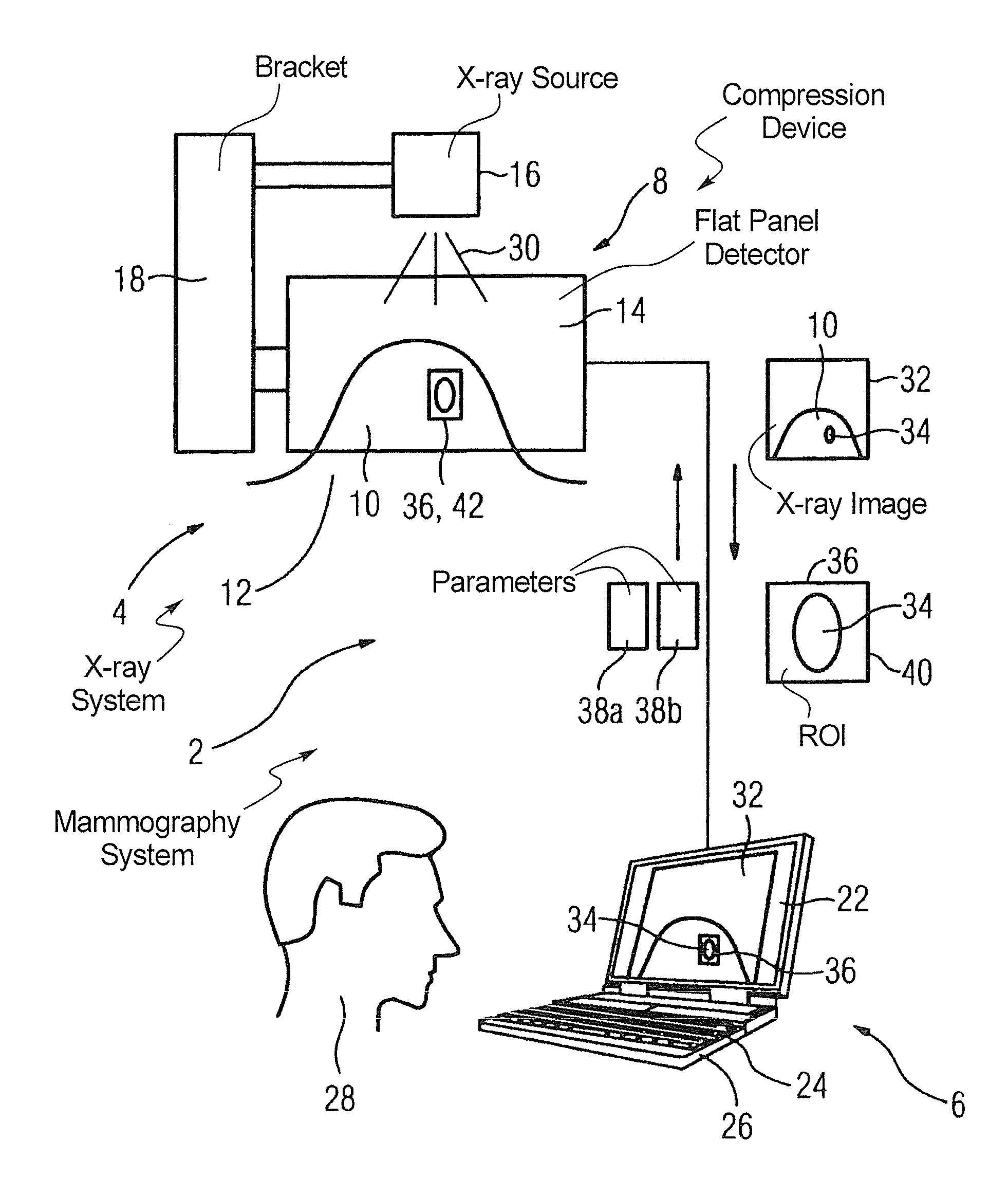
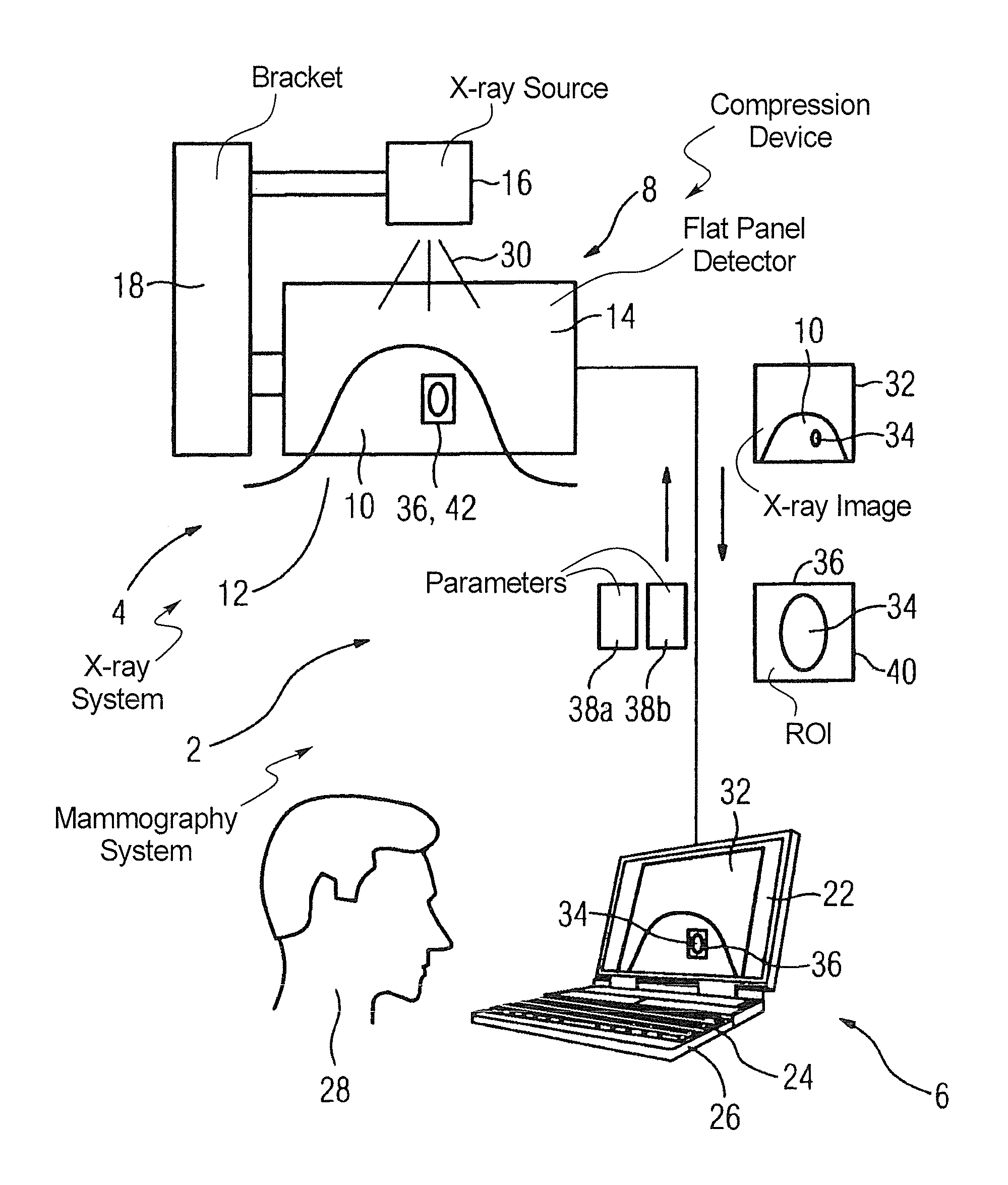
[0027]The FIGURE shows a mammography system 2 having an x-ray system 4 and an acquisition workstation 6. The x-ray system 4 has a bracket 8 or compression device for fixing a breast 10 of a patient 12 (symbolically shown). The bracket 8 carries an x-ray flat panel detector 14 and a compression plate (not shown in FIG. 1) interacting with the flat panel detector.

[0028]The flat panel detector 14 is attached on a bearing device 18 together with an x-ray source 16. The x-ray system 4 is connected via a data line 20 with the workstation 6, which essentially comprises a monitor 22, a keyboard 24 and a computer 26.

[0029]A physician would like to now conduct a mammography of the patient 12. For this he brings the breast 10 of said patient 12 into the bracket 8 and starts the x-ray source 16 via the workstation 6, which x-ray source 16 emits x-ray radiation 30 through the breast 10 towards the flat panel detector 14. This supplies a first x-ray image 32 to the workstation 6, which graphicall...


PUM


Abstract

In a continuous mammography procedure, the breast of a subject is fixed in place in a retention device, and a first x-ray image is generated by irradiating the breast in the retention device. While the breast is still held in the retention device, the first x-ray image is evaluated to define a condition for generating a second x-ray image. The second x-ray image is then generated according to the defined condition, with the breast still in the same position in the retention device.

Description

BACKGROUND OF THE INVENTION[0001]1. Field of the Invention[0002]The invention concerns a method for generating an x-ray image during a mammogram of a breast, wherein the breast is fixed in a retention device.[0003]2. Description of the Prior Art[0004]Mammography, thus x-ray radioscopy of the breast of a (normally) female patient, has permeated medical practice as an examination method. Mammography today is normally conducted digitally, meaning that the corresponding x-ray images are generated by a digital x-ray radiator (for example a flat panel detector).[0005]In known mammography systems today it is typical to fix or compress the breast in a retention device and to thereby generate a single x-ray exposure with static acquisition parameters. The breast is then released from the compression device again. If in the scope of mammography a second x-ray exposure is required, for example for a biopsy or a detail acquisition for fine diagnosis, the patient or the breast is re-fixed in the...

Claims


Application Information

Patent Timeline
Patent Type & Authority Patents(United States)
IPC IPC(8): G06K9/00
CPCA61B6/469A61B6/488A61B6/502
Inventor HANKE, WILHELMMERTELMEIER, THOMAS
Owner SIEMENS HEALTHCARE GMBH
Learn More