Laparoscopic vascular access

a vascular access and laparoscopic technology, applied in the field of medical devices, to achieve the effect of providing strength and suppor

Active Publication Date: 2013-01-22
WILLIAM A COOK AUSTRALIA +1
View PDF8 Cites 9 Cited by
  • Summary
  • Abstract
  • Description
  • Claims
  • Application Information

AI Technical Summary

Benefits of technology

[0024]Preferably the sheath is retractable to release the laparoscopic conduit and the pusher catheter extension prevents distal movement of the laparoscopic conduit during the retraction.
[0040]In one embodiment the elongate curved hollow needle can comprise a reinforcing body surrounding the needle. The reinforcement can comprise a catheter around the needle extending along a substantial proportion of the needle. The reinforcement not only provides strength and support for the needle as it is extended out beyond the laparoscopic port sheath but it also assists in obtaining a seal between the needle and the hemostatic valve at the distal end of the laparoscopic port sheath.

Problems solved by technology

The Seldinger method of access to the aorta through a femoral and iliac artery has been well practised but there are problems when the femoral and iliac arteries are convoluted, too small for access or occluded and in such situations a physician must resort to open surgery.

Method used


Image

  • Laparoscopic vascular access
  • Laparoscopic vascular access
  • Laparoscopic vascular access

Examples


Embodiment Construction

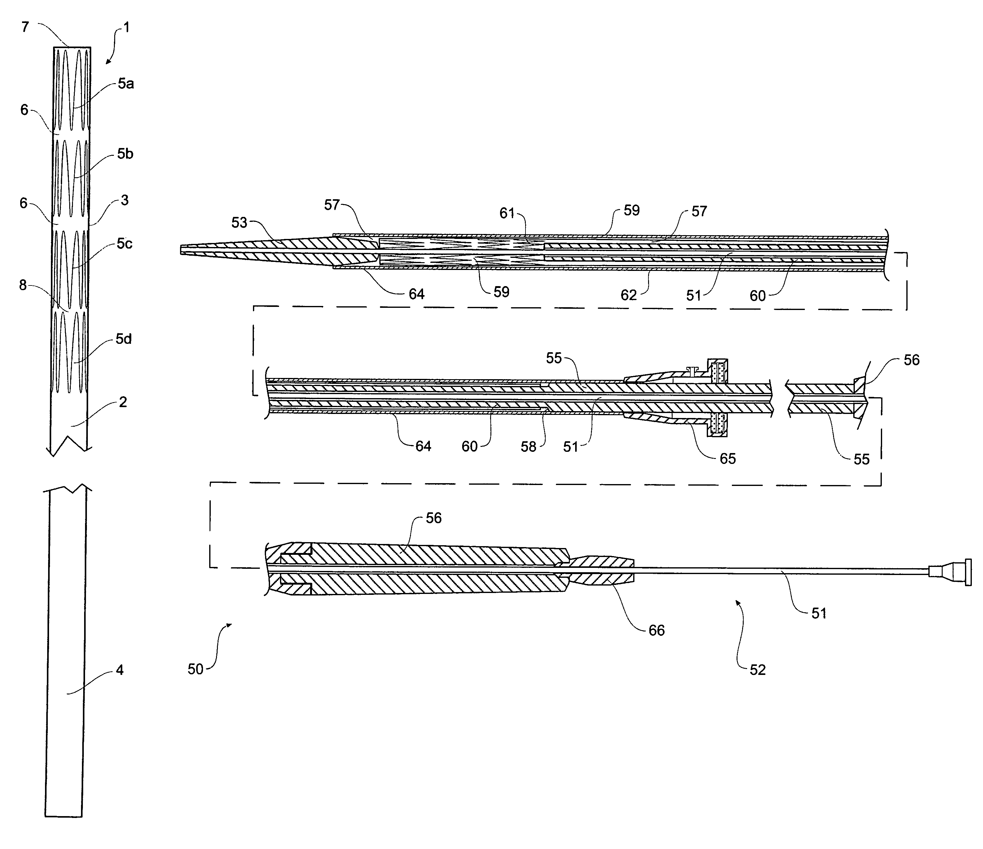
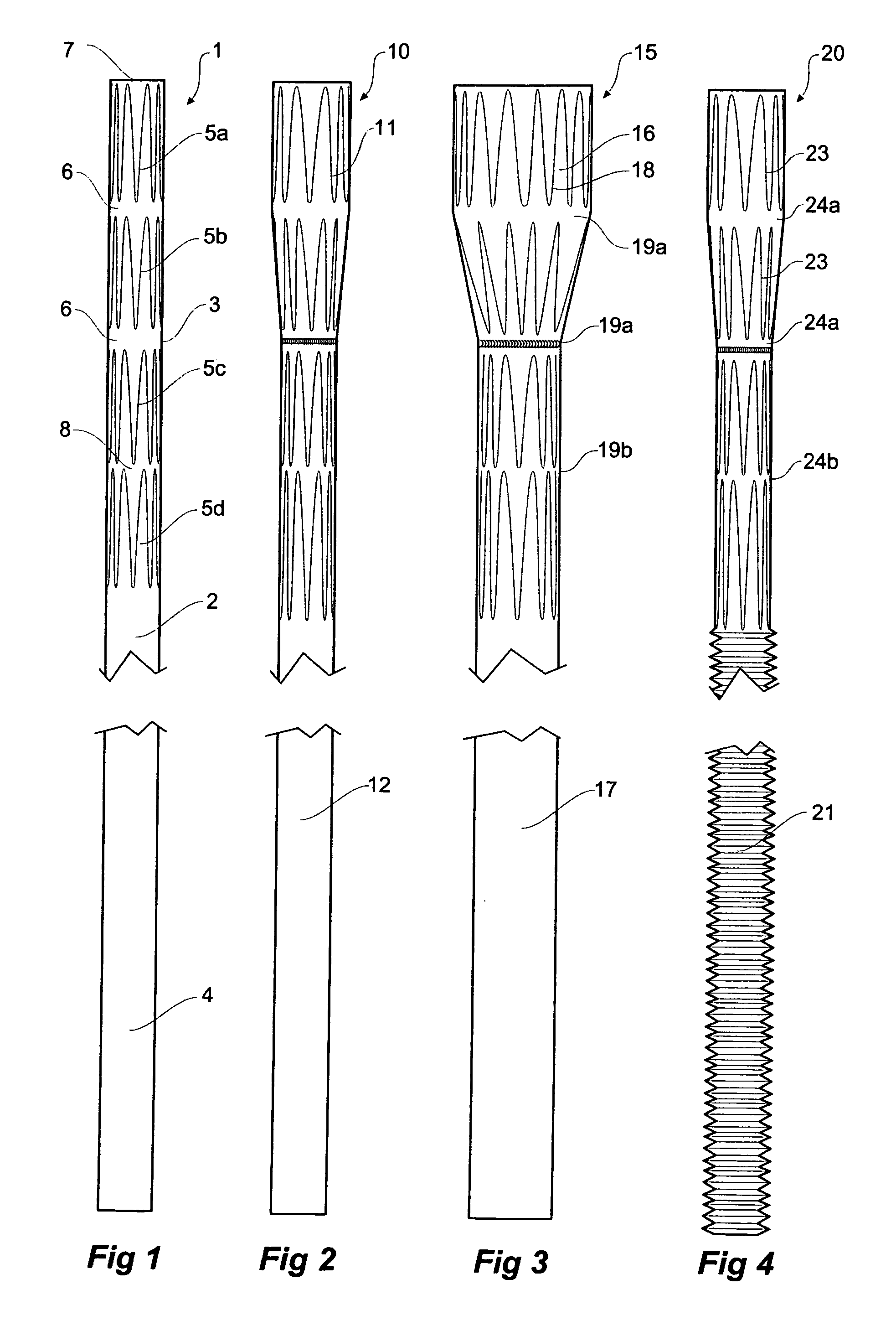
[0059]Now looking more closely to the drawings, and in particular FIG. 1, it will be seen that the laparoscopic conduit 1 according to one embodiment of the present invention comprises a elongate biocompatible graft material tube 2 which has a stented proximal portion 3 and an elongate unstented distal portion 4. There are four stents 5a to 5d in the proximal stented portion 3 with wide gaps 6 between the first and second stents 5a and 5b and second and third stents 5b and 5c from the proximal end 7 and a narrow gap 8 between the third and fourth stents 5c and 5d. In a preferred embodiment the tube has a diameter of 10 mm, the stents are 17 to 22 mm long each and the wider gaps are 3 mm and the narrower gap is 1 mm. The laparoscopic conduit 1 may have an overall length of about 400 mm.

[0060]When deployed, the laparoscopic conduit 1 fits into a vessel of the human or animal body with the wall of the vessel being received in one of the gaps 6 with the stents either side acting to seal...


PUM


Abstract

A laparoscopic vascular conduit arrangement has an elongate graft tube (1, 10) which is placed through a body cavity into a body vessel using a laparoscopic port sheath (70) with a distal valve (73) and a second sheath (80) with distal valve (86, 87). The graft tube (1, 10) extends through the laparoscopic port sheath so that the distal end (108) of the graft tube extends out of its valve and the proximal end of the graft tube extends out of the proximal end of the laparoscopic port sheath. The second sheath (80) is deployed into the distal end (108) of the graft tube (1, 10) such that the first valve (73) seals around the graft tube and the second sheath (84). The proximal end of the graft tube can be deployed into the vessel of the body to allow access into the vessel through the graft tube via the second valve. The conduit can be removed after use or used to provide vascular bypass.

Description

CROSS-REFERENCE TO RELATED APPLICATIONS[0001]This application claims priority of provisional application Ser. No. 60 / 679,612, filed May 10, 2005.TECHNICAL FIELD[0002]This invention relates to a medical device and more particularly to a medical device and arrangement for performing laparoscopic vascular access.BACKGROUND OF THE INVENTION[0003]Endovascular access using arteries close to the surface of the skin of a patient has been well practised in recent years. The Seldinger method of access to the aorta through a femoral and iliac artery has been well practised but there are problems when the femoral and iliac arteries are convoluted, too small for access or occluded and in such situations a physician must resort to open surgery.[0004]Endoscopic techniques to provide access into the various body cavities are also well known. It is an object of this invention to provide a system and equipment for endoscopic controlled and guided vascular access and in particular laparoscopic control...

Claims


Application Information

Patent Timeline
Patent Type & Authority Patents(United States)
IPC IPC(8): A61F2/06
CPCA61F2/95A61F2/966A61F2/07A61M39/06A61M39/0613A61M2039/0653A61M2039/0673A61M2039/0686A61B17/3423
Inventor FEARN, SHIRLEY JANEHARTLEY, DAVID ERNESTLAWRENCE-BROWN, MICHAEL
Owner WILLIAM A COOK AUSTRALIA
Why Eureka
Try Eureka