Apparatus and method for X-ray treatment

a radiation therapy apparatus and x-ray technology, applied in the direction of diagnostics, therapy, instruments, etc., can solve the problems of difficult to achieve rapidity and high resolution together, apparatus cannot track the moving treatment object part in real time, and it is difficult to improve the rapidity any further in the radiation therapy apparatus. to achieve the effect of accurate irradiation to the lesion

Inactive Publication Date: 2013-01-22
ACCUTHERA
View PDF24 Cites 3 Cited by
  • Summary
  • Abstract
  • Description
  • Claims
  • Application Information

AI Technical Summary

Benefits of technology

[0012]In a first aspect in accordance with the present invention, an X-ray treatment apparatus irradiates X-rays which intersect and penetrate a lesion from different directions to a patient who lies on a couch. The X-ray treatment apparatus includes a low energy X-ray generator and a marker sensor which detect a position of a marker fixed in the patient to the couch while keeping the predetermined position, the marker sensor is composed of an image sensor corresponding to the low energy X-ray generator, a couch operative means to make change the position and posture of the couch, an X-ray source for treatment controlling the profile and the dosage of the X-ray beam by deflecting the electron beam, an X-ray sensor for treatment detecting the X-ray which is irradiated from the X-ray source for treatment and penetrates the lesion, an X-ray source operative means to support the X-ray source and to control the position and posture of the X-ray source for treatment, a central processing unit which outputs the control signal to the X-ray source for treatment and other operative means after calculating the profile and the dosage of the X-ray source for treatment according to the position of the lesion specified by the output of the marker sensor based on the treatment plan, moreover the central processing unit modifies the treatment plan according to the output data of the X-ray sensor for treatment and outputs the next control signal to the X-ray source for treatment and other operative means. According to this X-ray treatment apparatus, because the marker is fixed in the patient while keeping the predetermined position to the lesion, the position of the lesion is specified by detecting the marker, the X-ray source for treatment can accurately irradiate to the lesion.
[0016]In a fifth aspect in accordance with the present invention, an X-ray treatment apparatus includes preferably an X-ray source for treatment which the activity of an X-ray target tube in the X-ray source is selected according to a treatment plan. The accurate treatment to the complicated shaped lesion is enabled by the X-ray treatment apparatus because a profile and condition of the X-ray for treatment which can be freely changed in real time by selecting the condition of the activity of each the X-ray target tube. Moreover, the X-ray treatment apparatus can improve the quality of treatment and shorten the treatment time.
[0020]In a ninth aspect in accordance with the present invention, an X-ray treatment method using the X-ray treatment apparatus includes preferably the steps of modifying a beam profile, a dosage and an radiation direction of an X-ray for treatment according to the latest data of the image data of the X-ray sensor for treatment or / and the image data of marker sensor. By this, the cyclic treatment is enabled accurately while changing the condition of the radiation, and reduces the patient's mental and physical burdens.

Problems solved by technology

In this apparatus, because the geometric accuracy of the radiation field depends on the setting accuracy of the aperture shape of the collimator, it is difficult to realize both rapidity and high resolution together.
However, the apparatus cannot track the moving treatment object part in real time.
However, it is difficult to improve the rapidity any further in this radiation therapy apparatus, because the rapidity of the continuous radiation is limited by the place recognition of the tumor markers and the settling time of the collimator.
Even more particularly, in the case of radiation to the edge part of the tumor, it is difficult to avoid the normal tissues because the collimator is used, and normal tissues are damaged.
However, it is difficult to improve the rapidity any further in this radiation therapy apparatus, because the rapidity of the continuous radiation is limited by the position recognition of the tumor markers and the settling time of the collimator.
Even more particularly, in the case of radiation to the edge part of the tumor, it is difficult to avoid normal tissues because the collimator is used, and normal tissues are damaged.
The patient's mental and physical burdens are heavy because it is not possible to irradiate continuously and it takes time for the treatment.
The accuracy of verification of the consequences of the X-ray treatment is inferior and complicated.
Because the diameter of the X-ray beam cannot be thin, it is difficult to completely remove tumors without damaging normal tissue.

Method used


Image

  • Apparatus and method for X-ray treatment
  • Apparatus and method for X-ray treatment
  • Apparatus and method for X-ray treatment

Examples


Embodiment Construction

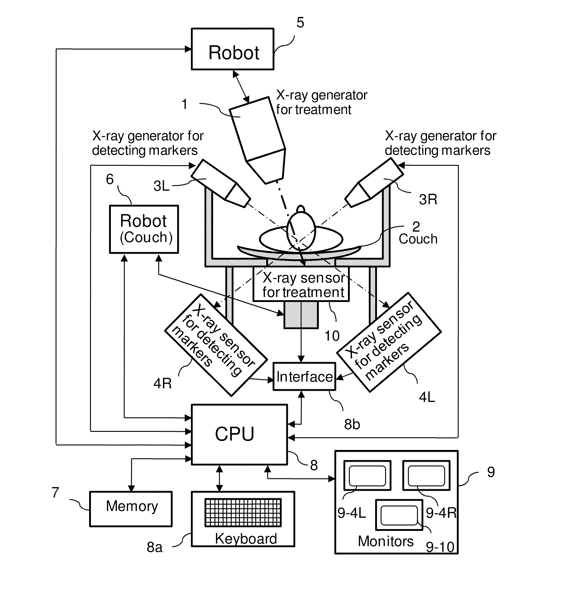
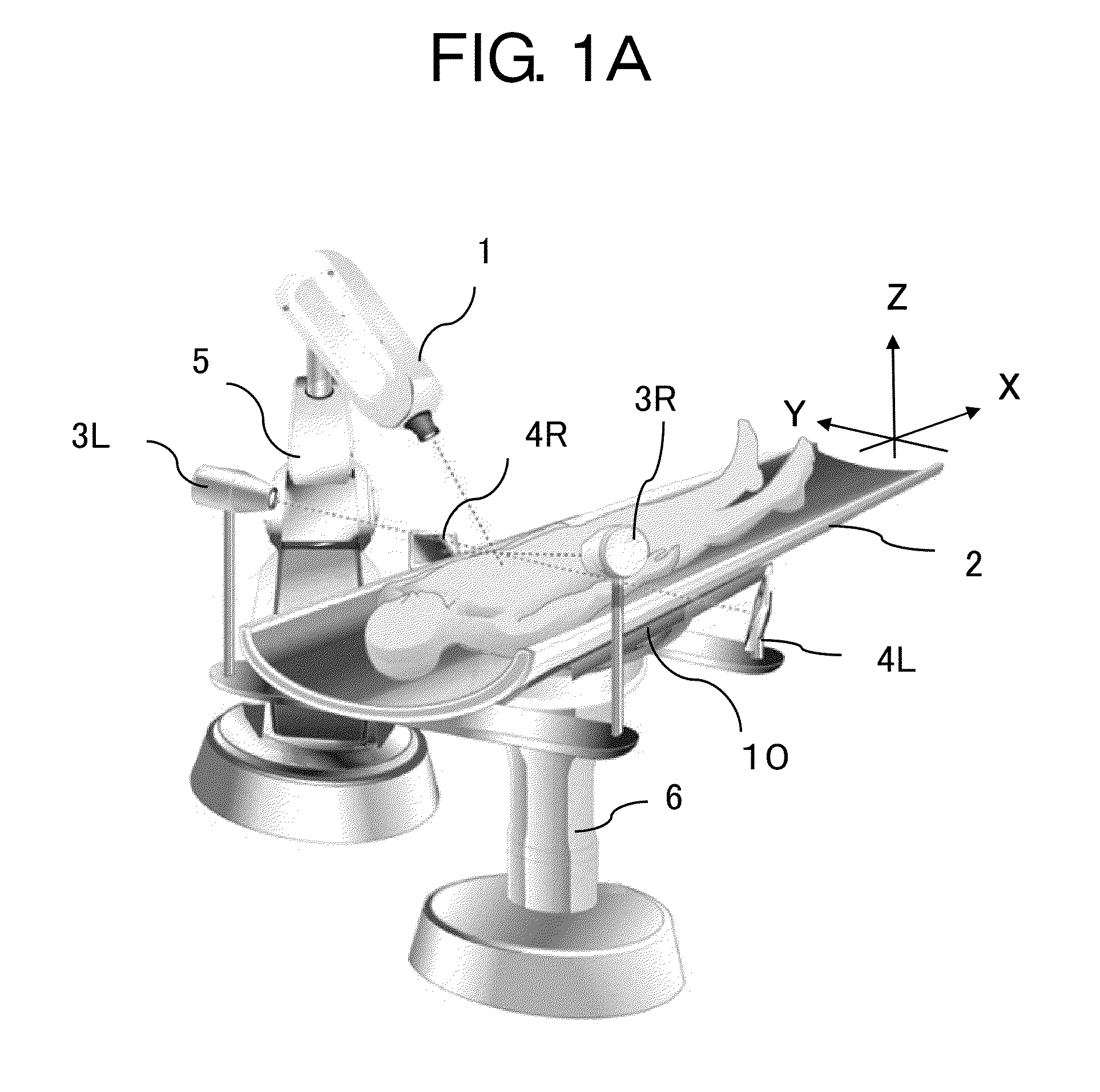
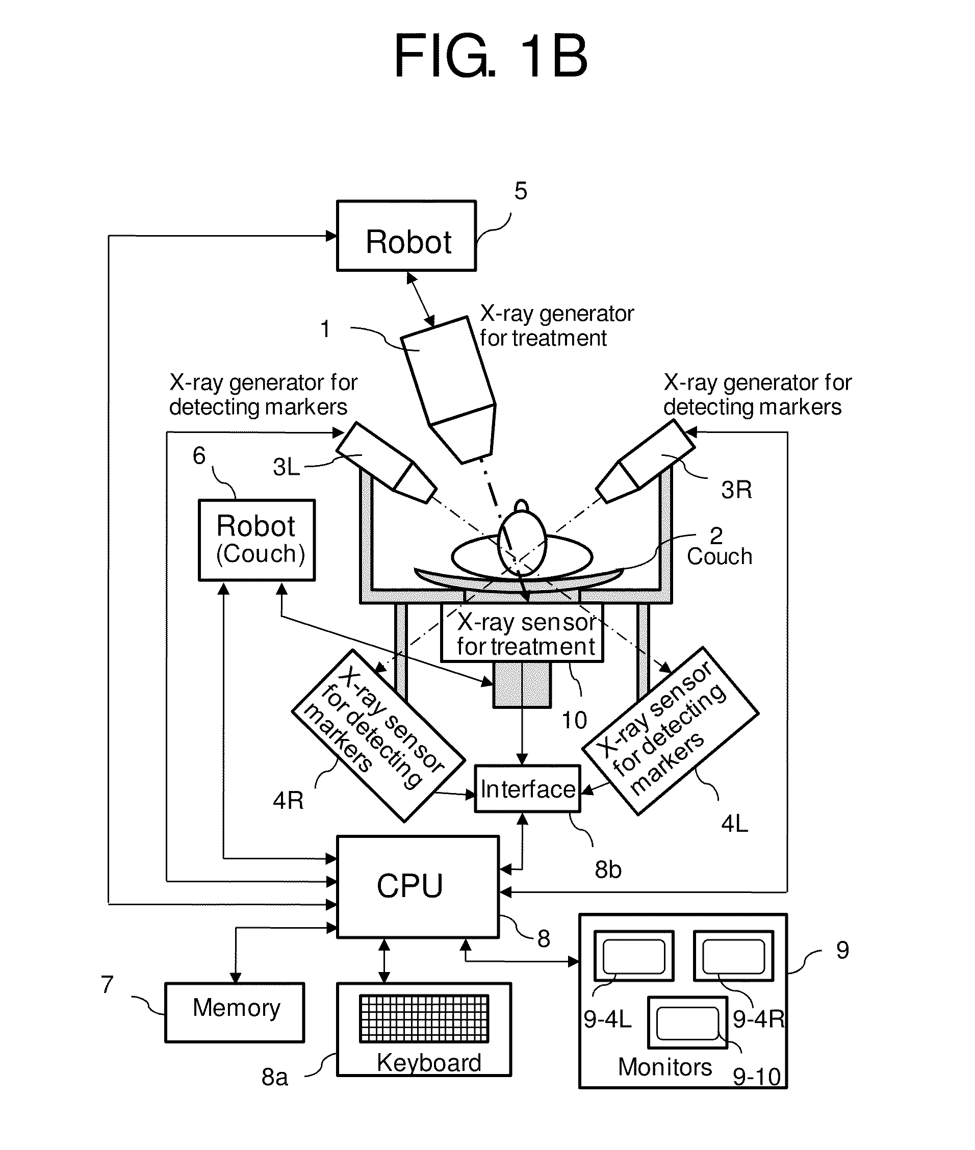
[0033]FIG. 1A shows a perspective view showing the X-ray treatment apparatus in accordance with an embodiment of the present invention, and FIG. 1B shows a block diagram of the X-ray treatment apparatus. The following detailed description is of a case in which the lesion is a tumor.

[0034]The marker is fixed in the vicinity of a tumor in the patient while maintaining a predetermined position relative to the couch. The material of the marker is a heavy metal such as Au or Pt which is safe for the human body and absorbs X-rays well.

[0035]The marker sensor includes two low energy X-ray generators 3L, 3R, which are installed in the couch 2 and are turned to the tumor to detect the position of a marker, and two image sensors 4L, 4R corresponding to each low energy X-ray generator. The marker sensor collects the information about the tumor, which keeps the predetermined position relative to the marker and the three-dimensional position of the marker relative to the couch 2.

[0036]The positi...


PUM


Abstract

An X-ray treatment apparatus comprises a low energy X-ray generator for detecting a marker, a marker sensor detecting a position of the marker fixed in the patient to a couch, and both low energy X-ray generator and the marker sensor are installed in the couch, a high energy X-ray generator for treatment, a X-ray sensor for treatment detecting the high energy X-ray for treatment. An X-ray treatment method using the X-ray treatment apparatus comprises the steps of detecting a position of a marker by the marker sensor, irradiating to a lesion the high energy X-ray for treatment, detecting the penetrated high energy X-ray for treatment by the X-ray sensor for treatment, modifying the beam profile, the dosage or / and the radiation direction of the X-ray for treatment according to the latest data of the sensors, performing the next radiation for the lesion.

Description

BACKGROUND OF THE INVENTION[0001]1. Field of the Invention[0002]The present invention relates to an apparatus for X-ray treatment and a treatment method using the apparatus. More particularly, the present invention relates to an apparatus and a method which can prevent the destruction of normal tissue with a new technique called MMRT (Microbeam Modulated Radiation Therapy).[0003]2. Description of the Related Art[0004]Disadvantageously, X-rays in an X-ray treatment are radiated to any normal tissue other than a lesion because the X-rays are radiated to the lesion through the normal tissue and the lesion moves by the breathing and other motion of the patient also. Various kinds of measures are effected for this problem.[0005]Japanese Patent No. 3,790,481 discloses a radiation treatment apparatus which comprises a radiation generator radiating X-rays for treatment, two radiation sources irradiating X-rays for inspecting, two or more detectors, an image forming apparatus, and an analyze...

Claims


Application Information

Patent Timeline
Patent Type & Authority Patents(United States)
IPC IPC(8): A61N5/01A61N5/10H05G1/70A61B6/04G21K5/10
CPCA61N5/1049A61N2005/1061A61N5/1083
Inventor TANABE, EIJI
Owner ACCUTHERA
Why Eureka
Try Eureka