Compression device for enhancing normal/abnormal tissue contrast in MRI including devices and methods related thereto

a compression device and contrast technology, applied in the field of magnetic resonance imaging, can solve the problems of cancer, potentially life-threatening, and difficult to detect tumors that are too deep or too small, and achieve the effects of improving contrast-to-noise ratio, prolonging scan times, and improving resolution

Active Publication Date: 2013-02-19
THE JOHN HOPKINS UNIV SCHOOL OF MEDICINE
View PDF4 Cites 7 Cited by
  • Summary
  • Abstract
  • Description
  • Claims
  • Application Information

AI Technical Summary

Benefits of technology

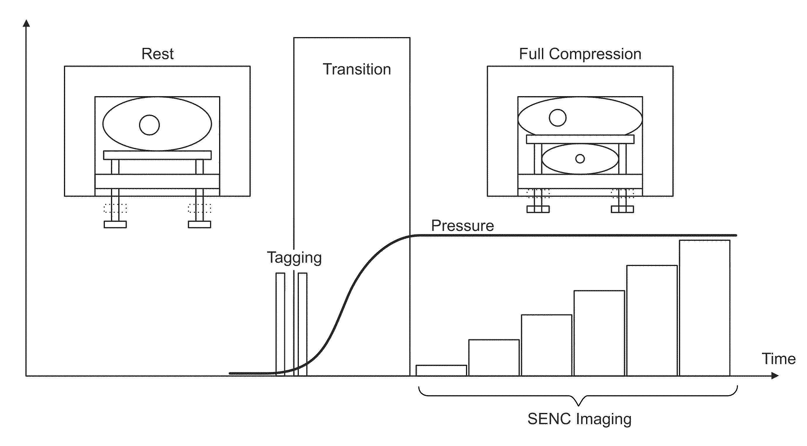
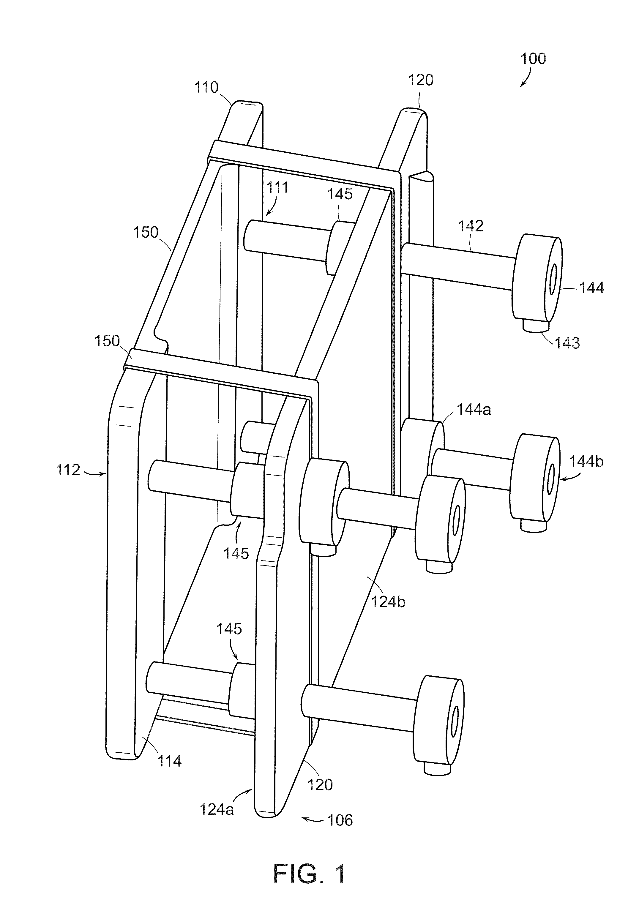
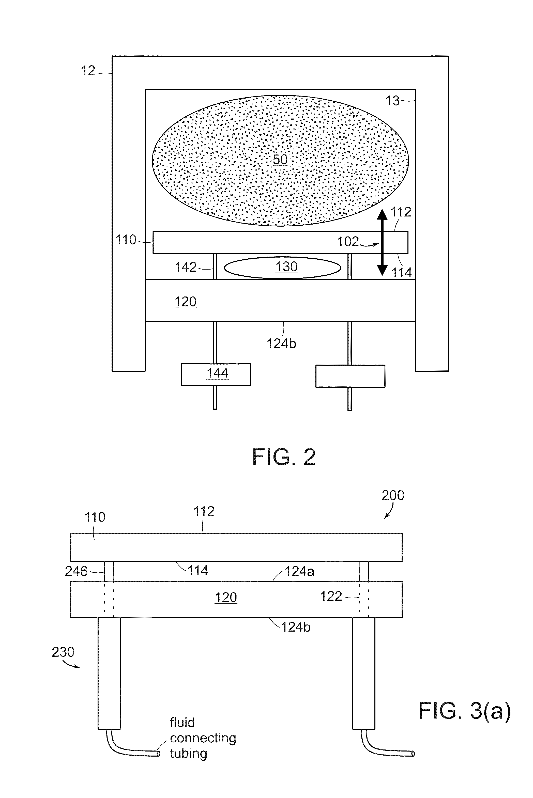
[0023]In particular embodiments, the controller controls flow of fluid to each of the one or more expandable members so as to thereby control the expansion of the one or more expandable members. More particularly, the controller is configured to cause fluid to flow to each of the one or more expandable members to move the moveable member with respect to the stationary member and to terminate fluid flow when a desired expansion of the one or more expandable members is achieved and so as to maintain the pressure in each of the one or more expandable members. Also, the controller is configured so as to cause fluid to flow from each of the one or more expandable members, so as to thereby reduce the compressive force on the target tissue. Also, it is within the scope of the present invention for a switch to be interconnected to the controller, which switch is operable by the patient and provides an output signal to the controller when so activated. In this way, if the compressive pressure on the tissue is creating excessive discomfort or the like to the patient, an output signal is provided to the controller which in turn can stop flow of fluid to the one or more expandable members and / or also cause fluid to flow from each of the one or more expandable members, so as to thereby reduce the compressive force on the target tissue.
[0025]According too yet a further aspect of the present invention there is featured a compression device for use in combination with MRI imaging techniques and MRI apparatuses as is known to those skilled in the art. Such a compression device according to this aspect that can periodically and repetitively compress the target tissue (e.g., breast tissue) whereby MRI image data using the Strain Encoded MRI technique (SENC). Such a compression device allows one to prolong scan times because a scan can be done for each of the plurality or multiplicity of periodic compressions. Such a device and the methods related thereto should lead to higher resolutions and better signal-to-noise ratio (SNR), and contrast-to-noise ratio (CNR). Moreover, such a device and related methods allow one to measure the compression and relaxation response of tissues in the non-linear region by compressing the tissues in the range of from about 10% to about 30%.

Problems solved by technology

The ability of the cancer cells to multiply continuously and spread from the breast to other organs identifies it as “malignant,” and potentially life threatening.
Nevertheless, palpation is still a subjective procedure, and detecting tumors that are too deep or too small is still problematic.
Ultrasound techniques, however, suffer from a number of drawbacks.
In addition, the off-axis and elevational tissue motion can severely corrupt the axial-strain estimation by inducing decorrelation noise which requires sophisticated correction. F. Kallel, T. Varghese, J. Ophir and M. Bilgen, “The non-stationary strain filter in elastography.
MRI can provide features that are difficult or even impossible to implement in ultrasound.
In addition to the unavoidable prolonged scan times, multiple compressions require a special device to produce exactly the same compression in every cycle, which may hinder the clinical use of these techniques.
Although the acquisition of a phase reference map is not troublesome when using multiple compression cycles, it constitutes a barrier towards the use of a single compression.
The MR tagging techniques do not require the correction of the background phases, however, because the k-space of a tagged image has a large bandwidth (due to the modulation of the intensity with a highly alternating pattern), acquisition of such images takes longer time than phase contrast or stimulated echo based techniques.
Unfortunately, the specificity (wrong-positive cases) of MRI is reported to be low and highly dependent on the imaging, processing, and interpretation technique.
Therefore, the challenge in MR breast imaging is to develop methods to minimize false positives and to more easily evaluate and / or localize malignant tumors. L. Esserman, “Integration of imaging in the management of breast cancer,”J Clin Oncol, vol.
In addition to the unavoidable prolonged scan times, multiple compressions require a special device to produce exactly the same compression in every cycle, which may hinder the clinical use of these techniques.

Method used


Image

  • Compression device for enhancing normal/abnormal tissue contrast in MRI including devices and methods related thereto
  • Compression device for enhancing normal/abnormal tissue contrast in MRI including devices and methods related thereto
  • Compression device for enhancing normal/abnormal tissue contrast in MRI including devices and methods related thereto

Examples


example 1

SENC MRI

[0167]The Strain Encoded (SENC) MRI technique was introduced to measure local strain distribution of deforming tissues. In SENC MRI, the magnetization of the object under examination at point p and time t is modulated in the z-direction with a sinusoidal pattern with the spatial frequency, ω(p,t). The z-direction here is defined as the direction orthogonal to the imaging plane. Once induced, this pattern lasts for a fraction of a second, during which, if the tissue is deformed, the frequency ω(p,t) proportionally changes with the degree of deformation at the pixel p. The resulting image intensity at this pixel is given by

[0168]I⁡(p_,t)≈12⁢ρ⁡(p_,t)⁢S⁡(ωT-ω⁡(p_,t)),(1)

[0169]where ρ(p,t) is a term representing the proton density of the voxel including the T1 relaxation effect, S(ω) is the Fourier transform of the slice profile determined by the envelope of the applied slice selection RF pulse and ωT is called the tuning frequency, which is determined during the image acquisitio...

example 2

[0190]A phantom experiment was used in order to test the tissue compression device of the present invention. The phantom was made of a silicon gel material prepared by mixing two compounds, A and B (kit 3-1450, Dow Corning). The ratio in the mixture determines the stiffness of the resultant gel. The phantom was composed of a small stiff sphere 1 cm in diameter immersed in a cube made from softer gel material with dimensions 8×8×10 cm3. A top-view of the phantom is shown in FIG. 19. It is worth noting that the stiff inclusion was intentionally placed near the corner of the phantom to test the ability of the system to detect lesions at the periphery of the breast (difficult situation). The stiffness of the inclusion was made very close to that of the surrounding soft gel. In fact, manual palpation on the surface of the phantom did not detect the existence of the inclusion.

Image Acquisition

[0191]The phantom experiment was done on a 3.0 T MR scanner (Achieva, Philips Medical Systems, Be...

example 3

[0254]Breast cancer is one of the most common causes of cancer related mortality. Early detection of breast lesions using mammography and ultrasound has resulted in lower mortality-rates. However, some breast lesions are mammography occult and the use magnetic resonance imaging (MRI) is recommended. MRI has high sensitivity and moderate specificity, thus, there is a need to increase specificity so that unnecessary biopsies procedures can be reduced. Higher specificity can be achieved by incorporating the stiffness property, as cancer tumors are 3-13 times stiffer than both normal tissue and benign tumors. Previously, fast strain-encoded (FSENC) MR with simple hardware was introduced to detect different stiffness by measuring the strain directly in a simple way; however, the simple hardware limited the scanning time thus limiting the imaging resolution. In this work, a new apparatus or device is introduced that is capable of periodically compressing the breast, which allows for longe...


PUM


Abstract

Featured are devices for compression of target tissue while magnetic resonance imaging the target tissue and methods and systems related thereto. The method includes disposing target tissue between the fixed surface and the moveable member of a compression device and compressing the target tissue between the fixed surface and the moveable member. The method also includes acquiring one or more, more specifically a plurality, of sequences of image data of the compressed target tissue using an MRI imaging technique (MRI). In particular embodiments, the MRI technique is a SENC MRI technique, where tissue encoding is done prior to compressing the tissue and acquiring includes adding a gradient moment in the slice-selection direction to cause demodulation with a specific frequency. In further embodiments, the sequences of image data are acquired one of during successive periodic compressions of the tissue or successive periodic relaxation of the tissues.

Description

FIELD OF INVENTION[0001]The present invention relates in general to techniques for magnetic resonance imaging and devices related thereto, in particular to techniques and devices used to enhance contrast, between normal and abnormal tissue, such as for example, tissue of a mammalian breast, and more particularly to compression devices that enhance contrast between normal and abnormal tissue.BACKGROUND OF THE INVENTION[0002]Breast cancer is the leading cause of death among women 40 to 44 years old, and is one of the leading causes of death in women of age 30 or more. According to the American Cancer Society, it is anticipated that more than 200,000 new cases of breast cancer will be developed in 2006 with approximately 40,000 estimated deaths. “Cancer Facts & Figures 2006, surveillance research,” American Cancer Society, Ed.: http: / / www.cancer.org / , 2006, pp. 4. Breast cancer results from uncontrolled growth of abnormal cells in the breast. The ability of the cancer cells to multiply...

Claims


Application Information

Patent Timeline
Patent Type & Authority Patents(United States)
IPC IPC(8): A61B5/05
CPCA61B5/702G01R33/30G01R33/56333G01R33/56358
Inventor OSMAN, NAEL F.JACOBS, MICHAEL A.HAROUNI, AHMED ELHOSSAIN, JAKIR
Owner THE JOHN HOPKINS UNIV SCHOOL OF MEDICINE
Why Eureka
Try Eureka