Radiographic imaging apparatus and processing method therefor

a technology of radiographic imaging and processing methods, applied in the field of radiographic imaging apparatus, can solve the problems of not being able to execute the intended imaging, not being able to change to a pre-scheduled imaging protocol, and not being able to consider the effect of the imaging device, so as to achieve the effect of easy adjustmen

Active Publication Date: 2013-07-23
CANON KK
View PDF3 Cites 6 Cited by
  • Summary
  • Abstract
  • Description
  • Claims
  • Application Information

AI Technical Summary

Benefits of technology

[0009]The present invention provides a technique which can easily cope with even a situation in which imaging based on an examination order has been executed by using a wrong imaging protocol.

Problems solved by technology

In this case, since the set parameters are not appropriate, the apparatus cannot execute the intended imaging.
This technique, however, takes no consideration of a situation in which an examination proceeds based on an examination order, and hence does not allow changing to a pre-scheduled imaging protocol.
Even if, therefore, an examination order includes an imaging protocol as a change destination, it is not possible to change to that imaging protocol.

Method used


Image

  • Radiographic imaging apparatus and processing method therefor
  • Radiographic imaging apparatus and processing method therefor
  • Radiographic imaging apparatus and processing method therefor

Examples


first embodiment

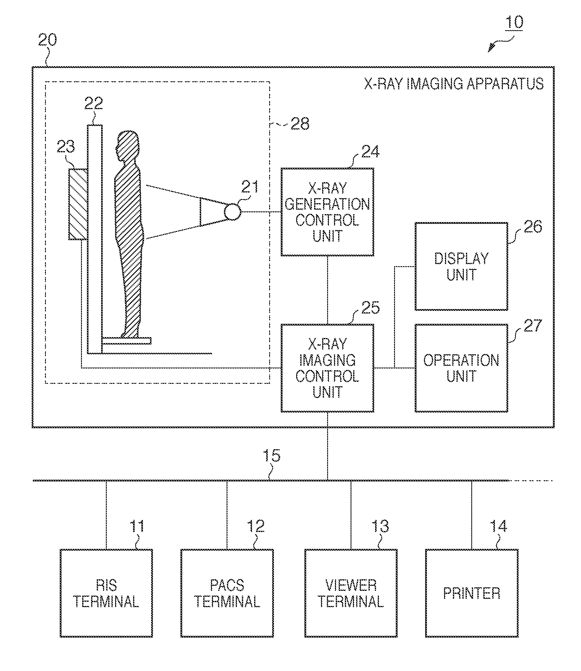
[0034]FIG. 1 is a block diagram showing an example of an X-ray imaging system 10 to which a radiographic imaging system according to an embodiment of the present invention is applied.

[0035]An X-ray imaging system 10 includes an RIS terminal 11, a PACS terminal 12, a viewer terminal 13, a printer 14, and an X-ray imaging apparatus 20. These apparatus are connected to each other via a communication 15 such as a network.

[0036]The RIS terminal 11 is an information system within a radiology department. The X-ray imaging apparatus 20 captures an X-ray digital image (to be referred to as a captured image hereinafter). The PACS terminal 12 stores and manages the images captured by the X-ray imaging apparatus 20. The viewer terminal 13 and the printer 14 output (display or print) a diagnosis image.

[0037]The X-ray imaging apparatus 20 executes an examination (imaging) based on an examination order constituted by a plurality of imaging protocols. In each imaging protocol, imaging conditions, t...

second embodiment

[0062]The second embodiment will be described next. The second embodiment will exemplify a case in which change conditions are provided at the time of changing an imaging protocol.

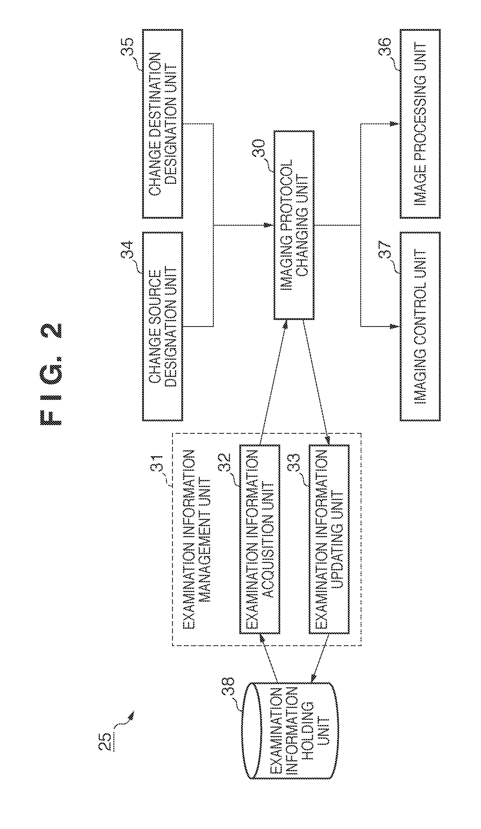
[0063]FIG. 5 is a view for explaining an example of the functional arrangement of an X-ray imaging control unit 25 according to the second embodiment. Note that the same reference numerals as in FIG. 2 explaining the first embodiment denote the same parts in FIG. 5.

[0064]A change condition input unit 52 inputs a change condition based on an instruction from the operator via an operation unit 27. A change condition holding unit 56 holds the change condition in correspondence with an imaging protocol. A change condition management unit 53 manages change conditions corresponding to each imaging protocol. The change condition management unit 53 includes a change condition registration unit 54 and a change condition acquisition unit 55. The change condition registration unit 54 registers the change condition in...

third embodiment

[0072]The third embodiment will be described next. The third embodiment will exemplify a case in which after an imaging protocol is changed, the information of the change source imaging protocol is handed over to the change destination imaging protocol.

[0073]FIG. 7 explains an example of the functional arrangement of an X-ray imaging control unit 25 according to the third embodiment. Note that the same reference numerals as in FIGS. 2 and 5 explaining the first and second embodiments denote the same parts in FIG. 7.

[0074]A handover information setting unit 62 makes setting to determine which information in the information associated with an imaging protocol is to be handed over. This setting is made based on an instruction from the operator via an operation unit 27. Note that handover information includes information associated with the generation of X-rays (for example, a tube current, tube voltage, and irradiation period), sensor information indicating the type of sensor (for exam...


PUM


Abstract

A radiographic imaging apparatus performs imaging based on an examination order including a plurality of imaging protocols, executes image processing for the captured image based on the imaging protocol used at the time of the imaging, designates a change source imaging protocol and a change destination imaging protocol from the examination order based on an instruction of an operator, and changes the imaging protocol corresponding to the image captured based on the change source imaging protocol from the change source imaging protocol to the change destination imaging protocol. When the change of protocol is made, the apparatus executes image processing based on the change destination imaging protocol for the image before the image processing which is captured based on the change source imaging protocol.

Description

BACKGROUND OF THE INVENTION[0001]1. Field of the Invention[0002]The present invention relates to a radiographic imaging apparatus and a processing method for the apparatus.[0003]2. Description of the Related Art[0004]Diagnoses and examinations based on imaging using radiation (for example, X-rays) have been actively practiced. Such an examination proceeds based on an examination order including a plurality of imaging protocols. In this case, an imaging protocol is manually created by, for example, a radiological technician in accordance with an examination order, and includes information such as the region of the patient's body to be imaged (the imaging region), imaging method, and image processing which are requested by the doctor.[0005]An increasing number of hospitals have been adopting an online system using an HIS (Hospital Information System), RIS (Radiology Information System), and the like. In accordance with this trend, an externally created examination order is sometimes t...

Claims


Application Information

Patent Timeline
Patent Type & Authority Patents(United States)
IPC IPC(8): G06K9/00
CPCA61B6/00A61B6/464G06T7/0012A61B6/465A61B6/501G16H40/60
Inventor WATANABE, YUKAKO
Owner CANON KK
Why Eureka
Try Eureka