Methods and systems for functional imaging of cardiac tissue

a functional imaging and cardiac tissue technology, applied in the field of imaging modalities, can solve the problems of not providing a fundamental understanding of the dynamics underlying the patient's cardiac rhythm, not being able to say with certainty, and current technology not allowing us to see action potential propagation

Active Publication Date: 2014-03-04
CORNELL UNIVERSITY
View PDF9 Cites 8 Cited by
  • Summary
  • Abstract
  • Description
  • Claims
  • Application Information

AI Technical Summary

Problems solved by technology

Current technology does not allow us to see action potential propagation deep within the walls of the heart.
While these tests do certainly yield a wealth of valuable information, they often do not provide a fundamental understanding of the dynamics underlying the patient's cardiac rhythm.
At the research level, this lack of a deep, panoramic view of action potential activity means that it can not be said with certainty that ventricular fibrillation (VF) is caused by multiple reentrant action potential waves, although of course it is strongly suspected.
The lack of clarity on these issues has greatly complicated the development of effective therapies for the prevention and treatment of several types of tachyarrhythmias, including VF.
This method does not as yet provide transmural information, however.
The spatial resolution of the plunge electrode method is, however, relatively low; furthermore, it is likely that the presence of these electrodes creates electrical heterogeneities that strongly modify the patterns of propagation.
Both transillumination and optical tomography techniques have some light penetration problems, thus potentially making them difficult to apply to thicker myocardial walls such as those found in canine or human ventricles.
Additionally, optical tomography cannot be applied in-vivo.

Method used


Image

  • Methods and systems for functional imaging of cardiac tissue
  • Methods and systems for functional imaging of cardiac tissue
  • Methods and systems for functional imaging of cardiac tissue

Examples


Embodiment Construction

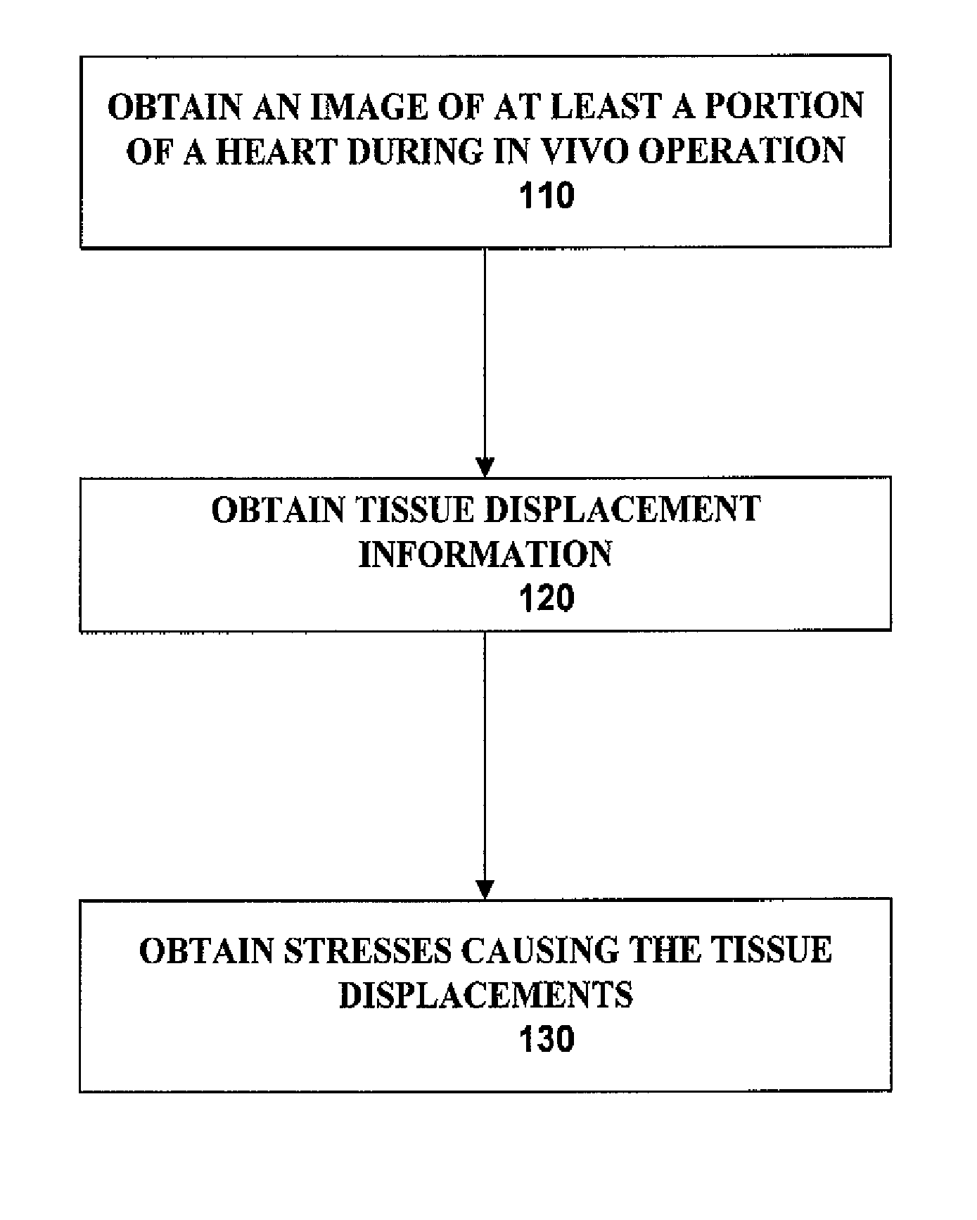
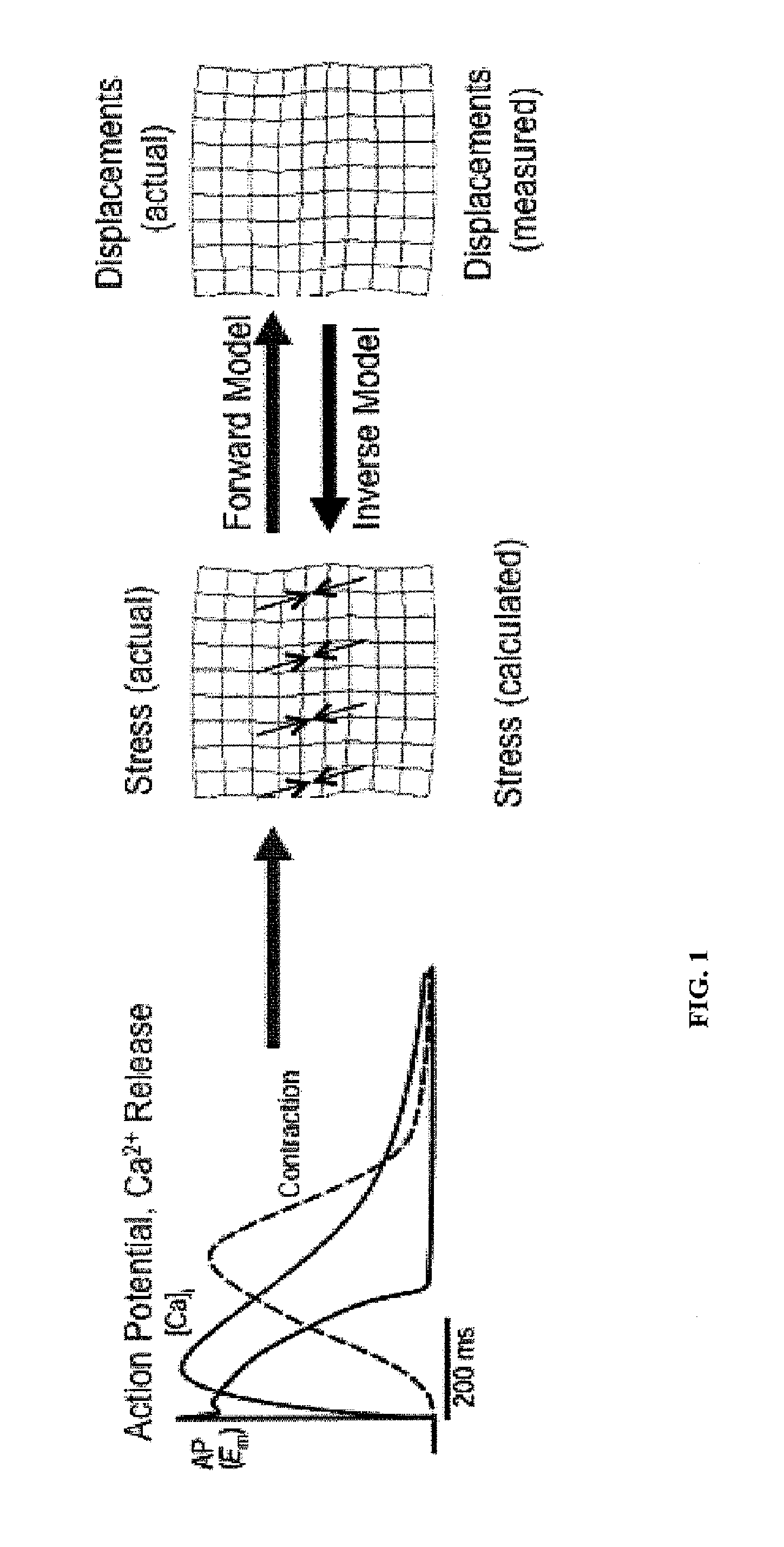
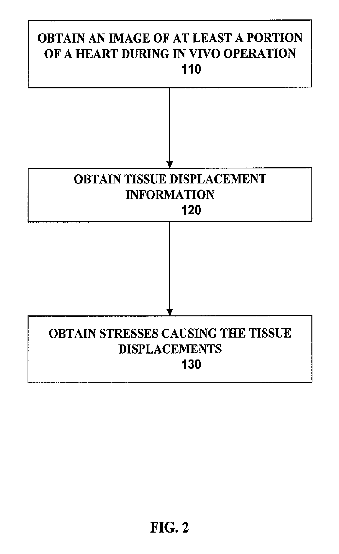
[0015]In one embodiment, the method of these teachings includes obtaining an image of at least a portion of a heart during in vivo or in vitro operation, obtaining, from the image, tissue displacement information, and obtaining, from the tissue displacement information, stresses causing the tissue displacements, whereby information on action potential inducing the stresses can be obtained. In one instance, the determination of stresses for each pixel or nodal point in a group of pixels or nodal points is obtained by operating on displacements for each pixel or nodal point in a group of pixels or nodal points rather than by operating on each pixel or nodal point separately.

[0016]One embodiment of these teachings includes a new imaging modality that is based on the ability of present day ultrasound to detect wave-induced tissue deformation at depth, that will allow the viewing of the propagation of action potentials deep within myocardial tissue, thereby helping to clarify these clini...


PUM


Abstract

One embodiment of these teachings includes an imaging modality that is based on the ability of imaging technologies to detect wave-induced tissue deformation at depth, that allows viewing the propagation of action potentials deep within myocardial tissue, thereby helping to clarify clinical and physiological dynamical issues.

Description

CROSS REFERENCE TO RELATED APPLICATIONS[0001]This application is the U.S. national phase entry under 35 U.S.C. §371 of International Patent Application No. PCT / US10 / 47767 filed Sep. 3, 2010, entitled METHODS AND SYSTEMS FOR FUNCTIONAL IMAGING OF CARDIAC TISSUE, which in turn claims priority from U.S. Provisional Application Ser. No. 61 / 239,982, filed Sep. 4, 2009, entitled FUNCTIONAL ULTRASOUND IMAGING OF CARDIAC TISSUE, both of which are incorporated by reference herein in their entirety for all purposes.BACKGROUND[0002]These teachings relate generally to imaging modalities based on the ability of imaging technologies to detect wave-induced tissue deformation.[0003]Some cells, referred to as excitable cells, can reverse their resting membrane potential from negative values to slightly positive values. This rapid change in membrane potential is referred to as an action potential. Typically, the action potential is the result of a rapid change in membrane permeability to certain ions...

Claims


Application Information

Patent Timeline
Patent Type & Authority Patents(United States)
IPC IPC(8): A61N1/00G06K9/00
CPCA61B8/0883A61B8/08A61B8/485
Inventor OTANI, NIELS F.LUTHER, STEFANGILMOUR, JR., ROBERT F.SINGH, RUPINDER
Owner CORNELL UNIVERSITY
Why Eureka
Try Eureka