
Method for prediction of the progression risk of tumors

a tumor progression and tumor technology, applied in the direction of instruments, biochemistry apparatus and processes, material analysis, etc., can solve the problems of difficult to predict which particular lesion is prone to regress and which will progress to invasive cancer, and impose problems on the diagnosing physician and the patien

Active Publication Date: 2014-05-20
VENTANA MEDICAL SYST INC
View PDF9 Cites 12 Cited by
  • Summary
  • Abstract
  • Description
  • Claims
  • Application Information

AI Technical Summary

Benefits of technology

The present invention provides a method for predicting the potential for aggressive growth and the risk to progress to high grade cancer for tumors, by detecting overexpression of cyclin-dependent kinase inhibitor gene products in a cell based detection procedure. The method may also involve detecting specific staining patterns of overexpression in immuno-histochemistry. Preparations of probes for diagnosis are also provided.

Problems solved by technology

Generally the detection of low grade lesions imposes problems to the diagnosing physician and on the patient.
The dilemma with low grade lesions in practice is that it is hard to predict which particular lesion is prone to regress and which will progress to invasive cancer.

Method used


Image

  • Method for prediction of the progression risk of tumors

Examples


example 1

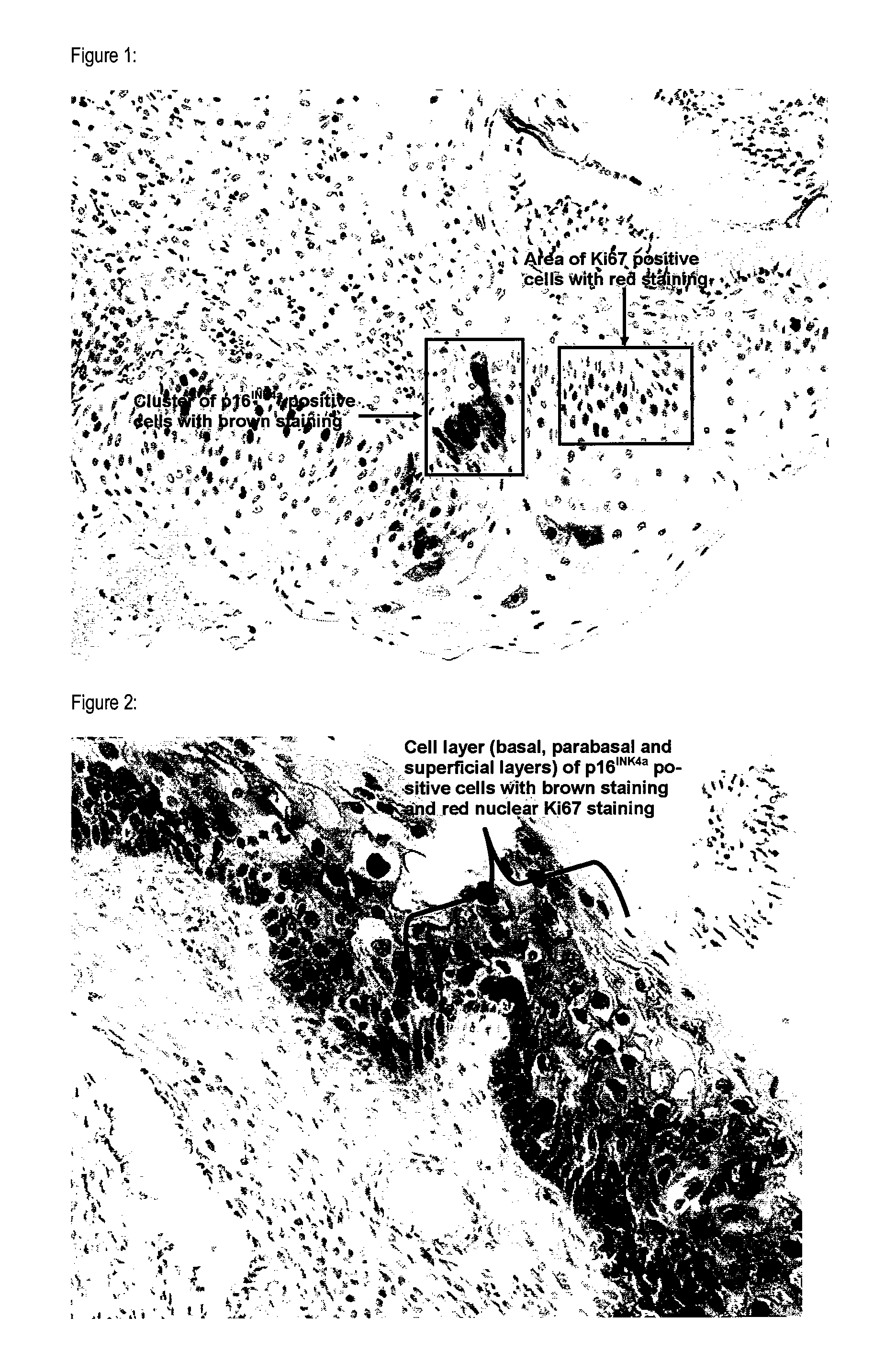
Prediction of the Progression Risk of Histological Specimens Obtained from the Cervix Uteri Classified as CIN 1 by Detection of p16INK4a Protein and Ki67 Protein

[0071]Archival histological samples (sections generated from neutral buffered formalin fixed, paraffin embedded punch biopsies) of the cervix uteri were immuno-histochemically stained using antibodies specific for p16INK4a and Ki-67. In total 10 specimens have been stained. Specimens with available follow up data have been chosen. For 6 specimens follow up examinations revealed regression of the CIN 1. For 4 specimens follow up examinations revealed progression of the CIN1 lesions to high grade dysplastic lesions. For all 10 specimens also HPV data were available or were generated during the experiments performed.

[0072]All specimens were treated as follows.

[0073]Tissue blocks were sectioned into slices of 4 μm thickness. Prior to deparaffinization slides have been placed in a drying oven at a temperature of 60° C. for a time...

example 2

Prediction of the Progression Risk in Breast Biopsies by Detection of p16INK4a Protein and Different Cell Proliferation Marker Proteins

[0094]In total 15 archival biopsy specimens of breast tumors (ductal carcinoma in-situ and lobular carcinoma in situ specimens) were used for the present experiment. 10 of the tested specimens were of less aggressive tumors, 5 stem from more aggressive tumor types. Progression risk and aggressiveness of the tumors were proven by follow up data on the individual cases. For each sample five sections were subjected to different double staining experiments. In each experiment p16INK4a staining was combined with one of the following cell proliferation markers: Ki67, MCM2, KiS2, MCM5, topoisomerase-2-alpha.

[0095]Specimen preparation and staining was performed as described above for Example 1.

[0096]Also in this experiment microscopic inspection revealed, that double-staining of single cells was detected only in those specimens that had more aggressive chara...


PUM

PropertyMeasurementUnit
temperatureaaaaaaaaaa
timeaaaaaaaaaa
fluorescenceaaaaaaaaaa

Abstract

The present invention concerns a method for predicting the potential for aggressive growth and / or the risk to progress to high grade cancer for tumors in cell based detection procedures. In one aspect the invention concerns the detection of overexpression of cyclin-dependent kinase inhibitor gene products as a tool for predicting the progression risk and / or potential for aggressive growth of tumors. In a second aspect the invention concerns predicting the progression risk and / or potential for aggressive growth in tumors on the basis of the simultaneous co-detection of the presence of overexpression of cyclin-dependent kinase inhibitor gene products together with the expression of markers for active cell proliferation. Further the invention concerns preparations of probes for diagnosis namely for predicting the progression risk and / or the potential for aggressive growth of tumors.

Description

[0001]This application is a National Stage of International Application PCT / EP2009 / 006415, filed Sep. 4, 2009, published Mar. 11, 2010, under PCT Article 21(2) in English; which claims the priority of European Patent Application No. 08105235.9, filed Sep. 4, 2008.[0002]The present invention concerns a method for predicting the potential for aggressive growth and / or the risk to progress to high grade cancer for tumors in cell based detection procedures. In one aspect the invention concerns the detection of overexpression of cyclin-dependent kinase inhibitor gene products as a tool for predicting the progression risk and / or potential for aggressive growth of tumors. In a second aspect the invention concerns predicting the progression risk and / or potential for aggressive growth in tumors on the basis of the simultaneous co-detection of the presence of overexpression of cyclin-dependent kinase inhibitor gene products together with the expression of markers for active cell proliferation....

Claims


Application Information

Patent Timeline
Patent Type & Authority Patents(United States)
IPC IPC(8): G01N33/574
CPCG01N33/57496G01N33/57415G01N2800/50G01N2800/56C12Q2600/118C12Q1/6886G01N2333/4706G01N2333/4704
Inventor MARTIN, PETERRIDDER, RUDIGER
Owner VENTANA MEDICAL SYST INC
Learn More