Ophthalmologic imaging apparatus, method of controlling the same, and program

a technology of ophthalmologic imaging and control method, applied in the field of ophthalmologic imaging apparatus, can solve the problems of increased apparatus size, lack of flexibility of technique, high cost, etc., and achieve the effect of favorable manual focus operability

Inactive Publication Date: 2015-05-05
CANON KK
View PDF10 Cites 7 Cited by
  • Summary
  • Abstract
  • Description
  • Claims
  • Application Information

AI Technical Summary

Benefits of technology

[0011]The present invention provides an inexpensive ophthalmologic imaging apparatus having favorable manual focusing operability for anterior ocular segment imaging and fundus imaging.
[0013]The present invention can provide an inexpensive ophthalmologic imaging apparatus having favorable manual focusing operability for anterior ocular segment imaging and fundus imaging.

Problems solved by technology

According to the technique disclosed in Japanese Patent No. 4430378, however, the use of different focus areas for anterior ocular segment imaging and fundus imaging makes it necessary to ensure a large drive area for the focus lens, resulting in an increase in apparatus size.
In addition, this technique requires the barrel cam, resulting in high cost.
Furthermore, this technique lacks flexibility in terms of focus lens drive amount corresponding to the unit rotational angle of the barrel at the time of anterior ocular segment imaging and at the time of fundus imaging, and hence has the problem of a low degree of freedom in operability setting when performing manual focusing.

Method used


Image

  • Ophthalmologic imaging apparatus, method of controlling the same, and program
  • Ophthalmologic imaging apparatus, method of controlling the same, and program
  • Ophthalmologic imaging apparatus, method of controlling the same, and program

Examples


first embodiment

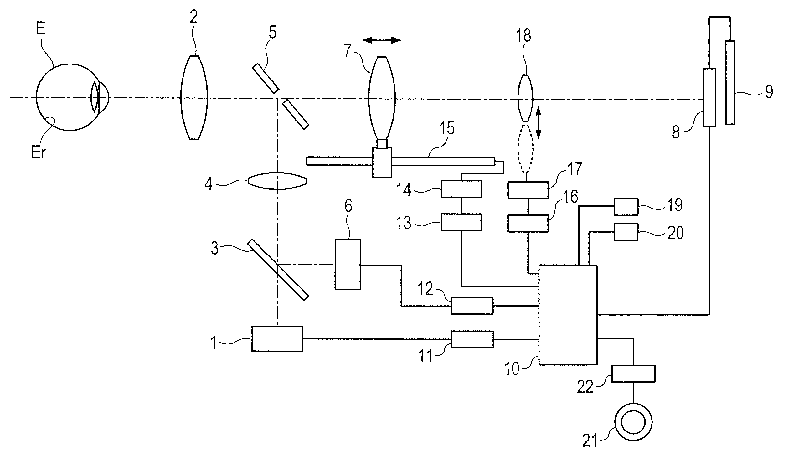
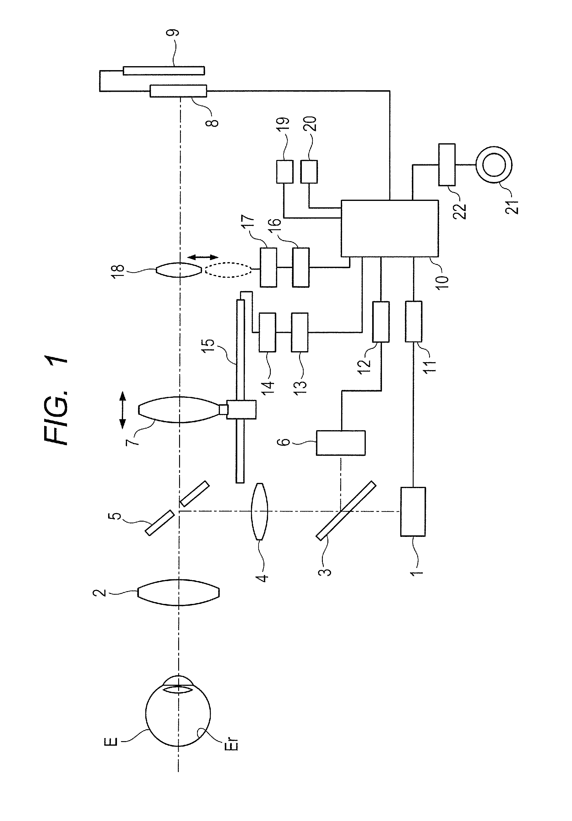
[0025]FIG. 1 is a view showing the arrangement of a fundus camera according to the first embodiment which is used as an ophthalmologic imaging apparatus. The fundus camera of this embodiment is a non-mydriatic fundus camera. The fundus camera located in front of an eye E to be inspected incorporates an observation illumination optical system ranging from an observation light source 1, which is formed from, for example f an infrared LED and emits infrared light, to an objective lens 2 located in correspondence with the eye E. In this observation illumination optical system, the observation light source 1, a dichroic mirror 3, a relay lens 4, and a perforated mirror 5 are sequentially arranged. An imaging light source 6 formed from a xenon tube is located as an imaging illumination optical system in the incident direction of the dichroic mirror 3.

[0026]A focus lens 7 which adjusts focus by moving in the optical axis direction is located as an imaging optical system behind the perforat...

second embodiment

[0058]The second embodiment of the present invention will be described next.

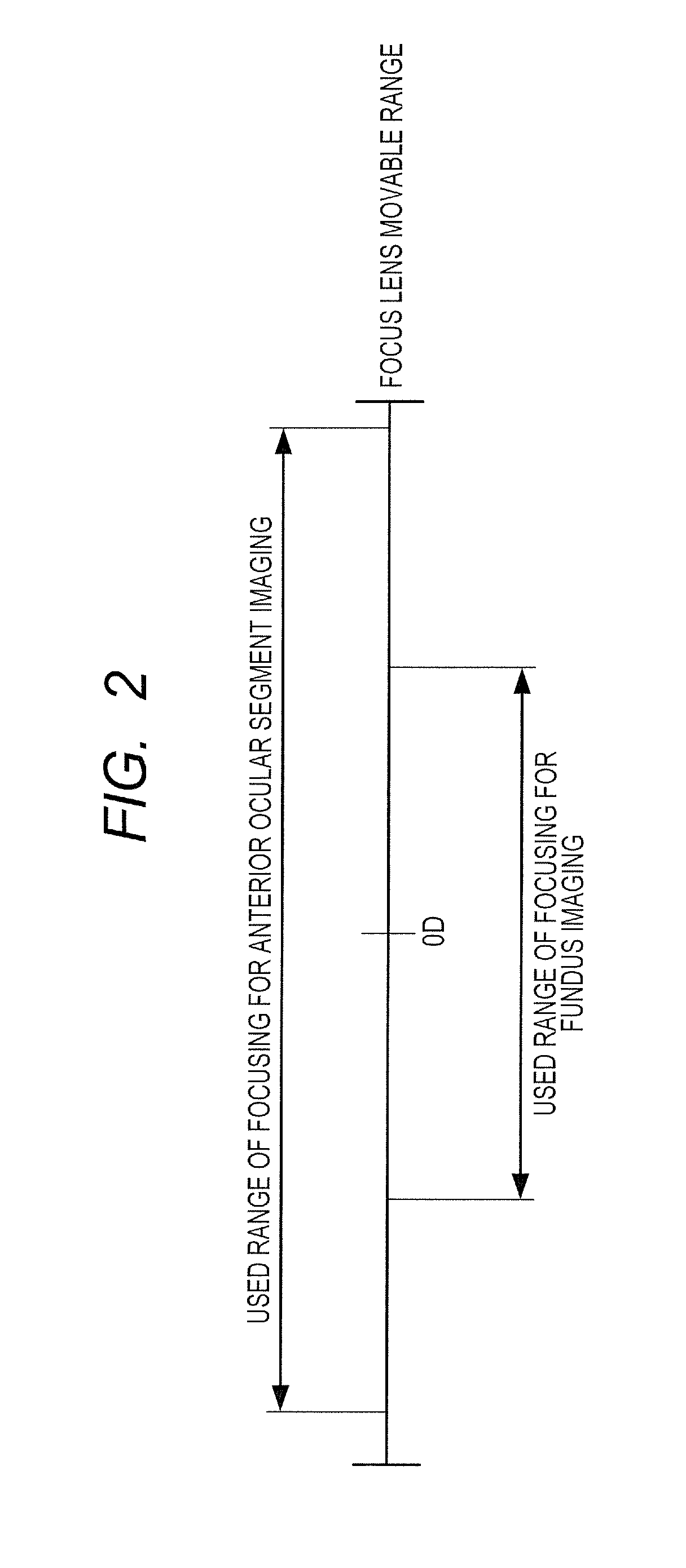
[0059]In anterior ocular segment imaging, operators differ in their regions of main interest, but a specific operator tends to perform imaging under predetermined conditions unique to him / her. In such a case, the operator does not always use the entire movable range of focusing for anterior ocular segment imaging, and hence allowing the operator to finely set a specific range of focusing will provide better usability.

[0060]The second embodiment has been made in consideration of this point and is configured to switch between at least two types of sensitivities for anterior ocular segment imaging operation, namely fine alignment and rough alignment.

[0061]The second embodiment will be described below with reference to FIG. 4. FIG. 4 is a view showing the arrangement of an ophthalmologic imaging apparatus according to the second embodiment. The same reference numerals as in FIG. 1 denote the same components in F...

third embodiment

[0073]The third embodiment of the present decent roc will be described next.

[0074]As described in the second embodiment, a specific operator tends to perform anterior ocular segment imaging under predetermined conditions unique to him / her. For this reason, enabling the operator to set a range of focusing for anterior ocular segment imaging by himself / herself can further improve the operability. The third embodiment is configured to nave such an arrangement.

[0075]The third embodiment will be described below with reference to FIG. 7. FIG. 7 is a view showing the arrangement of an ophthalmologic imaging apparatus according to the third embodiment. The same reference numerals as in FIGS. 1 and 4 denote the same components in FIG. 7, and a description of them will be omitted.

[0076]This embodiment adds a focusing position set switch 24 for anterior ocular segment and a focusing position return switch 25 for anterior ocular segment to a control unit 10.

[0077]When the operator operates the ...


PUM


Abstract

There is provided an inexpensive ophthalmologic imaging apparatus having favorable operability in anterior ocular segment imaging and fundus imaging. In an ophthalmologic imaging apparatus which includes a focus lens located in an optical system, a focus lens drive unit configured to drive the focus lens, and a focusing operation unit configured to designate the drive amount of the focus lens, and has a fundus imaging mode of imaging a fundus and an anterior ocular segment imaging mode of imaging an anterior ocular segment, the drive amount of the focus lens by the focus lens drive unit is changed in accordance with a selected imaging mode and a focusing operation amount in the focusing operation unit.

Description

BACKGROUND OF THE INVENTION[0001]1. Field of the Invention[0002]The present invention relates to an ophthalmologic imaging apparatus and, more particularly, to an ophthalmologic imaging apparatus which can image the anterior ocular segment of an eye to be inspected, a method of controlling the same, and a program.[0003]2. Description of the Related Art[0004]Conventionally there has been known a fundus camera which allows an operator to observe and image the fundus and anterior ocular segment of an eye to be inspected. The fundus camera disclosed in Japanese Patent Application Laid-Open No. H04-317628 is configured to cope with imaging of the anterior ocular segment by separating the eye to be inspected from the fundus camera and moving the focus lens, which focuses an imaging plane relative to the eye to be inspected, in the hypermetropic direction.[0005]In addition, the fundus camera disclosed in Japanese Patent Application Laid-open No. 2012-50592 is configured to insert a diopter...

Claims


Application Information

Patent Timeline
Patent Type & Authority Patents(United States)
IPC IPC(8): A61B3/14A61B3/00A61B3/12
CPCA61B3/12A61B3/14
Inventor SHIKAUMI, MASAO
Owner CANON KK
Why Eureka
Try Eureka