Generation of a multicolour image of an unstained biological specimen

a multicolour image and unstained technology, applied in image enhancement, instruments, television systems, etc., can solve the problems of digital pathology world's prejudice against imaging techniques that create black-and-white (b&w) or greyscale, and achieve the effect of improving the resemblence of multicolor image to a conventional staining-based imag

Inactive Publication Date: 2015-05-26
KONINK PHILIPS ELECTRONICS NV
View PDF16 Cites 23 Cited by
  • Summary
  • Abstract
  • Description
  • Claims
  • Application Information

AI Technical Summary

Benefits of technology

[0011]generating the multicolour image on the basis of the substance images. Each substance image is thus a map of the distribution of the respective substance in the specimen. The substance images can be obtained by any suitable method. In particular it is proposed to measure the absorbance of light of multiple wavelengths by the specimen and to translate this measurement data into RGB image data. The RGB image data may be a close approximation of an RGB image of the specimen if the latter had been stained with e.g. the popular staining method Hematoxylin & Eosin (H&E) and viewed with a traditional microscope. Thus staining can be completely avoided. The invention may be applicable to the entire field of histopathology. Staining is customary for almost all tissues / cells based diagnoses (in the order of 300.000+ samples per hospital per year). Avoiding the staining procedure could dramatically increase the throughput time and facilitate a (partial) automation of diagnosis by computers.
[0022]The multicolour image may be obtained from the superimposed single-colour images according to a subtractive colour model. That is, each of the colours assigned to the various substances of interest acts as a filter transmitting only the assigned colour. The multicolour image may then be produced by superimposing the single-colour images on a white background. Superimposing, for example, blue and red would produce a grey tone or black, rather than purple. Thereby the resemblance of the multicolor image to a conventional staining-based image can be improved.

Problems solved by technology

It may be noted here that there is a strong prejudice in the digital pathology world against any imaging technique that would create black-and-white (B&W) or greyscale images.

Method used


Image

  • Generation of a multicolour image of an unstained biological specimen
  • Generation of a multicolour image of an unstained biological specimen
  • Generation of a multicolour image of an unstained biological specimen

Examples


Embodiment Construction

[0048]Unless specified otherwise, identical or similar reference numerals appearing in different Figures label identical or similar components.

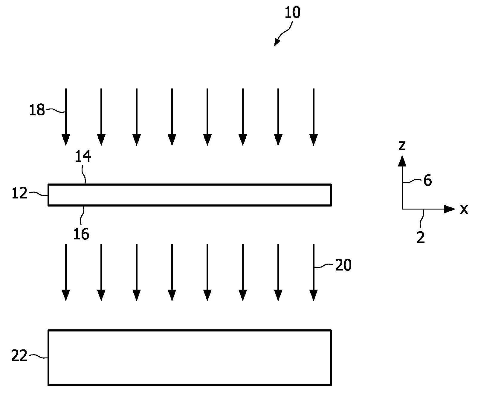
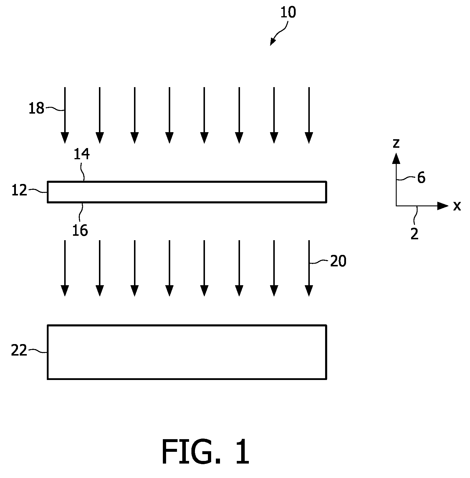
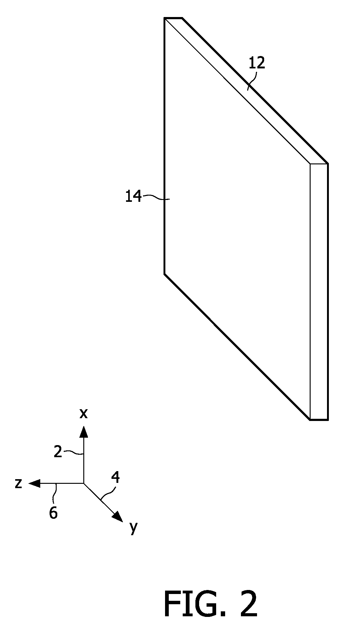
[0049]FIG. 1 illustrates schematically a system 10 for generating a two-dimensional multicolour image of a specimen 12 while FIG. 2 provides a perspective view of the specimen 12. The specimen 12 may, for example, be a liquid layer containing organic material such as individual cells, or a slice of paraffin in which biological material has been embedded. The specimen 12 extends principally along an x-y-plane perpendicular to the plane of the Figure. The specimen 12 comprises at least a first substance (e.g. protein) and a second substance (e.g. nucleic acid). Ultraviolet light 18 of a first frequency is shone onto an upper surface 14 of the specimen 12. Transmitted ultraviolet light 20 of the same frequency leaves the specimen 12 via a lower surface 16 and reaches a detector 22. The detector 22 is coupled to a computer (not shown), for exampl...


PUM


Abstract

At least two chemically different substances of interest of an unstained biological specimen that for each a substance image is generated, indicating for every region of the image an amount of the substance. A multicolor image is generated on the basis of the substance images.

Description

FIELD OF THE INVENTION[0001]In a first aspect, the invention relates to a method of generating a multicolour image of an unstained biological specimen, the specimen comprising at least two chemically different substances of interest.[0002]In a second aspect, the invention relates to a data carrier.[0003]In a third aspect, the invention relates to a system for generating a multicolour image of an unstained biological specimen, the object comprising at least two chemically different substances of interest.BACKGROUND OF THE INVENTION[0004]In histopathology and cytopathology a pathologist routinely analyses microscopic images taken from tissues or cell smears. The specimens are typically analysed with a standard microscope using visible light. Due to the fact that cells and tissues hardly absorb visible light it is a common practice to stain the specimens. The staining chemical can be observed in visible light. It indicates the presence and generally also the amount of the structure it ...

Claims


Application Information

Patent Timeline
Patent Type & Authority Patents(United States)
IPC IPC(8): G06K9/00H04N7/18A63B43/02G06T5/50G06T7/40G06T11/00
CPCA63B43/02G06T5/50G06T7/408G06T11/001G06T2207/10056G06T2207/10152G06T2207/30024G06T7/90
Inventor VAN LEEUWEN, MARINUS B.KUIPER, STEIN
Owner KONINK PHILIPS ELECTRONICS NV
Why Eureka
Try Eureka