

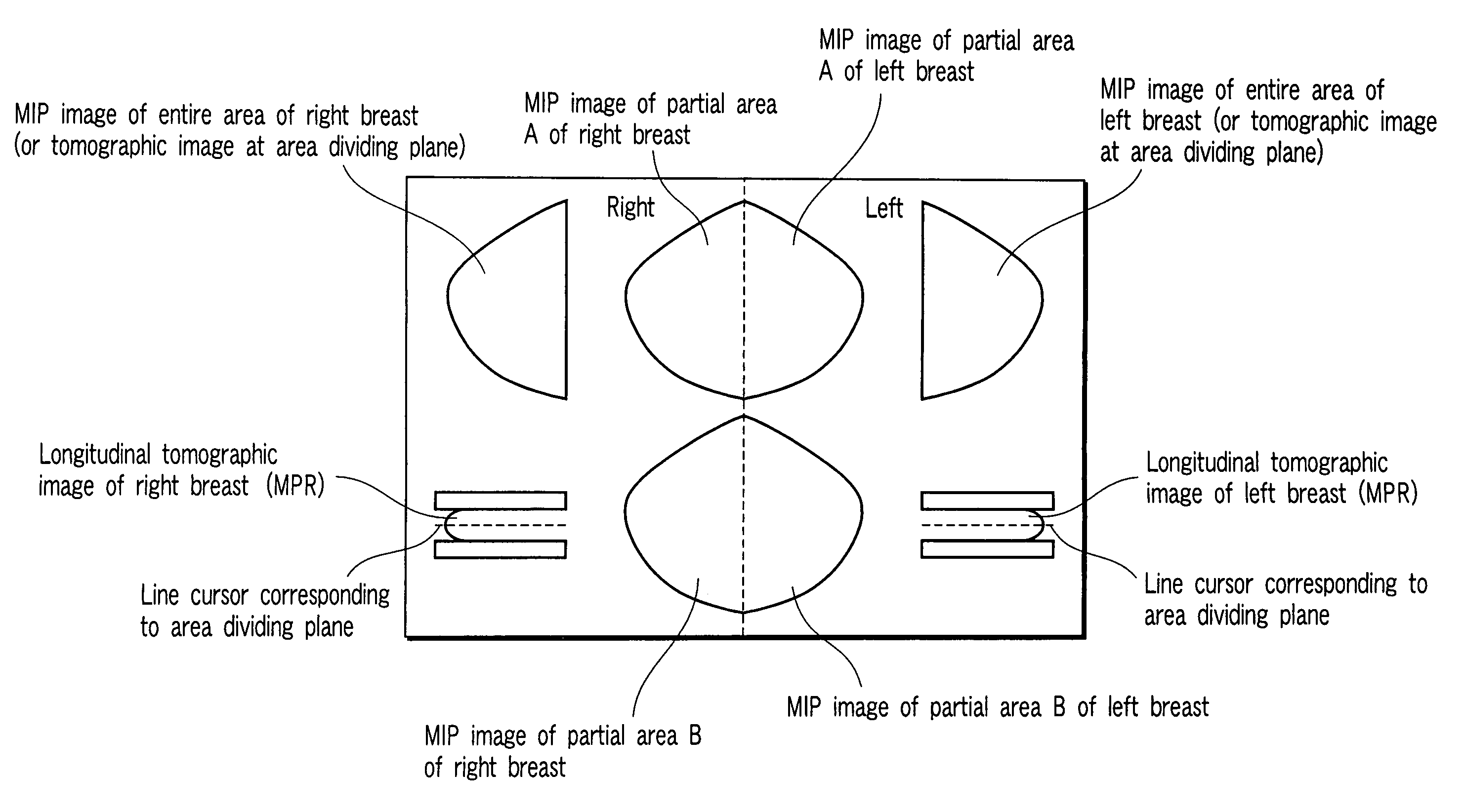
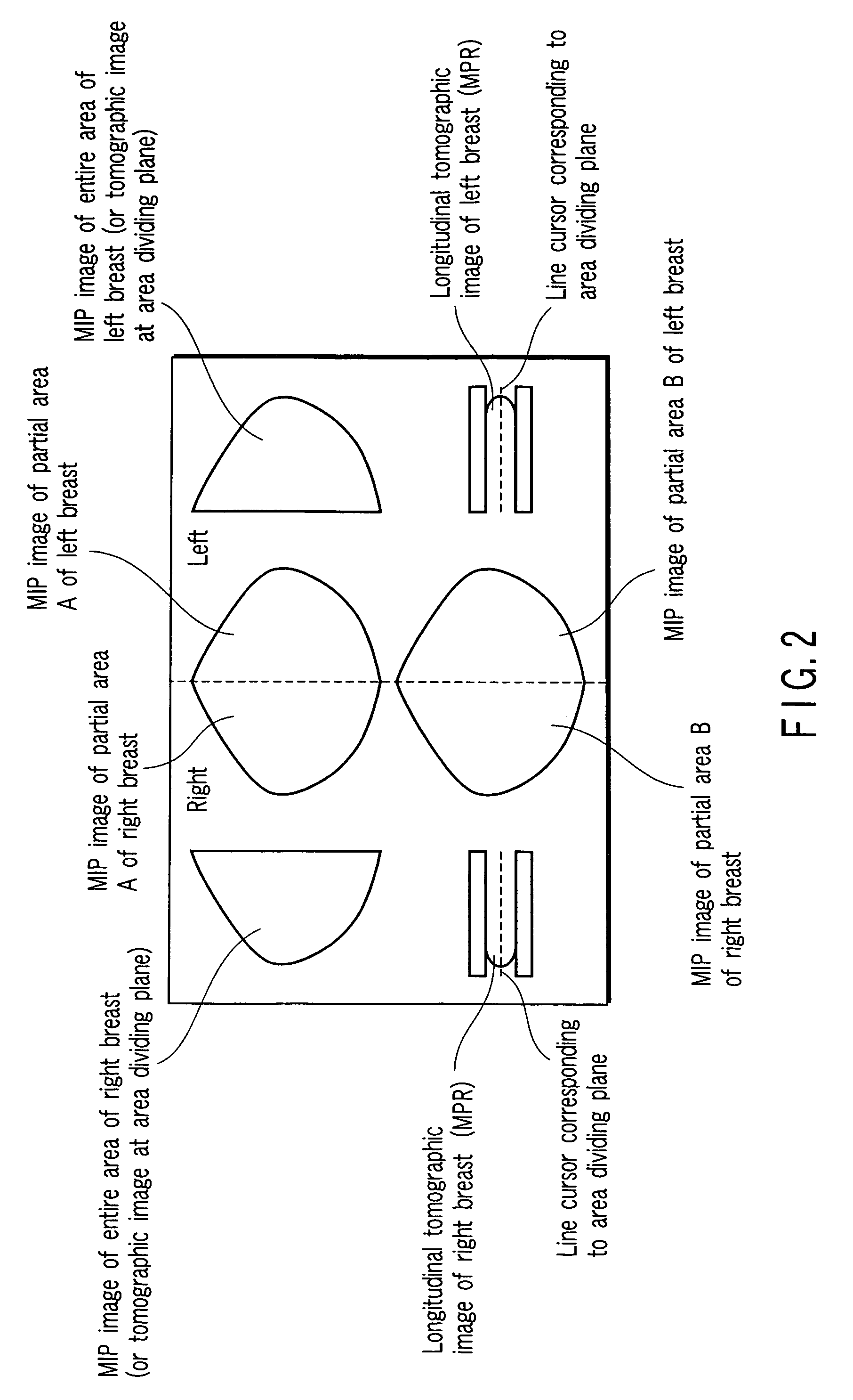
Image displaying apparatus, image displaying method, and computer readable medium for displaying an image of a mammary gland structure without overlaps thereof
a mammary gland and image technology, applied in the field of image displaying apparatus, image displaying method, computer program product, can solve the problems of difficult discrimination/diagnosis of tumors, inability to make detailed evaluation, etc., and achieve the effect of eliminating overlaps of mammary glands and allowing easy comprehension of mammary gland structures
- Summary
- Abstract
- Description
- Claims
- Application Information
AI Technical Summary
Benefits of technology
Problems solved by technology
Method used
Image

Examples
Embodiment Construction
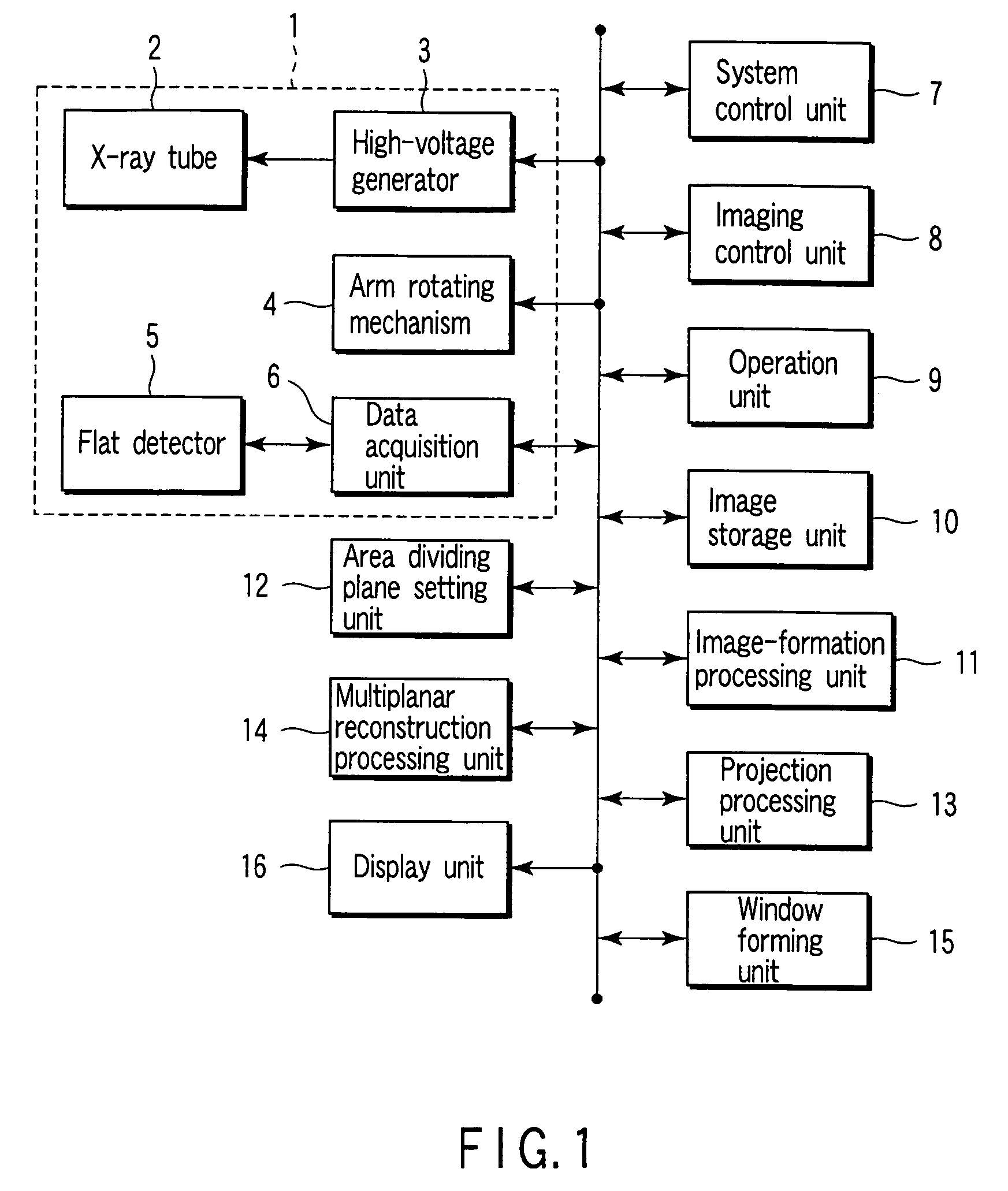
[0044]An embodiment of the present invention will be described below with reference to the views of the accompanying drawing. According to this embodiment, the present invention can be provided as an X-ray three-dimensional imaging apparatus as an X-ray diagnostic apparatus, an image displaying apparatus incorporated in the X-ray three-dimensional imaging apparatus, an image displaying method, a computer program product for causing a computer to realize a combination of means for image display, and a computer readable medium which records the computer program product.
[0045]This embodiment uses an X-ray three-dimensional imaging method. According to the X-ray three-dimensional imaging method, a tomographic image is reconstructed from a plurality of X-ray transmission images obtained by imaging from many directions (this operation will be referred to as “image formation” hereinafter to discriminate it from “reconstruction” in X-ray CT). In actual processing, a plurality of rays passin...
PUM
 Abstract
Description
Claims
Application Information