3D-2D image registration for medical imaging

a 3d-2d, medical imaging technology, applied in image enhancement, image analysis, instruments, etc., can solve problems such as geometric calibration limits the general application of conventional 3d-2d registration methods to x-ray imaging systems involving an unconstrained source-detector geometry, and the geometric relationship of x-ray source and detector must be known and fixed

Active Publication Date: 2015-10-20
THE JOHN HOPKINS UNIV SCHOOL OF MEDICINE
View PDF7 Cites 15 Cited by
  • Summary
  • Abstract
  • Description
  • Claims
  • Application Information

AI Technical Summary

Benefits of technology

[0013]It is, therefore, an object of the invention, to provide a method of 3D-2D registration for medical imaging, which does not require a known and fixed relative position between the x-ray source and the detector, thereby making no assumption on the geometric relationship of the x-ray source and detector.

Problems solved by technology

Such registration and visualization may assist a surgeon in localizing the target anatomy, for example a specific vertebral level, and offer numerous advantages with regard to, for example, time, dose, cost and accuracy) in comparison to conventional methods, for example manual level counting and pre-operative fiducial screw placement, which are prone to error and may potentially result in “wrong-level” surgery.
However, the geometric calibration limits general application of the conventional 3D-2D registration methods to x-ray imaging systems involving an unconstrained source-detector geometry, for example a mobile x-ray radiography system in which the x-ray source (x-ray tube) is freely positionable above the patient in a manner that is largely unconstrained in angle and distance from the patient and detector and, thus, placement of the x-ray source with respect to the detector changes case by case.
Thus, a disadvantage of the conventional methods is that the geometric relationship of the x-ray source and detector must be known and fixed.

Method used


Image

  • 3D-2D image registration for medical imaging
  • 3D-2D image registration for medical imaging
  • 3D-2D image registration for medical imaging

Examples


Embodiment Construction

[0055]The invention offers at least the following advantages.

[0056]3D-2D Registration Method With 9 DoF (9 degrees of freedom)

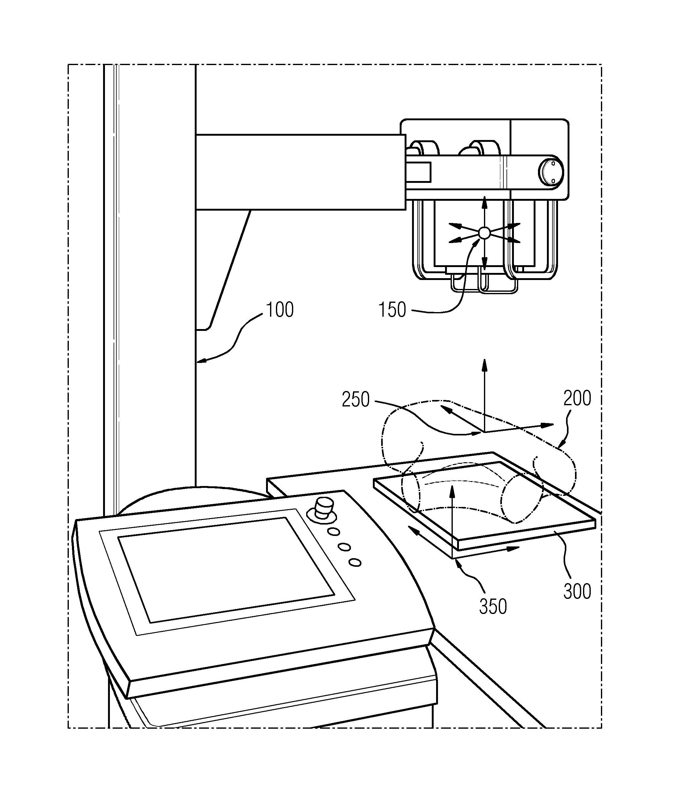
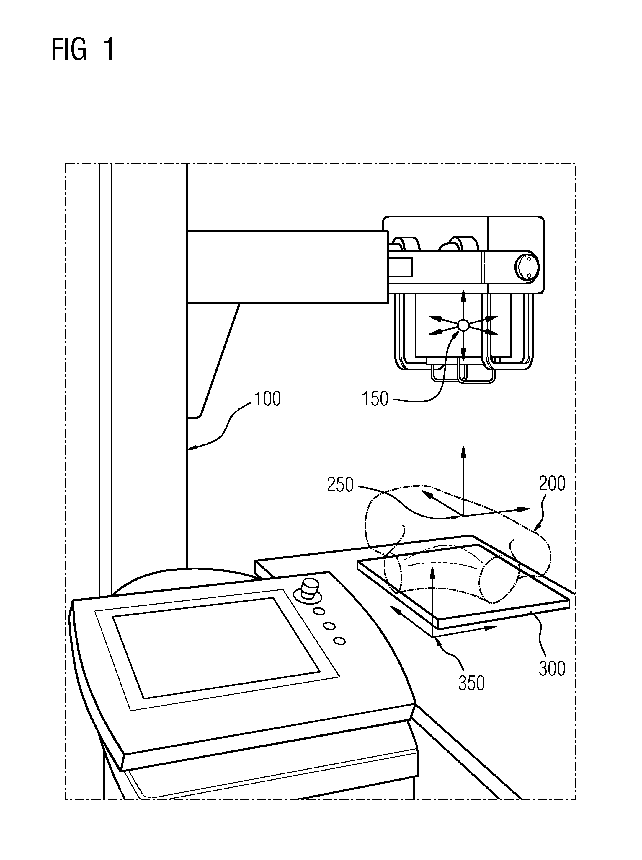
[0057]The 3D-2D registration method according to the invention may process 2D images, for example 2D projection x-ray images, acquired with an imaging system having an unconstrained source-detector geometry. It includes parameters, for example additional 3 DoF parameters, representing the source-detector geometry, such that 2D images can be accommodated without knowing the calibration. Thus, it is well suited to imaging systems, for example mobile radiography systems, where the x-ray source may be freely positioned with respect to the (patient and) detector.

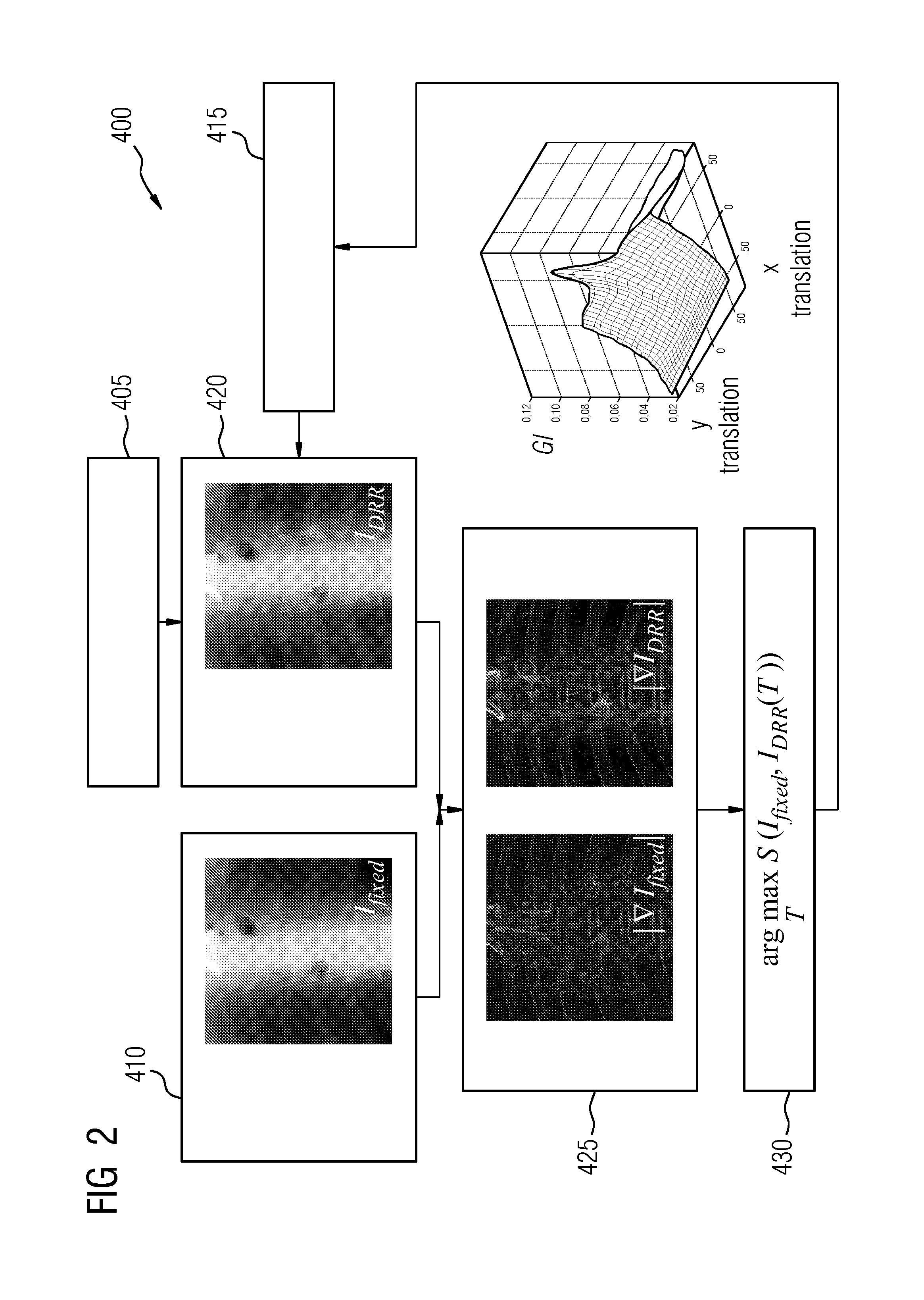
[0058]The 3D-2D registration method according to the invention may be feature-based or intensity-based. Feature-based methods and intensity-based methods are distinguished by the way that they determine, for example measure or quantify, the similarity between two images. The feature-based method uses extra...


PUM


Abstract

A method of 3D-2D registration for medical imaging includes the following steps: providing a first input interface for acquiring a three-dimensional image; providing a second input interface for acquiring a fixed two-dimensional image using an imaging system that includes a source and a detector and that has an unknown source-detector geometry; initializing image transformation parameters and source-detector geometry parameters; generating a reconstructed two-dimensional image from the three-dimensional image using the image transformation parameters and the source-detector geometry parameters; determining an image similarity metric between the fixed two-dimensional image and the reconstructed two-dimensional image; and updating the image transformation parameters and the source-detector geometry parameters using the image similarity metric, and a corresponding non-transitory computer-readable medium and apparatus.

Description

BACKGROUND OF THE INVENTION[0001]1. Field of the Invention[0002]The present invention relates generally to 3D-2D image registration and, more particularly, to 3D-2D image registration with nine degrees of freedom (9 DoF).[0003]2. Description of Related Art[0004]Three-dimensional (3D) pre-operative images are ubiquitously used for surgical planning and intra-operative guidance. In surgical planning, a variety of structures, for example surgical devices, needles, prosthetics, intended incisions and anatomical structures, such as a surgical target or an adjacent structure, e.g. a critical structure, are geometrically defined in these 3D images. The intended trajectory as well as desired location and orientation of a structure are defined in a 3D coordinate system of a pre-operative image. More recent advances bring such 3D imaging capability into an operating room (OR) as well as interventional environment, for example interventional radiology (IR) or vascular and interventional radiol...

Claims


Application Information

Patent Timeline
Patent Type & Authority Patents(United States)
IPC IPC(8): G06K9/00G06T7/00
CPCG06T7/0026G06T2207/10072G06T2207/10116G06T2207/10121G06T2207/20016G06T7/32
Inventor SIEWERDSEN, JEFFREYOTAKE, YOSHITOWOLINSKY, JEAN-PAUL
Owner THE JOHN HOPKINS UNIV SCHOOL OF MEDICINE
Why Eureka
Try Eureka