Unlock instant, AI-driven research and patent intelligence for your innovation.

Method for detecting interactions between two and more biological macromolecules

a biomolecule and interaction technology, applied in the field of new biomolecule interaction detection methods, can solve the problems of high probability of false positive, difficult investigation of membrane proteins or nuclear proteins such as transcriptases, and inability to find a substance capable of regulating protein-protein interactions

Active Publication Date: 2015-11-24
KOREA BASIC SCI INST
View PDF1 Cites 0 Cited by
  • Summary
  • Abstract
  • Description
  • Claims
  • Application Information

AI Technical Summary

Benefits of technology

[0010]The inventors of the present invention have researched to develop a method enabling the real-time detection of binding and interactions of materials in living cells. As a result, we have found that the interaction of a bait and a prey in a living cell can be detected in real time by using a first construct which includes a translocation module moving to a specific region in a cell in response to an external signal or via an internal signaling mechanism and a bait which is a target of an interaction, and a second construct which includes a prey which is another target of the interaction. The first construct and the second construct are labeled with a labeling material, so that the interaction of the bait and the prey in a living cell can be detected in real time by tracing their movements in the cell.
[0011]Accordingly, the present invention provides a novel method enabling the detection of bait and a prey which interact with each other.

Problems solved by technology

These methods require the production, isolation and purification of protein and are disadvantageous in that an information different from the actual interaction may be obtained depending on the buffer condition in the test tube, the secondary modification of extracted proteins, or the like.
It is advantageous in that large-scale screening is possible using a gene library, but is disadvantageous in that investigation of membrane proteins or nuclear proteins such as transcriptase is difficult and there is a high probability of false positive.
Besides, this method is inappropriate to find a substance capable of regulating protein-protein interactions.
Further, since the detection itself is somewhat ambiguous, the technique is not suitable for general drug screening.
The FRET method provides good accuracy, but it is disadvantageous in that positioning of fluorescent proteins or fluorescent materials, which is required for the fluorescence resonance energy transfer to occur, is difficult, thereby having low rate of experimental success.
However, like the FRET method, it is disadvantageous in that relative positioning of proteins for complementary binding is difficult, thereby having low rate of success.

Method used


Image

  • Method for detecting interactions between two and more biological macromolecules
  • Method for detecting interactions between two and more biological macromolecules
  • Method for detecting interactions between two and more biological macromolecules

Examples


example 1

Animal Cell Line and Transformation

[0135] Animal Cell Line and Culturing

[0136]CHO-k1 (ATCC CCL-61, Cricetulus griseus, hamster, Chinese), HEK293 (ATCC CRL-1573, Homo sapiens, human), HeLa (ATCC CCL-2, Homo sapiens, human) and SH-SY5Y (ATCC CRL-2266, Homo sapiens, human) cell lines were used. The animal cells were cultured according to the instructions of ATCC (American Type Culture Collection) for the individual cells. CHO-k1 cells were cultured using F-12 medium, and HEK293, HeLa and SH-SY5Y cells were cultured using DMEM medium. Other culturing condition was the same. The cells were cultured as follows (Those skilled in the art may modify the specific conditions depending on purposes.). The cells were cultured in pH 7.4 medium (F-12 and DMEM) containing 25 mM HEPES, 10% fetal bovine serum (FBS, v / v), 100 units / ml penicillin and 100 μg / ml streptomycin in a 5% CO2 incubator maintained at 37° C.

[0137] Transformation of Cell Line

[0138]In the Examples of the present invention, genes we...

example 2

Design and Preparation of First Construct and Second Construct

[0139] Design and Preparation of First Construct

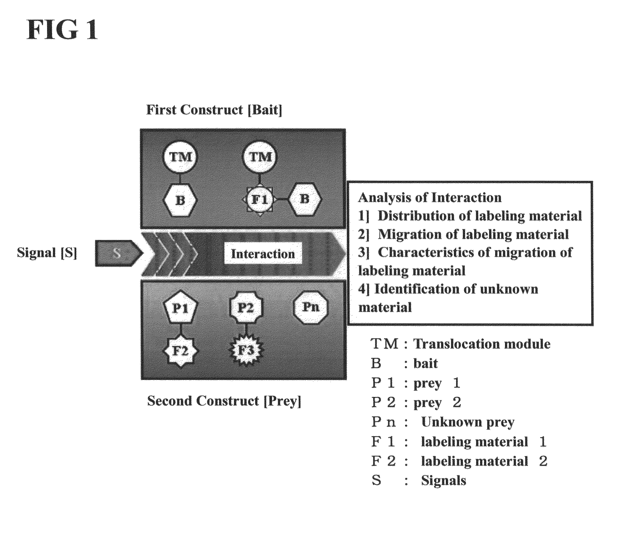
[0140]The first construct may be fused construct comprising a translocation module capable of moving the protein uniformly expressed in the cytoplasm toward the cell membrane, a fluorescent protein analyzable using a microscope, and a bait.

[0141]Vectors expressing the first construct (see A of FIG. 4) were prepared as follows. Translocation modules were prepared by PCR using the following templates and primers and inserted at the NheI / AgeI site of a pEGFP-C3 vector (GenBank Accession No. U57607; Clontech Catalog No. #6082-1, SEQ ID NO: 21) and a pmRFP-C3 vector (mRFP; GenBank Accession No. DQ903889, SEQ ID NO: 22), thereby completing the vectors.

[0142]The TMD translocation module was prepared by PCR using a pCMV-SPORT6-PRKCD vector [GenBank Accession No. BC043350; purchased from Open Biosystems (see the world wide web at (www)openbiosystems.com); Catalog No. EHS1001-410108-B...

example 3

Verification of Translocation Characteristics of First Construct and Second Construct

[0156] Verification of Expression of Constructs and Analysis of Translocation Characteristics

[0157]A cover slip containing the cells in which the first construct and second construct vectors had been introduced was fixed to a perfusion chamber and mounted on the object stage of a confocal laser fluorescence microscope (Carl Zeiss LSM510). Images of the construct vectors were taken before and after external stimulation (treatment with 1 μM PMA).

[0158]488 nm argon laser (EGFP or AzG), 543 nm HeNe laser (mRFP) or 561 nm DPSS laser (HcR) of the confocal laser fluorescence microscope was used to excite the fluorescent label, and the fluorescence signal generated by each fluorescent label was filtered through the band path filter BP505-530 (EGFP or AzG), long path filter LP560 or BP560-630 (mRFP) or long path filter LP650 (HcR). Images were taken after completely removing the interference between the fluo...


PUM

PropertyMeasurementUnit
Green Fluorescent Proteinaaaaaaaaaa
Red Fluorescent Proteinaaaaaaaaaa
Fluorescentaaaaaaaaaa

Abstract

Disclosed is a novel method for detecting interactions of biomolecules. More particularly, the disclosed method includes (a) preparing a cell comprising (i) a first construct comprising a bait, a first labeling material and a translocation module; and (ii) a second construct comprising a prey and a second labeling material; (b) detecting the distribution of the first construct and the second construct in the cell. the present invention provides a method capable of detecting bindings and interactions occurring in a living cell in real time, and a method for screening a material that alters the binding and the interaction. The method of the present invention overcomes the disadvantages including inaccuracy and complexity of existing biomaterial interaction detection techniques. By labeling both constructs to promote accuracy, the present invention provides a novel real-time, antibody-free analysis.

Description

CROSS-REFERENCE TO RELATED APPLICATIONS[0001]This is a Continuation-In-Part application of U.S. application Ser. No. 12 / 547,043 filed Aug. 25, 2009, now abandoned, which claim priority to Korean Application No. 10-2008-0112354 filed Nov. 12, 2008, patented Mar. 12, 2010, which applications are incorporated herein by reference.SEQUENCE LISTING[0002]The instant application contains a Sequence Listing which has been submitted in ASCII format via EFS-Web and is hereby incorporated by reference in its entirety. Said ASCII copy, created on Jan. 2, 2013, is named 84724-CIP-307161_ST25.txt and is 57,284 bytes in size.TECHNICAL FIELD[0003]Disclosed is a novel method for detecting interactions of biomolecules. More particularly, the disclosed method includes (a) preparing a cell comprising (i) a first construct comprising a bait, a first labeling material and a translocation module; and (ii) a second construct comprising a prey and a second labeling material; (b) detecting the distribution of...

Claims


Application Information

Patent Timeline
Patent Type & Authority Patents(United States)
IPC IPC(8): G01N33/53C12Q1/02G01N33/50G01N33/542G01N33/557C12N15/10
CPCC12Q1/025C12N15/1055G01N33/5035G01N33/542G01N33/557C07K2319/01C07K2319/60
Inventor LEE, ZEE WONKIM, SOO HYUN
Owner KOREA BASIC SCI INST