Method for determining an artifact-reduced three-dimensional image data set and X-ray device

a three-dimensional image and data set technology, applied in the field of method for determining an artifact-reduced three-dimensional image data set and x-ray device, can solve the problems of artifacts in the image data set, which are for the most part extremely expensive, and occur in such investigations, and achieve the effect of improving the image quality of the image data s

Active Publication Date: 2015-12-22
SIEMENS HEALTHCARE GMBH
View PDF31 Cites 0 Cited by
  • Summary
  • Abstract
  • Description
  • Claims
  • Application Information

AI Technical Summary

Benefits of technology

[0009]It is an object to specify a correction method which may be employed in particular both for truncated and also for non-truncated projection images and enhances the image quality of the image data set.
[0011]A correction in the projection images is consequently proposed which is based on determining the attenuation components originating from the patient, thus specifically the head, by using a mask data set which shows either only the neurosurgical apparatus or only the head of the patient. By using a mask data set showing the neurosurgical apparatus, it is for example possible for portions of the attenuation values of the projection images originating from the neurosurgical apparatus to be removed in the primary data set, while in the other case the mask data set can ultimately be employed as a type of “attenuation map” for regions in which the neurosurgical apparatus is visible in the primary data set. In both cases it is therefore proposed to generate a comparison data set using comparison projection images which make it possible to ultimately remove the neurosurgical apparatus from the projection images and consequently to determine an image data set of the head alone.
[0013]The described method has a multitude of advantages. In particular in comparison with methods which utilize an interpolation, measured data is used in order to avoid the artifacts occurring during the reconstruction on account of the neurosurgical apparatus, which contributes towards maintenance of the resolution and of the image quality. The proposal described here permits a correction of artifacts from the neurosurgical apparatus both for truncated and also for non-truncated data. The method is simple to use and to implement. Both capture operations, in other words that for the primary data set and that for the mask data set, can be carried out independently, and if an additional patient dose occurs this can be adjusted accordingly.
[0015]In this situation it is particularly expedient if movements between the capture of the primary data set and of the mask data set are avoided as far as possible, which means it is advantageously provided that the head or the neurosurgical apparatus is held essentially motionless between the capture of the projection images of the mask data set and the capture of the projection images of the primary data set. If no appreciable movement whatsoever takes place it can be assumed that because the projection images already correspond to one another in their projection directions a registration naturally results, in which case in the event of smaller movements and / or adjustments a registration, which will be described in more detail in the following, can lead to a successful outcome in a particularly simple manner.
[0017]The particular advantage of this first alternative embodiment is that no additional patient dose occurs during the capture of the mask data set because the latter does not actually show the patient, here specifically the head of the patient, but it is only the neurosurgical apparatus which is captured. Compared with the conventional methods in which the primary data set is captured as the single date set of projection images, no increase in the patient dose is consequently required.
[0027]In a development of this second embodiment, provision can be made that mask pixels of the captured projection images of the primary data set, associated with the neurosurgical apparatus, are determined and the image data of these pixels is replaced by attenuation data of the projection images of the mask data set. In this situation the mask pixels can in turn be derived from a segmentation of the neurosurgical apparatus, which however preferably does not take place within the projection images of the primary data set, but provision is made in a particularly advantageous embodiment that the segmentation takes place in subtraction data obtained from the subtraction of projection image data of the primary data set and of the mask data set of corresponding projection directions or from three-dimensional reconstruction data sets derived from the primary data set and the mask data set, in which case when reconstruction data sets are used the mask pixels are obtained in the context of a forward projection of the segmentation result. The position of the neurosurgical apparatus in relation to the projection images can therefore easily be determined through subtraction of the two data sets, which means that the neurosurgical apparatus remains the only remaining object and a segmentation is consequently possible in a simple manner.

Problems solved by technology

Problems occur with regard to such investigations for example in the situation when a supporting apparatus, in particular a stereotactic frame, is employed in the region of the neurosurgery.
Because in particular frames and markers for the optical registration frequently consist of a very dense material, in particular exhibiting a high atomic number, they can cause artifacts in the image data sets.
Although neurosurgical apparatuses which are improved in this respect, for example consisting of materials having a low atomic number, are also known, for example stereotactic frames made of carbon, these are however for the most part extremely expensive.
These approaches have the disadvantage that they are for the most part not suitable for handling effects of truncated projection images which in particular do not show the entire neurosurgical apparatus.
The algorithms have a strong noise dependence and can only be employed scarcely meaningfully in the case of low 3-D image quality since they operate in image-based fashion.
A further disadvantage is the fact that the interpolation, in particular a linear interpolation, has too great an influence on the resolution and the image quality.

Method used


Image

  • Method for determining an artifact-reduced three-dimensional image data set and X-ray device
  • Method for determining an artifact-reduced three-dimensional image data set and X-ray device
  • Method for determining an artifact-reduced three-dimensional image data set and X-ray device

Examples


first embodiment

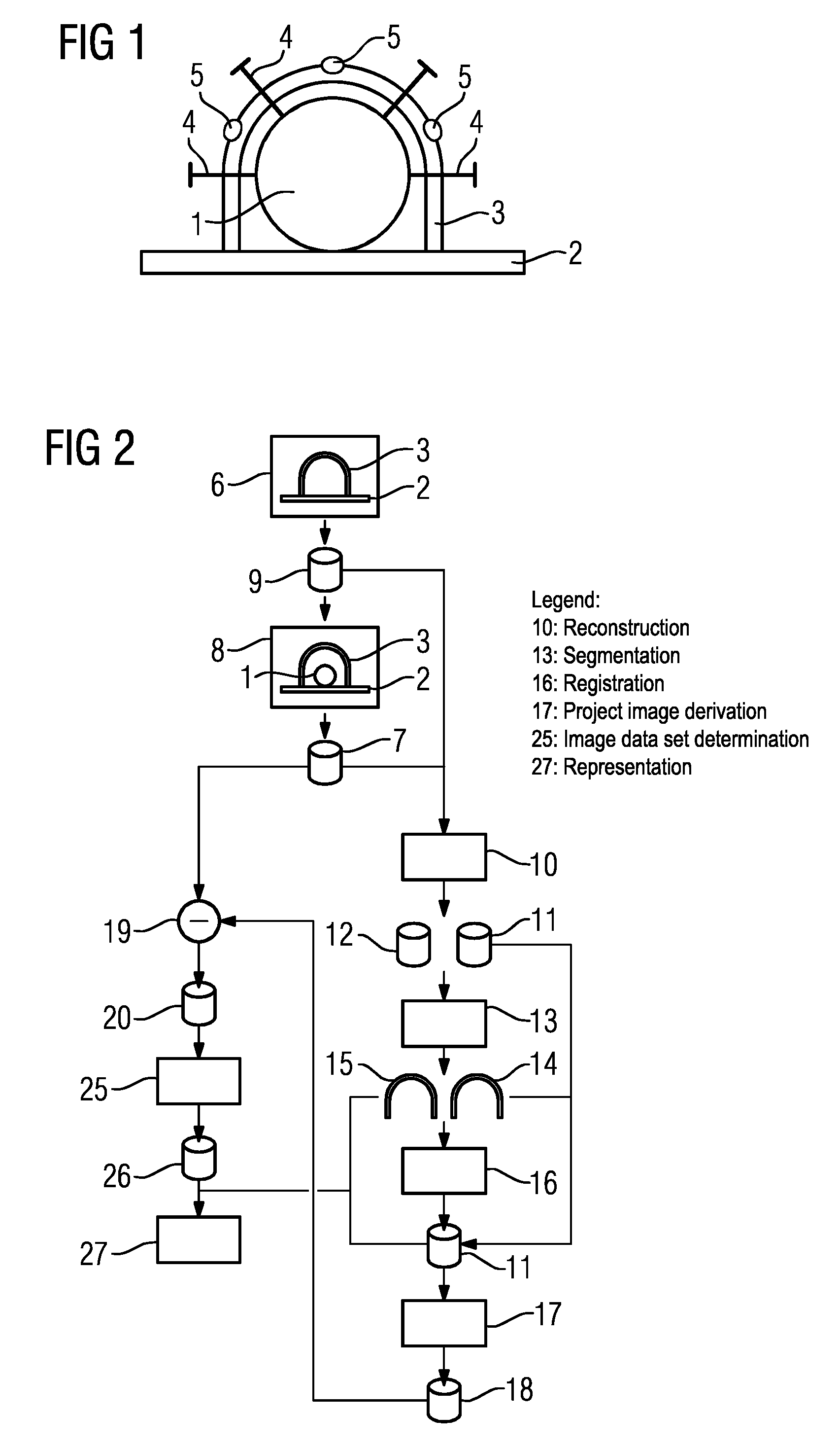
[0046]FIG. 2 shows a basic flowchart of a first exemplary embodiment according to the method.

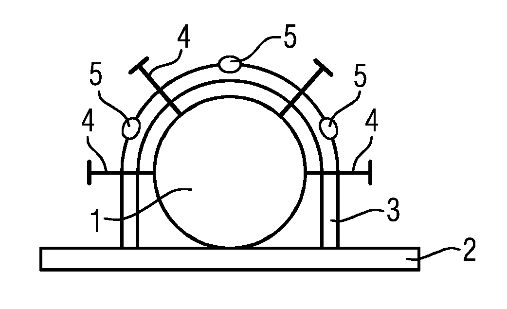
[0047]The stereotactic frame 3 which is already adjusted as well as possible to the head 1 is initially arranged on the patient table 2 without the patient. In a step 6, under the same capture conditions and using the same capture parameters as later when capturing the projection images of the primary data set7 in a step 8, capture then takes place of two-dimensional projection images of a mask data set 9, in which projection images consequently only the stereotactic frame 3 (with the markers 5) is contained.

[0048]Then in step 8 the patient is firstly positioned on the patient table 2, during which the stereotactic frame 3 remains motionless. Merely minor settings, consequently adjustments, of the stereotactic frame 3 to the head 1 are carried out before the capture of the primary data set which then shows the head 1 and the stereotactic frame 3 (with the markers 5) on its projection images ...

second embodiment

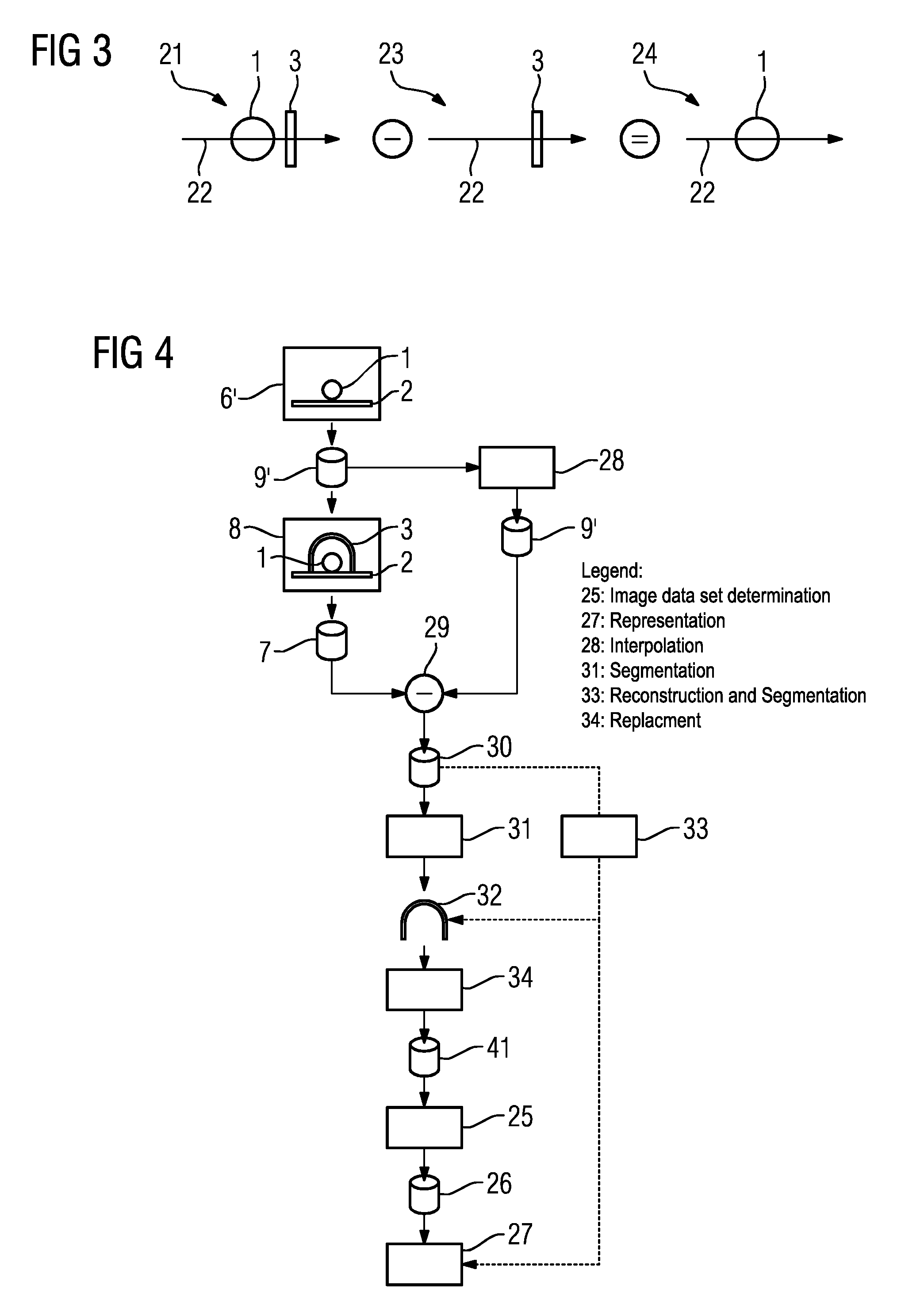
[0056]FIG. 4 shows a flowchart of the second exemplary embodiment of the method which corresponds to the In this situation the same elements are identified by the same reference characters for the sake of simpler presentation.

[0057]A mask data set 9′ is also captured in the second exemplary embodiment in a step 6′, except that here at the time of capture in step 6′ solely the head 1 is situated on the patient table 2 but not the stereotactic apparatus 3 with the markers 5. The projection images of the mask data set 9′ consequently show only the head 1.

[0058]The patient is now not moved and the stereotactic frame 3 is attached and appropriately adjusted so that in a step 8 the primary data set 7 can be captured again, the projection images of which show the head 1 and the stereotactic frame 3 (with the markers 5).

[0059]In this situation it should be noted here that for the sake of simplified presentation a registration to be established where applicable on the basis of the head 1 or...


PUM


Abstract

A method of determining an artifact-reduced three-dimensional reconstructed image data set includes a plurality of projection images of a primary data set, which show a head of a patient together with at least one neurosurgical apparatus generating artifacts in a three-dimensional reconstruction. The projection images of the primary data set are captured using different projection directions by an X-ray device having a C-arm. With regard to a reduction of artifacts, a projection image based correction is based upon projection images of a mask data set which show the neurosurgical apparatus without the head of the patient or the head of the patient without the neurosurgical apparatus.

Description

CROSS REFERENCE TO RELATED APPLICATIONS[0001]This application claims priority of German Application No. 10 2012 205 222.4 DE filed Mar. 30, 2012. All of the applications are incorporated by reference herein in their entirety.FIELD OF INVENTION[0002]A method for determining an artifact-reduced three-dimensional image data set reconstructed taking into consideration a plurality of projection images of a primary data set which show a head of a patient together with at least one neurosurgical apparatus generating artifacts in the case of a three-dimensional reconstruction is provided, said projection images being captured using different projection directions by an X-ray device, in particular an X-ray device having a C-arm, and an X-ray device is provided.BACKGROUND OF INVENTION[0003]The generation of three-dimensional image data sets from two-dimensional projection images captured using different projection directions is already widely known and forms a basis for three-dimensional tomo...

Claims


Application Information

Patent Timeline
Patent Type & Authority Patents(United States)
IPC IPC(8): G06K9/00G06T5/50A61B6/00G06T5/00G06T7/00
CPCG06T7/0012A61B6/5258G06T5/005G06T5/50G06T2207/30016G06T2207/10116G06T2207/20221G06T2207/20224
Inventor KYRIAKOU, YIANNIS
Owner SIEMENS HEALTHCARE GMBH
Why Eureka
Try Eureka