
Terminal tissue attachment and repair device

a tissue and endoscope technology, applied in the field of endoscopes, can solve the problems of putting the tissue at risk of mechanical degradation, affecting the operation of the joint under repair, and unable to fully grasp the endoscope, so as to facilitate the fixation of the proximal componen

Active Publication Date: 2016-02-09
SUDEKUM IP HLDG LLC
View PDF2 Cites 1 Cited by
  • Summary
  • Abstract
  • Description
  • Claims
  • Application Information

AI Technical Summary

Benefits of technology

[0025]The distal component of the device is composed of a suture or woven fibrous material firmly attached to the proximal component. The distal component is smaller and narrower dimension than the proximal component. The distal component has a firm secure attachment to a tapered terminal end of the proximal component through adhesion, welding, weaving, mechanical connection and the like. The distal component serves as means of fixation and / or anchorage for the device so that the tendon, tendon graft, ligament or other elongated body tissue can be securely fixed to bone, such as a bony insertion or a skeletal point, or other rigid tissue.
[0026]The distal component serves to facilitate fixation of the proximal component of the device with the attached tendon, to bone or skeletal component. The suture or woven fibrous material (“tail” or “tails”) which makes up the distal end of the device is fixed to the bone or skeletal component by passing the tails through the bone and knotting them together or used in conjunction with suture anchors, interference screws, buttons and like means to provide secure, permanent fixation of the device and attached tendon or other elongated body tissue to bone.
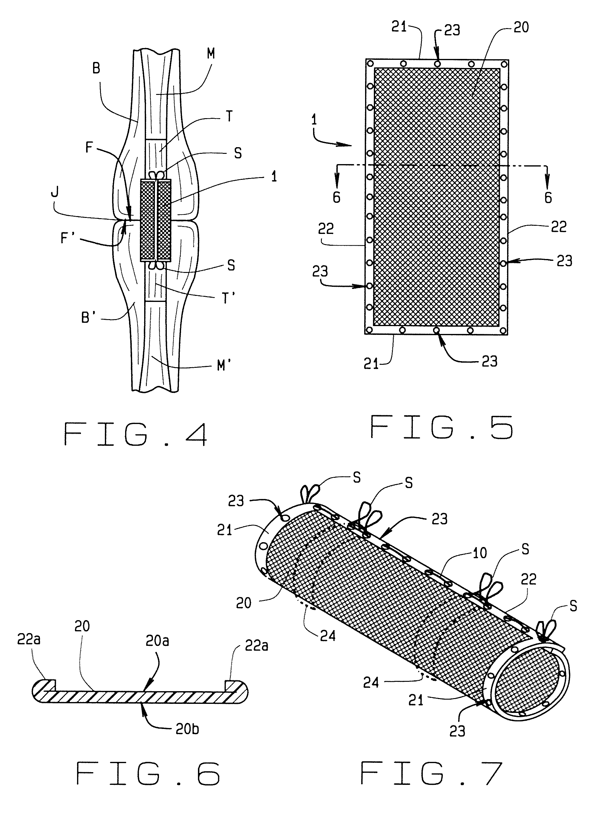
[0031]A surgeon, during use of the invention, places the end of healing elongated tissue, such as a severed tendon, inside, that is, within the proximal component of the invention through the wider end of the conical shape and advances the tendon distally to the tapered tip of the proximal component. Upon containing the tendon within the proximal component, the surgeon then laces the suture material through the plurality of holes on either side of the slit and pulls the ends of the suture material simultaneously, similar to tightening a shoelace, to compress and to constrict the mesh material upon and around the elongated tissue for securement of the invention and for healing of the tissue. Upon pulling the suture, the surgeon secures it using a knot. Alternatively, the surgeon laces the sides of the slit before inserting the elongated tissue into the invention. Having used the invention which is compressed circumferentially around the elongated tissue, the surgeon then places additional transfixion mattress sutures through the invention and also through the tendon, perpendicular to the long axis of the tendon and the invention to further secure the tendon to the invention. The transfixion sutures provide secure anchorage of the proximal component of the invention to the tendon and also compress the device along its anterior / posterior dimension. This compression further reestablishes the normal anatomic shape and form of a repaired tendon or other elongated tissue. The unilateral closure device with its mesh attains a generally ovoid, or elliptic or elliptical, shape that approximates the natural cross section of a tendon.
[0035]Another object is to provide such a terminal tissue attachment and repair device that allows for passage of the invention installed upon tissue, through the adjacent bony tunnel to facilitate secure attachment of the device and tendon to the distal bony insertion.
[0037]Another object is to provide such a terminal tissue attachment and repair device that minimized the instance of bone grafting.
[0039]The terminal tissue attachment and repair device described in this application reaches the goals of 1) withstanding the forces of muscle contraction early active motion immediately after the repair; 2) optimizing healing, blood flow and vascularity between the repaired tendon ends or between the tendon and bone; and 3) approximating the normal anatomic shape of the tendon at the repair interface.

Problems solved by technology

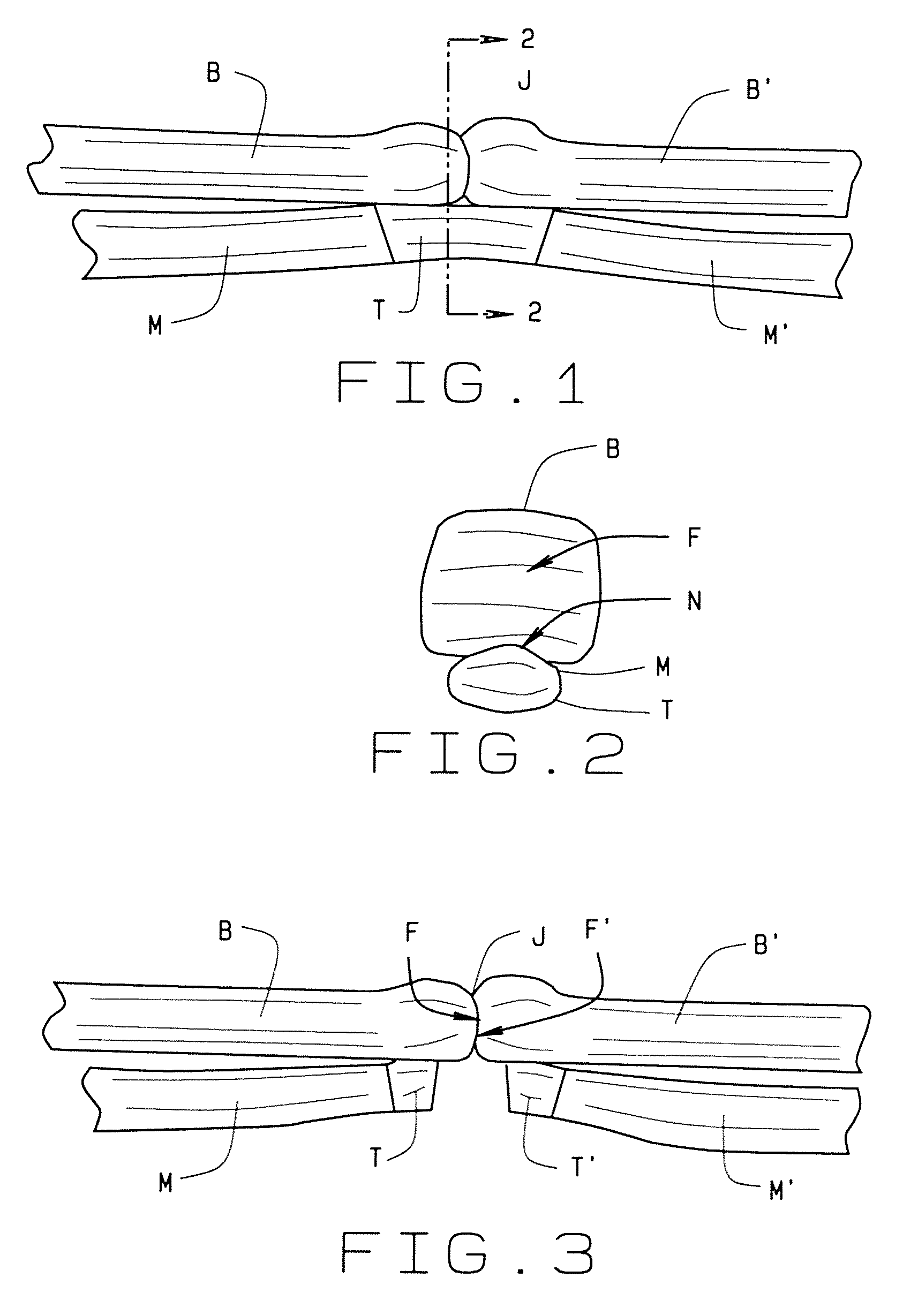
This metallic tendon repair becomes a permanent part of the patient and has some risk of rejection or working outwardly from the tendon and interfering with the operation of the joint under repair.
Akin to the helical anchors, the spines insert deep into tendon tissue putting that tissue at risk for mechanical degradation.

Method used


Image

  • Terminal tissue attachment and repair device
  • Terminal tissue attachment and repair device
  • Terminal tissue attachment and repair device

Examples


Embodiment Construction

[0067]The present invention overcomes the prior art limitations by providing a terminal tissue attachment and repair device also suitable for use upon other elongated body tissue such as ligaments, nerves, and the like. The “Tendon Trap,”“Finger Trap,” or tendon repair device is composed of a resorbable or non-absorbable suture material. The invention has a construction of a diagonal crisscrossing, interlocked sheet of mesh like construction which has the overall shape of an open or slit cylinder, or alternatively a conical shape. The longitudinal slit in the mesh cylinder or cone has a longitudinal opening to allow the tendon end(s) to be easily placed within the cylinder, whether the tendon has already been approximated or the cut ends remain unattached. The cylinder thus formed is generally hollow and the device attains an overall tubular form.

[0068]The grossly approximated tendon is then placed inside the open “Tendon Trap” cylinder or cone which wraps around and envelops the te...


PUM


Abstract

A terminal soft tissue attachment and repair device has a sheet of mesh with two longitudinal sides and two lateral ends angled to the sides. One end has a smooth surface where the healing tissue rests and the sides mutually overlap. The device attains a frusto-conical shape of a proximal component and a distal component. The two sides and one end of the proximal component have suture holes. The other end of the proximal component merges with both sides and joins them to the distal component. From the joint, the two sides extend in a V shape spanned by laced suture. A surgeon places an end of tissue within the device. The surgeon then laces suture and then pulls it from one location to secure the invention upon the tissue and the distal component to a bony insertion. The invention attains a shape like the natural tissue.

Description

CROSS-REFERENCE TO RELATED APPLICATION[0001]This non-provisional application claims priority to the provisional application 61 / 915,574 filed on Dec. 13, 2013, and the pending non-provisional application Ser. No. 13 / 946,607 filed on Jul. 19, 2013, and the provisional application 61 / 673,827 filed on Jul. 20, 2012, which are all owned by the same inventor.BACKGROUND OF THE INVENTION[0002]The terminal tissue attachment and repair device generally relates to orthopedic devices and more specifically to an elongated tissue enwrapping and repair device. More particularly, the invention wraps one end of an elongated tissue and secures that end to a bony insertion or fixed anatomic point.[0003]Effective repair of elongated tissue, such as flexor tendons, extensor tendons, ligaments, muscles and the like, has long been a significant problem that remains unresolved within the realms of hand surgery, orthopedic surgery and reconstructive musculoskeletal surgery. The pursuit of a secure, relative...

Claims


Application Information

Patent Timeline
Patent Type & Authority Patents(United States)
IPC IPC(8): A61F2/08A61B17/06A61F2/00A61B1/06
CPCA61F2/0811A61B17/06166A61F2/0063A61F2/08A61F2002/087
Inventor SUDEKUM, ANTHONY, E.
Owner SUDEKUM IP HLDG LLC
Learn More