
Human monoclonal antibodies to human nucleolin

a technology of human nucleolin and monoclonal antibodies, which is applied in the field of cell biology and immunology, can solve the problems of limited success, limited specificity of certain targets, and untapped potential for therapeutic and diagnostic agents, and achieves the effects of reducing bcl-2 levels, inducting complement-dependent cytotoxicity, and reducing bcl-2 levels

Active Publication Date: 2016-02-16
MUSC FOUND FOR RES DEV
View PDF184 Cites 5 Cited by
  • Summary
  • Abstract
  • Description
  • Claims
  • Application Information

AI Technical Summary

Benefits of technology

[0012]In another aspect the invention provides an immortalized human B-cell that expresses an IgG antibody or fragment thereof that binds to a protein encoded by SEQ ID No. 3. In one embodiment, the IgG antibody is an IgG1, IgG2, IgG3, or IgG4 antibody. In another embodiment, the IgG antibody is an IgG1 antibody. In another embodiment, the IgG antibody comprises a kappa light chain. In another embodiment, the IgG antibody comprises a lambda light chain. In another embodiment, the B-cell is EBV immortalized. In another embodiment, the antibody or fragment thereof is a monoclonal antibody or fragment thereof. In another embodiment, the antibody or fragment thereof is substantially non-immunogenic to a human. In another embodiment, the antibody or fragment thereof is a human antibody or fragment thereof. In another embodiment, the antibody or fragment thereof kills at least 20% of a population of MCF-7 cells, when incubated with said MCF-7 cells and human AB serum for 48 hours. In another embodiment, the antibody or fragment thereof kills at least 20% of a population of MV4-11 cells, when incubated with said MCF-7 cells and human AB serum for 48 hours. In another embodiment, the antibody or fragment thereof binds to an RNA binding domain of human nucleolin. In another embodiment, the antibody or fragment thereof inactivates an RNA binding domain of human nucleolin. In another embodiment, the isolated antibody or fragment thereof induces complement-dependent cytotoxicity to a cancer cell. In another embodiment, the isolated antibody or fragment thereof induces complement-independent cytotoxicity to a cancer cell. In another embodiment, the isolated antibody or fragment thereof induces apoptosis in a cancer cell upon contact. In another embodiment, the isolated antibody or fragment thereof inhibits or kills an AML cancer cell, a CLL cancer cell or a breast cancer cell. In another embodiment, the isolated monoclonal antibody or fragment thereof reduces BCL-2 levels in a cancer cell.

Problems solved by technology

However, the difficulty of generating specific antibodies for certain targets has limited the successes, and the potential for therapeutic and diagnostic agents remains largely untapped.
One impediment to the development of monoclonal antibodies for human therapy is the need to “humanize” such antibodies, which are generally made in mice, rats and rabbits.
If human patients are administered such antibodies without humanized constant regions, they can suffer from “serum sickness,” meaning that an endogenous immune response is mounted by the recipient against the non-human antibody sequences.
However, the cost in time and expense for humanization of antibodies can be considerable.

Method used


Image

  • Human monoclonal antibodies to human nucleolin
  • Human monoclonal antibodies to human nucleolin
  • Human monoclonal antibodies to human nucleolin

Examples


example 1

Materials & Methods

[0357]Isolation and Culture of Tonsil B Cells.

[0358]To prepare B cells from tonsils, tonsil tissue was placed inside a sterile Petri dish (VWR International, cat. #25384-088) containing 20-30 ml Dulbecco's phosphate buffered saline (DPBS, without CaCl2 or MgCl2; Gibco / Invitrogen, Grand Island, N.Y. cat. #14190144) supplemented with 1× Antibiotic-Antimycotic (Gibco / Invitrogen cat. #15240-062). The tissue was chopped and minced with scalpels to approximately 1 mm3 pieces. Additional lymphocytes were released by gentle grinding of tonsil pieces between the frosted glass surfaces of two sterile microscope slides (VWR cat. #12-550-34), and single cell preparation was made by straining through 70 μm nylon strainer (BD Falcon, cat. #352350, BD Biosciences, Two Oak Park, Bedford, Mass.). This suspension was layered onto a Ficoll (Amersham Biosciences cat. #17-1440-03, Uppsala, Sweden) cushion (35 ml sample over 15 ml Ficoll) and resolved at 1500 G for 20 min. The boundary...

example 2

Results

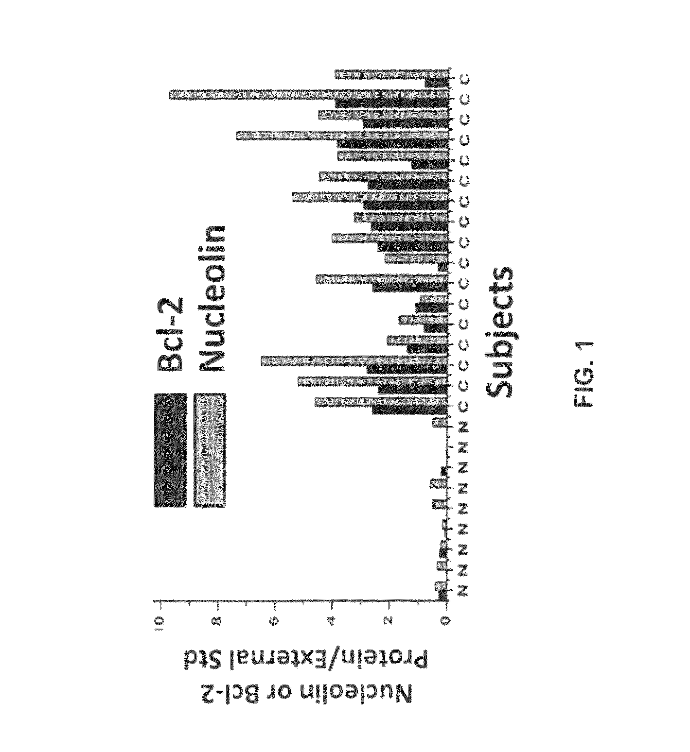
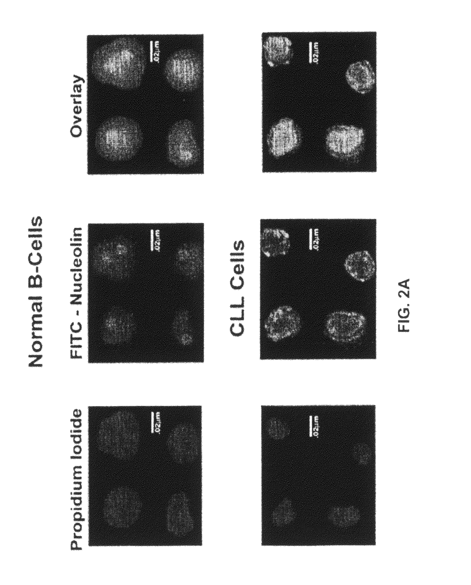
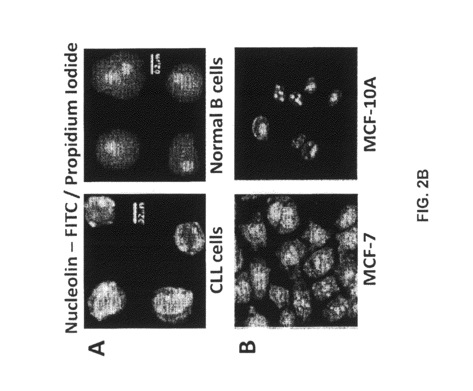
[0516]Nucleolin and Bcl-2 Protein are Overexpressed in the Plasma Membrane And Cytoplasm of B-CLL Cells Compared to B Cells from Normal Human Volunteers.

[0517]CLL is indolent during most of its clinical course and the clonal B cells accumulate in the bone marrow and circulation during the indolent phase by avoiding apoptosis (Klein et al., 2000). CLL cells circumvent apoptosis by over-expressing the anti-apoptotic protein Bcl-2. High-level expression of bcl-2 mRNA and protein is seen in the absence of gene rearrangements that are known to enhance bcl-2 transcription (Bakhshi et al., 1985; Robertson et al., 1996; Steube et al., 1995). One of the inventors discovered that bcl-2 mRNA is highly stabilized in CLL cells from patients compared to normal CD19+ B cells from healthy volunteers (Otake et al., 2007). In addition, the inventors showed that the enhanced stability of bcl-2 mRNA in CLL cells was a direct result of binding of the stabilizing protein nucleolin to an ARE elemen...


PUM

PropertyMeasurementUnit
concentrationsaaaaaaaaaa
temperatureaaaaaaaaaa

Abstract

The present invention provides for methods of producing human monoclonal antibodies to human nucleolin, cells producing such antibodies, and the antibodies themselves. Also provided are methods of using the antibodies in diagnosing and treating malignant and non-malignant diseases wherein cells that express nucleolin on the cell surface contribute to the pathophysiology of the disease.

Description

CROSS-REFERENCE[0001]This application is a national phase application under 35 U.S.C. 371 of International Application No. PCT / US2010 / 057046 filed Nov. 17, 2010 which claims benefit of priority to U.S. Provisional Application Ser. No. 61 / 261,909, filed Nov. 17, 2009, which is incorporated herein by reference in its entirety.STATEMENT AS TO FEDERALLY SPONSORED RESEARCH[0002]This invention was made with the support of the United States government under grant number NCI CA109254-04S1 awarded by the National Cancer Institute, National Institutes of Health. The U.S. Government may have rights in this invention.BACKGROUND OF THE INVENTION[0003]The present invention relates generally to the fields of cell biology and immunology. More particularly, it concerns methods and compositions relating to the production and use of human monoclonal antibodies to human nucleolin (NCL).[0004]Antibodies are a class of agents known as “biologicals.” The source of antibodies can be a polyclonal supply, su...

Claims


Application Information

Patent Timeline
Patent Type & Authority Patents(United States)
IPC IPC(8): A61K39/00C07K16/18C07K16/28C12N5/07C07K16/00C07K16/30A61K39/395
CPCC07K16/28C07K16/18C07K16/3015C07K2317/21C07K2317/73C07K2317/734C07K16/30C07K16/303C07K16/3038C07K16/3069A61P35/00A61P35/02A61P43/00A61K47/6843C07K2317/76
Inventor SUTKOWSKI, NATALIEFERNANDES, DANIELHOEL, BRIANRUBINCHIK, SEMYON
Owner MUSC FOUND FOR RES DEV
Learn More