
Methods and systems for evaluating bone lesions

a bone lesions and image data technology, applied in the field of medical diagnostics and treatment assessment, can solve the problems of manual segmentation of skeletal structure and manual detection of lesions, large inter-operator variability, and inaccurate quantitative analysis of skeletal structur

Active Publication Date: 2016-04-26
GENERAL ELECTRIC CO
View PDF13 Cites 2 Cited by
  • Summary
  • Abstract
  • Description
  • Claims
  • Application Information

AI Technical Summary

Benefits of technology

This patent describes a computer program that can access data from different types of imaging modalities to segment patient skeletal structures and calculate a total bone volume. The program can also detect and classify bone lesions, and calculate a lesion burden based on the total bone volume and the size of the bone lesion. This technology helps improve the accuracy and efficiency of diagnostic imaging.

Problems solved by technology

The manual segmentation of the skeletal structural and manual detection of lesions is very challenging due to the complexity of the skeletal structure.
For example, the anatomy and composition of bone material can vary significantly among patients, which can lead to significant inter-operator variability.
Further, the segmentation of the skeletal structure typically includes the brain, which can cause inaccurate quantitative analytics of the skeletal structure, and in turn affects a practitioner's ability to efficiently and accurately acquire measurements and data to assess the patient's condition.

Method used


Image

  • Methods and systems for evaluating bone lesions
  • Methods and systems for evaluating bone lesions
  • Methods and systems for evaluating bone lesions

Examples


Embodiment Construction

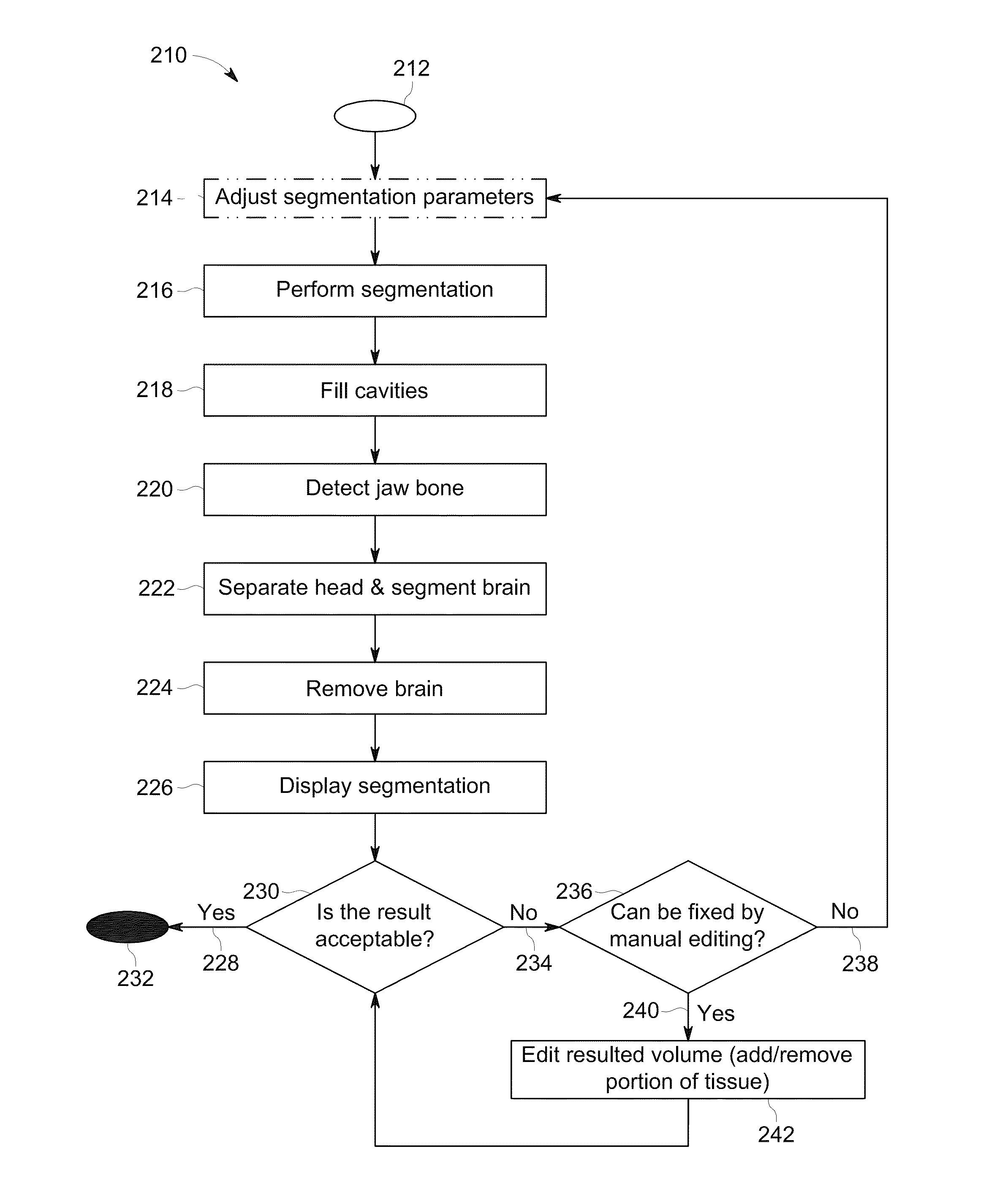
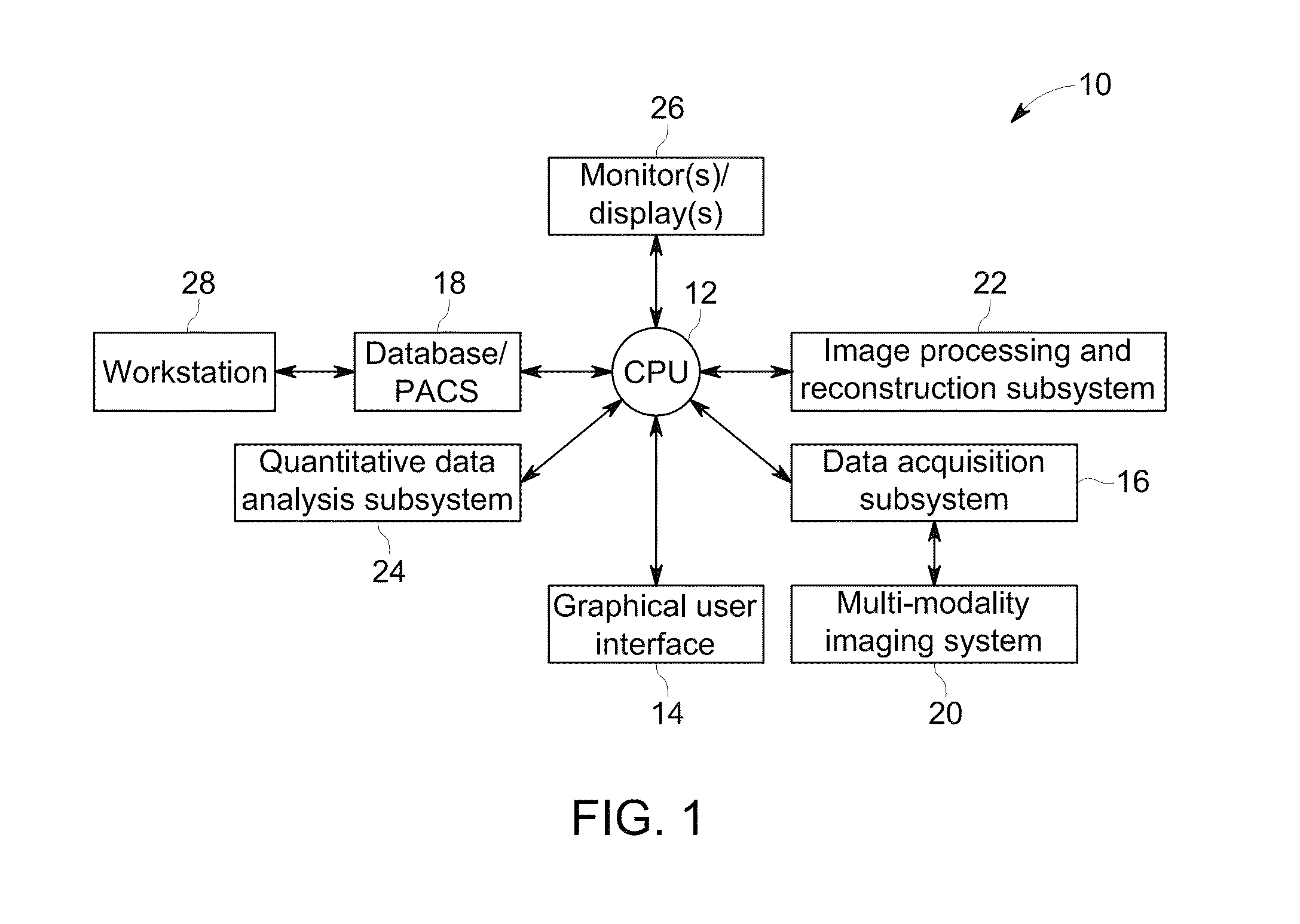
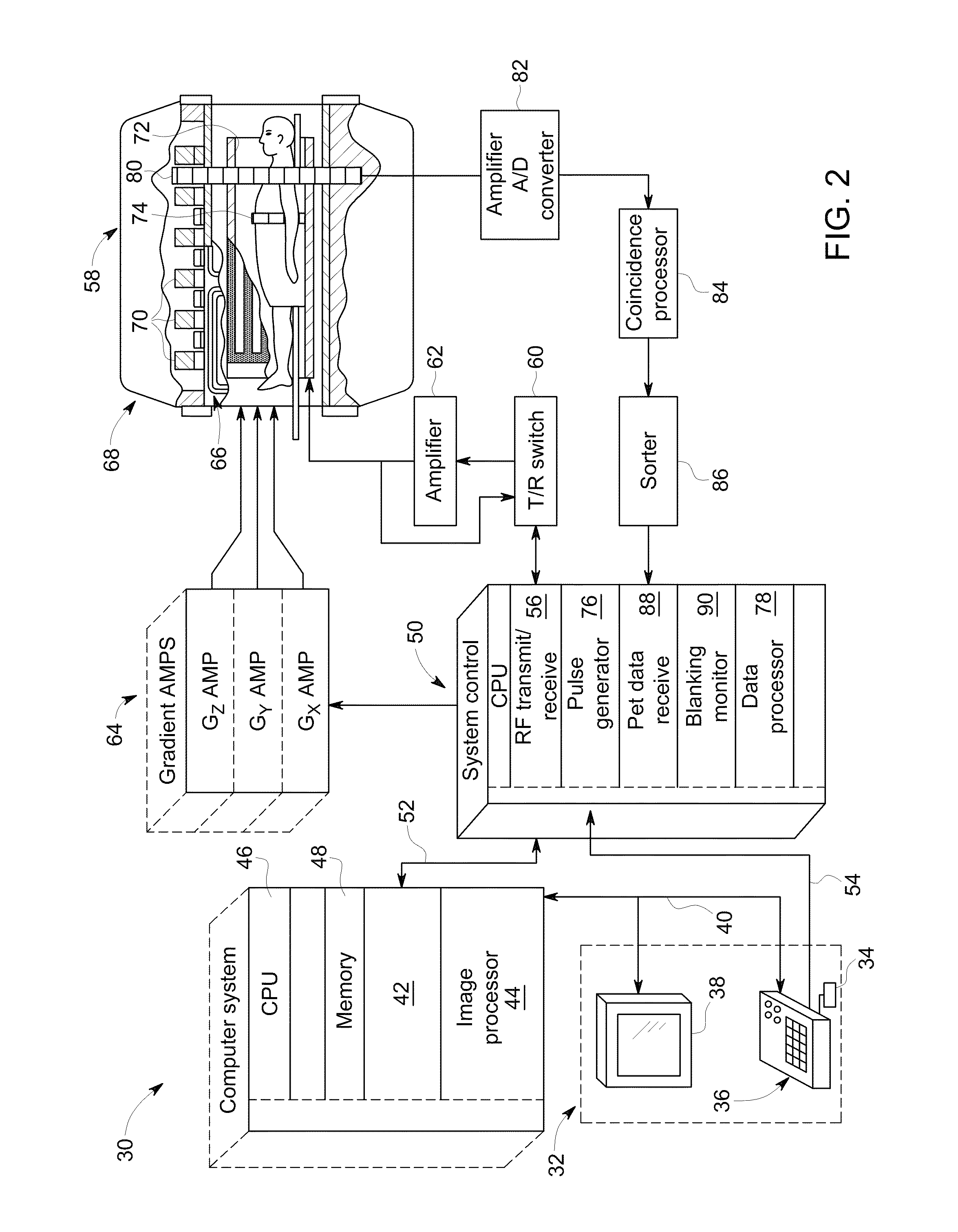
[0022]The operating environment of embodiments of the invention is described below with respect to a multi-modality imaging system that includes a positron emission tomography (PET) imaging system and one of an magnetic resonance (MR) imaging system and a sixty-four-slice, “third generationcomputed tomography (CT) imaging system. However, it will be appreciated by those skilled in the art that the invention is equally applicable for use with other multi-slice configurations and with other CT imaging systems. In addition, while embodiments of the invention are described with respect to techniques for use with MR or CT imaging systems and PET imaging systems, one skilled in the art will recognize that the concepts set forth herein are not limited to CT and PET and can be applied to techniques used with other imaging modalities in both the medical field and non-medical field, such as, for example, an x-ray system, a single-photon emission computed tomography (SPECT) imaging system, o...


PUM


Abstract

Methods and systems for evaluating bone lesions include accessing a first dataset acquired from a patient with a first imaging modality and a second dataset acquired from the patient with a second imaging modality. A segmentation is performed on the first dataset to identify a subset of the first dataset corresponding to a skeletal structure of the patient and a patient skeletal metric representing a total bone volume of the patient is automatically calculated from the subset of the first dataset. The methods and systems further include detection of at least one lesion in the second dataset, classification of the at least one lesion as a bone or non-bone lesion, automatic calculation of a bone lesion metric based on the classification, and calculation of a lesion burden as a ratio of the bone lesion metric and the patient skeletal metric.

Description

BACKGROUND OF THE INVENTION[0001]Embodiments of the invention relate generally to medical diagnostics and treatment assessment and, more particularly, to methods and systems for evaluating bone lesions using multi-modality image data.[0002]It is not uncommon for a single patient to undergo a multitude of medical imaging exams, whether in a single doctor's visit, in a hospital stay, or even over the course of a lifetime. This is particularly likely when a patient undergoes a series of “tests” and scans to investigate a recently onset or previously undetected condition. It is increasingly common for a patient to be subjected to multiple scans across multiple medical imaging modalities because each exam can provide different pieces of information. For example, during a single doctor's visit or hospital stay, a magnetic resonance (MR) imaging system, an x-ray imaging system, or a computed tomography (CT) imaging system can be used to acquire images that provide anatomical information, w...

Claims


Application Information

Patent Timeline
Patent Type & Authority Patents(United States)
IPC IPC(8): G06T7/00G06F19/00A61B6/03A61B6/00A61B5/00
CPCA61B6/505A61B6/5217A61B6/5235G06F19/321G06F19/345G06F19/3431G06F19/3437G06T7/0081G06T7/0012A61B5/0035A61B5/4504A61B6/032A61B6/037A61B6/461G06F19/3406G06K2209/055G06T2207/10081G06T2207/10088G06T2207/10104G06T2207/10108G06T2207/30008G06T7/11G06T7/136G16H40/63G16H50/30G16H50/50G16H50/20G16H30/40G06V2201/033G06T2200/24G06T2207/20224
Inventor SPRENCZ, PAL CSONGORJOHNSEN, ROBERT JOHNUBELHART, ISTVANPHILPS, ELIZABETHKRISTON, ANDRASFAZEKAS, ANDREA
Owner GENERAL ELECTRIC CO
Learn More