Sacroiliac joint exposure, fusion, and debridement

a technology of sacroiliac joints and fusion, applied in the field of sacroiliac joint exposure, fusion, debridement, can solve problems such as interference with complete healing and function restoration, and achieve the effect of facilitating closure and subsequent healing

Inactive Publication Date: 2016-09-27
HILL ROBERT CHARLES
View PDF17 Cites 43 Cited by
  • Summary
  • Abstract
  • Description
  • Claims
  • Application Information

AI Technical Summary

Benefits of technology

[0015]Briefly, a surgical method embodiment of the present invention comprises positioning the patient prone on a radiolucent surgical support table. Fluoroscopy or radiographs are used to confirm the pelvis, and lateral outline of the sacrum and Sacroiliac joint complex. A longitudinal incision is planned over the posterior iliac crest, centered on the posterior iliac spine. An incision is made down the iliac crest to expose the deep fascia. Holes which will be used to repair the iliac osteotomy are made obliquely from the medial to lateral to cross the plane of exposure. A series of holes or a bur cut are made lateral to the previous drill holes and centered over the sacroiliac complex with the aid of fluoroscope or radiograph imaging. The cut is parallel to the main axis of the spine and extends from the distal to the proximal away from the sciatic notch. The cut is guided by the imaging. An osteotome or side cutting burr is used to complete the osteotomy. The medial part of the iliac crest is freed from the back of the sacrum and displaced medially. This then exposes the back of the sacrum and sacroiliac joint complex for inspection, debridement, and curettage for fusion. Harvested bone or allograft can be applied respectively to the prepared fusion surfaces on the sacrum and ilium. The exposure within the joint may be used to guide a targeting device for sacroiliac joint fixation prior to closure. Such exposure of the sacroiliac joint can also be used advantageously to place a fusion device inside the joint proper. The closure and subsequent healing are facilitated by the predrilled oblique holes. These are used to re-attach the medial iliac bone to the remaining lateral ilium. Thereafter, the closure of the remaining soft tissues and skin are routine.

Problems solved by technology

For example, these all can cause wide injuries in varying degrees to the soft tissues and bones adjacent to the joint that will consequently interfere with complete healing and restoration of function.

Method used


Image

  • Sacroiliac joint exposure, fusion, and debridement
  • Sacroiliac joint exposure, fusion, and debridement
  • Sacroiliac joint exposure, fusion, and debridement

Examples


Embodiment Construction

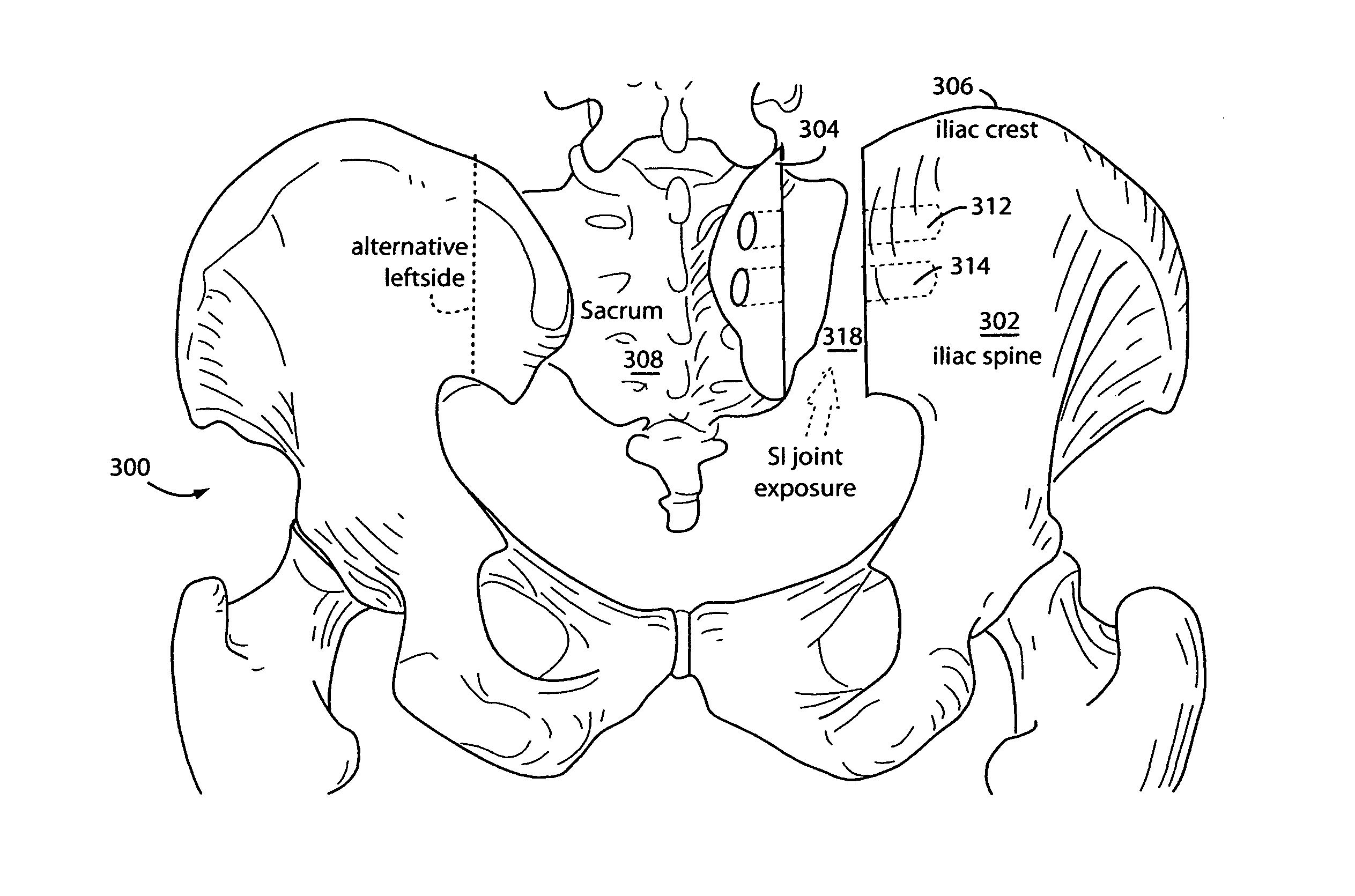
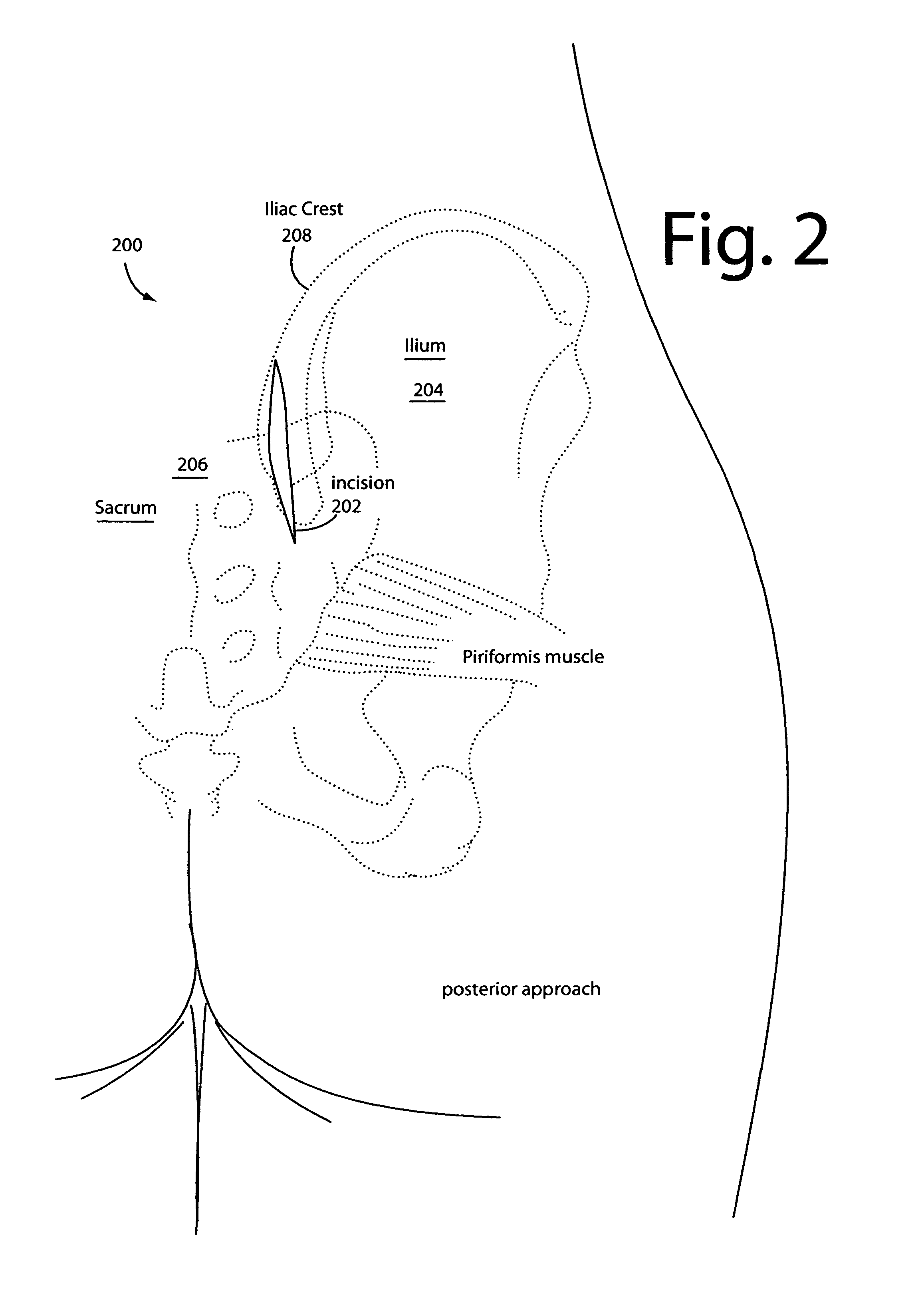
[0020]The posterior approach to exposing the sacroiliac joint is generally considered safe and simple because it does not endanger vital structures. This approach has been widely used to treat disruptions of the sacroiliac joint, fractures of the ilium near the joint, and infections of the sacroiliac joint and surrounding bones.

[0021]Surgical method embodiments of the present invention use a much smaller, vertical incision and iliac osteotomy (pelvis bone cutting) to expose the sacroiliac joint underneath.

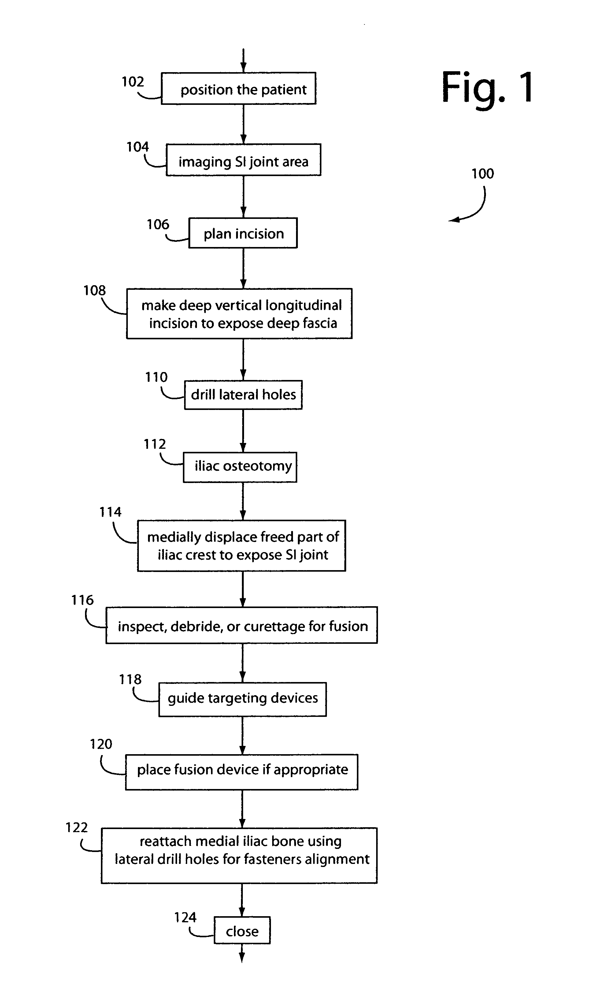
[0022]FIG. 1 illustrates a surgical method embodiment of the present invention for sacroiliac joint exposure, fusion, and debridement methods and the reliable restoration of nearby tissues, and such is referred to herein by the general reference numeral 100.

[0023]Method begins in a step 102 by positioning the patient prone on a radiolucent surgical support table. Fluoroscopy or radiographs are used in a step 104 to confirm the pelvis, and lateral outline of the sacrum and Sacroilia...


PUM


Abstract

A surgical method for exposure of the sacroiliac joint using a posterior approach begins with a longitudinal incision over the posterior iliac crest, centered on the posterior iliac spine. Holes which will be used to repair the iliac osteotomy are made obliquely from the medial to lateral to cross the plane of exposure. A cut through the bone is made lateral to the drill holes and centered over the sacroiliac complex with the aid of fluoroscope or radiograph imaging. The medial part of the iliac crest is freed from the back of the sacrum and displaced medially for the sacroiliac joint complex to be inspected, debrided, or curettaged for fusion. The closure and subsequent healing are facilitated by the predrilled oblique holes. These are used to re-attach the medial iliac bone to the remaining lateral ilium.

Description

BACKGROUND OF THE INVENTION[0001]1. Field of the Invention[0002]The present invention relates to medical and surgical methods and devices for sacroiliac joint exposure, fusion, and debridement methods and the reliable restoration of nearby tissues.[0003]2. Description of Related Art[0004]The human body has two sacroiliac joints in the pelvis. In between them is the sacrum which supports the spine at the fifth lumbar vertebrae. So one joint is on the left and the other is close by on the right. The outside of each sacroiliac joint meets and is overlapped by an ilium on each side of the pelvis. The ilium is the uppermost and largest bone of the pelvis. All of these bones are held together by strong ligaments.[0005]Transosseous, medial posterior and anterior exposures of the sacroiliac joint are conventional and each has its shortcomings. For example, these all can cause wide injuries in varying degrees to the soft tissues and bones adjacent to the joint that will consequently interfer...

Claims


Application Information

Patent Timeline
Patent Type & Authority Patents(United States)
IPC IPC(8): A61F2/44A61F2/46
CPCA61F2/4611A61F2/4455A61B17/56A61B17/84A61B2017/564
Inventor LAWSON, KEVIN JON
Owner HILL ROBERT CHARLES
Why Eureka
Try Eureka