Detecting tumorous breast tissue in a thermal image

a breast and thermal image technology, applied in the field of breast cancer screening software interface tool and breast thermal image detection method, can solve the problems of large equipment required for mammography, discomfort in personal physical contact and procedure, and inability to detect malignancies as effectively as examination,

Active Publication Date: 2016-11-08
NIRAMAI HEALTH ANALYTIX PVT LTD
View PDF15 Cites 4 Cited by
  • Summary
  • Abstract
  • Description
  • Claims
  • Application Information

AI Technical Summary

Problems solved by technology

In the mammography exam, the subject is subject to highly personal physical contact and a procedure which many subjects find discomforting.
In subjects with dense breast tissue, the exam may not be as effective in spotting malignancies as for other subjects.
Moreover, the equipment required for mammography is relatively large and relatively expensive.
Since mammography is an x-ray machine it cannot be used at homes without supervision.
The x-ray radiation itself may result in cancer.
It is also not effective for younger women due to the dense breast tissue.
There is also some evidence that the physical manipulation of the breast tissue during mammography could rupture the malignant cysts, thereby increasing risk of spreading the malignant cells to other tissues and into the subject's blood stream.
Moreover, since medical practitioners trained in thermography are not readily available in rural areas in emerging markets like India, automatic screening tools will help open up these market for software applications for breast cancer screening and detection.

Method used


Image

  • Detecting tumorous breast tissue in a thermal image
  • Detecting tumorous breast tissue in a thermal image
  • Detecting tumorous breast tissue in a thermal image

Examples


Embodiment Construction

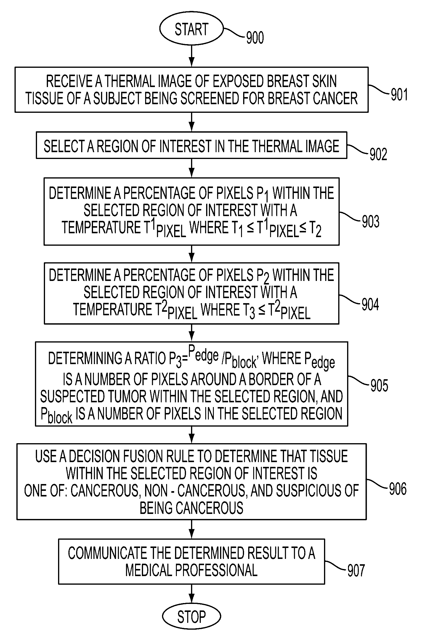
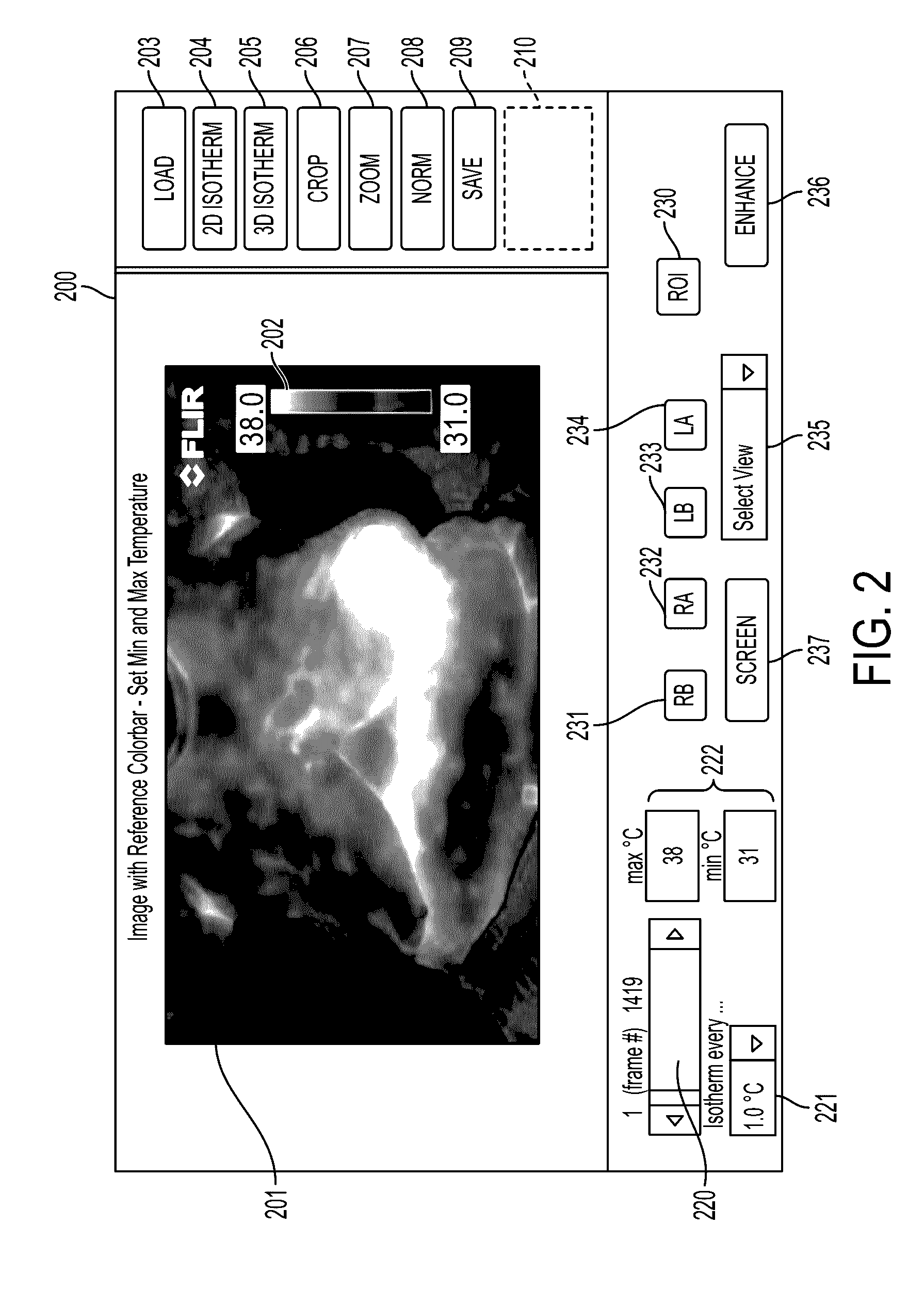
[0021]What is disclosed is a software interface tool for breast cancer screening and a method for detecting cancerous tissue in a thermal image of a breast.

NON-LIMITING DEFINITIONS

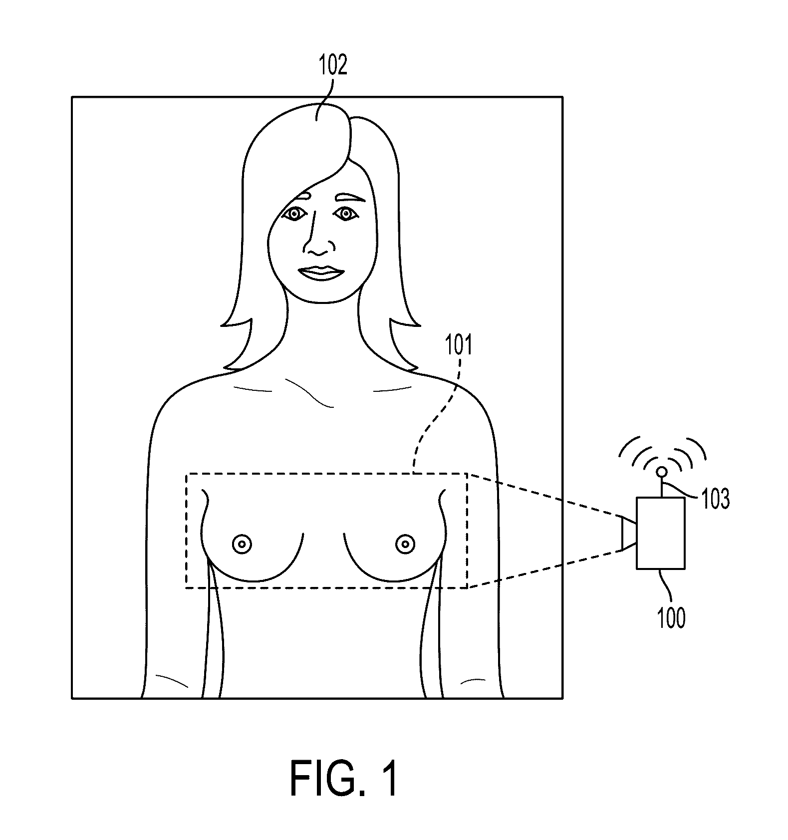
[0022]A “subject” refers to a living being. Although the term “person” or “patient” may be used throughout this disclosure, it should be appreciated that the subject may be something other than a human such as, for example, a primate. Therefore, the use of such terms is not to be viewed as limiting the scope of the appended claims strictly to humans. FIG. 1 shows an example human female patent 102.

[0023]A “thermal imaging system” is a camera with a lens that focuses infrared energy from objects in a scene onto an array of specialized sensors which convert infrared energy into electrical signals on a per-pixel basis and outputs a thermal image comprising an array of pixels with color values corresponding to surface temperatures of the objects in the image across a thermal wavelength band. FIG. 1 shows a the...


PUM


Abstract

What is disclosed is a system and method for detecting cancerous tissue in breast tissue using a thermal image. In one embodiment, the present tumor detection method involves selecting a region of interest in the thermal image to be processed for breast cancer screening. Thereafter, a percentage of pixels p1 in the selected region having a temperature Tpixel1, where T1≦Tpixel1≦T2, is determined. A percentage of pixels p2 in the selected region having a temperature Tpixel2, where T3≦Tpixel2, is determined. A ratiop3=Pedge / Pblockis also determined, where Pedge is a number of pixels around a border of a suspected tumor within the selected region, and Pblock is a number of pixels in a perimeter of the selected region. A decision fusion rule R, as more fully disclosed herein, is utilized to determine, based on these determination whether tissue within that region is cancerous, or non-cancerous, or is suspicious of being cancerous.

Description

CROSS REFERENCE TO RELATED APPLICATIONS[0001]This patent application is related to concurrently filed and commonly owned U.S. patent application Ser. No. 14 / 668,178, entitled: “Software Interface Tool For Breast Cancer Screening”, by Krithika Venkataramani et al., which is incorporated herein in its entirety by reference.TECHNICAL FIELD[0002]The present invention is specifically directed to a software interface tool for breast cancer screening and a method for detecting cancerous breast tissue in a thermal image of the breast.BACKGROUND[0003]Breast cancer incidence rates are relatively high in women. Nearly 1 in 8 women in the western world and nearly 1 in 11 women in India will have breast cancer. In the western world, it is the leading cancer in women. In India, for example, it is the second after cervical cancer. Early detection is key to survival as the mortality rates are high for advanced stages. Mammography is considered the gold standard for breast cancer screening. Screenin...

Claims


Application Information

Patent Timeline
Patent Type & Authority Patents(United States)
IPC IPC(8): G06K9/00A61B5/01A61B5/00H04N5/33G06T7/00
CPCA61B5/015A61B5/4312A61B5/746G06T7/0012H04N5/33G06T2207/10048G06T2207/10116G06T2207/20104G06T2207/30068G06T2207/30096A61B5/0075A61B5/0082A61B5/72A61B5/0008A61B5/748A61B10/0041A61B2576/02G06T7/136G16H30/40
Inventor VENKATARAMANI, KRITHIKA
Owner NIRAMAI HEALTH ANALYTIX PVT LTD
Why Eureka
Try Eureka