
Detection of epileptiform activity

a technology of epileptiform activity and detection field, which is applied in the field of detection of epileptiform activity, can solve the problems of high morbidity and mortality, and disturbed brain function and the resulting eeg, and achieves the effect of efficient computation power

Active Publication Date: 2017-02-28
GENERAL ELECTRIC CO
View PDF10 Cites 11 Cited by
  • Summary
  • Abstract
  • Description
  • Claims
  • Application Information

AI Technical Summary

Benefits of technology

The invention provides a way to detect epileptiform activity in brain wave data automatically and reliably, regardless of changes in the patient's level of consciousness. It is efficient in terms of the computation power needed and can detect specific epileptiform waveforms efficiently. The detection is based on the morphology of the brain wave data and does not require high computation power. This makes it useful for devices with limited computation power.

Problems solved by technology

Different derangements of internal system homeostasis disturb the environment in which the brain operates, and therefore the function of the brain and the resulting EEG are disturbed.
However, when brain activity is recorded from the scalp, the measured signal is a composition originating from multiple sources and methods indicative of the complexity of the signal show an increase during a seizure, cf.
CSE is a medical emergency and is associated with high morbidity and mortality.
As mentioned above, epileptiform activity commonly refers to signal waveforms or patterns which are typical in epilepsy and which may also be associated with an increased risk of seizures.
However, the EEG findings in encephalopathy have many similarities to those during sedation and anesthesia, which makes the detection of encephalopathy in sedated patients difficult.
Therefore, the methods are not enough specific to the epileptiform activity only.
One drawback related to the above techniques for automatic detection of epileptiform activity is the weak specificity to epileptiform activity.
Therefore, the methods cannot distinguish, for example, whether an increase in the wavelet entropy is caused by an epileptiform EEG of anesthetized patient or the arousal of the patient.
A further drawback of the prior art detection techniques is that they cannot indicate when a specific type of epileptiform activity is present in the EEG or which type of epileptiform waveforms are present in the EEG signal.
Many of the algorithms are also rather complex and require high computation power, which makes them unsuitable for ambulatory devices.

Method used


Image

  • Detection of epileptiform activity
  • Detection of epileptiform activity
  • Detection of epileptiform activity

Examples


Embodiment Construction

[0040]Below, different embodiments of the invention are discussed assuming that the brain wave signal data measured from the patient is EEG signal data and that either one or both of the entropy and kurtosis of the subband-specific output data is / are used as the indicator(s) of epileptiform activity.

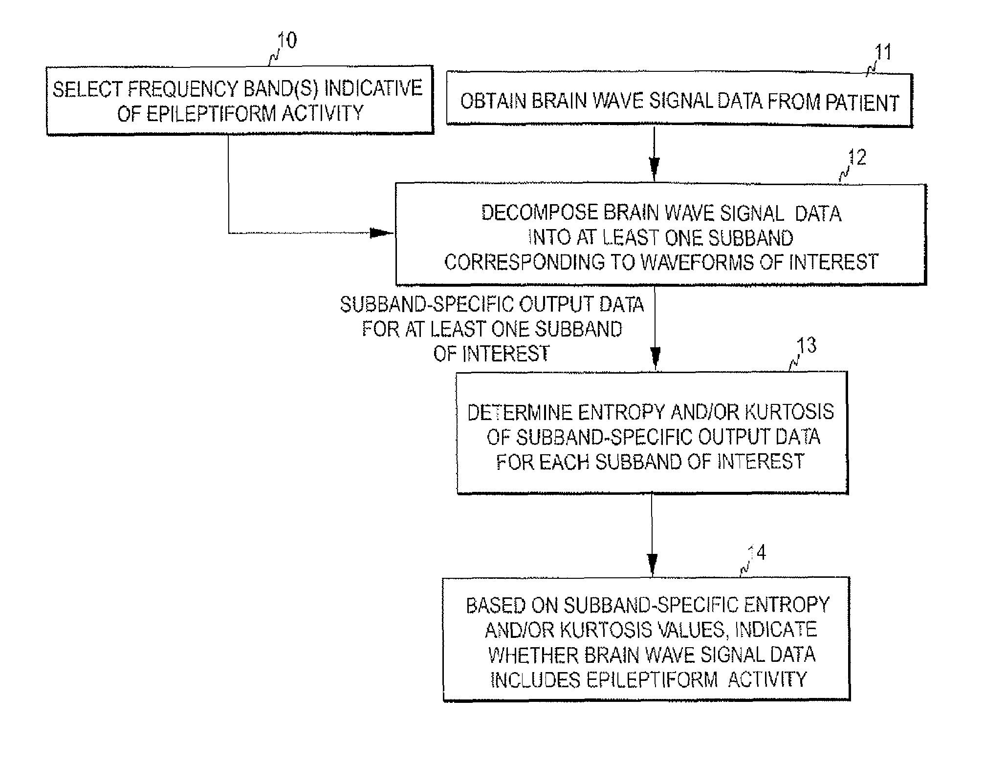
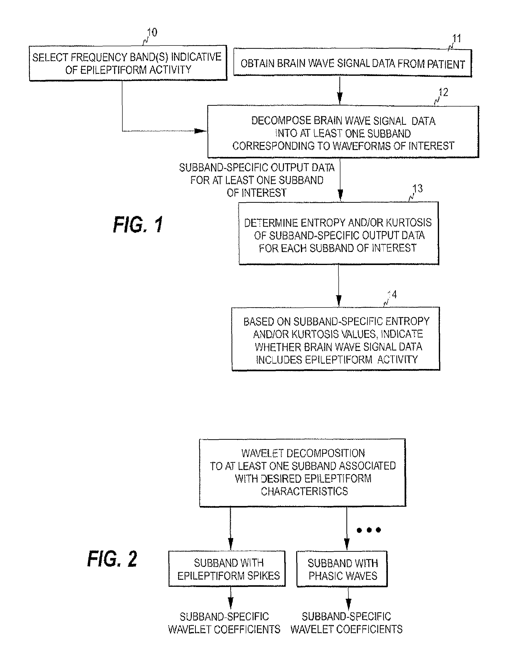
[0041]FIG. 1 is a flow diagram illustrating the detection mechanism of the invention. As discussed above, the epileptiform EEG activity may include spiky waveforms. Although the frequency contents of the spikes may reach up to about 70 Hz, epileptiform EEG components are typically below 30 Hz. In the present invention, at least one EEG frequency band is selected, which contains epileptiform activity (step 10) and the raw EEG signal data obtained from a patient is decomposed into at least one subband on which the waveforms of interest appear (steps 11 and 12) so as to obtain subband-specific output data for each of the at least one subband. For example, if epileptiform spikes are to be de...


PUM


Abstract

The invention relates to detection of epileptiform activity. In order to accomplish a mechanism with improved specificity to epileptiform activity and with the capability to detect specific type of epileptic patterns, brain wave signal data obtained from a subject is decomposed into at least one predetermined subband, each subband being indicative of a specific type of epileptiform activity. The subband-specific output data obtained represents a time series of a quantitative characteristic of the brain wave signal data. At least one measure is determined for any one or more of the at least one predetermined subband, the at least one measure belonging to a measure set comprising a first measure indicative of the entropy of the subband-specific output data and a second measure indicative of a normalized form of k:th order central moment of the subband-specific output data, where k is an integer higher than three. The presence of a specific type of epileptiform activity may be detected based on the at least one measure of the respective subband.

Description

FIELD OF THE INVENTION[0001]The present invention relates generally to the detection of epileptiform activity. More particularly, the present invention relates to a mechanism for automatic detection of epileptiform activity in subject's brain wave data. Epileptiform activity here refers to signal waveforms or patterns which are typical in epilepsy and which may also be associated with an increased risk of seizures.BACKGROUND OF THE INVENTION[0002]Electroencephalography (EEG) is a well-established method for assessing brain activity. When measurement electrodes are attached on the skin of the skull surface, the weak biopotential signals generated in brain cortex may be recorded and analyzed. The EEG has been in wide use for decades in basic research of the neural systems of the brain as well as in the clinical diagnosis of various central nervous system diseases and disorders.[0003]The EEG signal represents the sum of excitatory and inhibitory potentials of large numbers of cortical ...

Claims


Application Information

Patent Timeline
Patent Type & Authority Patents(United States)
IPC IPC(8): A61B5/04A61B5/048G06K9/00A61B5/00A61B5/374
CPCA61B5/048A61B5/4094A61B5/726G06K9/00523A61B5/374G06F2218/08
Inventor SARKELA, MIKA
Owner GENERAL ELECTRIC CO
Learn More