Endoscopic stapler

a stapler and endoscope technology, applied in the field of endoscope staplers, can solve the problems of difficulty in obtaining a purchase on the sac wall, and achieve the effect of facilitating the inserting of at least one stapl

Active Publication Date: 2017-07-25
THE CLEVELAND CLINIC FOUND
View PDF11 Cites 648 Cited by
  • Summary
  • Abstract
  • Description
  • Claims
  • Application Information

AI Technical Summary

Benefits of technology

[0006]In accordance with an embodiment of the present invention, an endoscopic stapler comprises a first jaw and a second jaw. The first jaw includes structure for retaining a plurality of staples. The first jaw and second jaw are mounted to permit pivotal movement of the first and second jaws relative to one another. The endoscopic stapler also comprises at least one clamp member for clamping tissue and pulling the tissue into a position between the first jaw and the second jaw to facilitate inserting at least one staple of the plurality of staples into the tissue. A first handle is operably coupled with the first jaw such that movement of the first handle effects delivery of the at least one staple of the plurality of staples to the tissue. A second handle is operably coupled with the second jaw and the at least one clamp member such that movement of the second handle effects movement of the second jaw and movement of the at least one clamp member.
[0007]In accordance with another embodiment of the present invention, an endoscopic stapler for suturing tissue comprises a first jaw and a second jaw. The first jaw includes structure for retaining a plurality of staples. The first jaw and second jaw are mounted to permit pivotal movement of the first and second jaws relative to one another. The endoscopic stapler also comprises two clamp members for clamping tissue and pulling the tissue into a position between the first jaw and the second jaw to facilitate inserting at least one staple of the plurality of staples into the tissue. The two clamp members are laterally spaced from one another. The first jaw and the second jaw are disposed laterally between the two clamp members. A handle is operably connected with the second jaw and with the clamp members such that movement of the handle effects pivotal movement of the second jaw relative to the first jaw and also effects clamping movement of the clamp members.

Problems solved by technology

Due to the limited field of view, obtaining purchase on the sac wall is technically challenging, but is essential for adequate correction of the deformity.

Method used


Image

  • Endoscopic stapler
  • Endoscopic stapler
  • Endoscopic stapler

Examples


Embodiment Construction

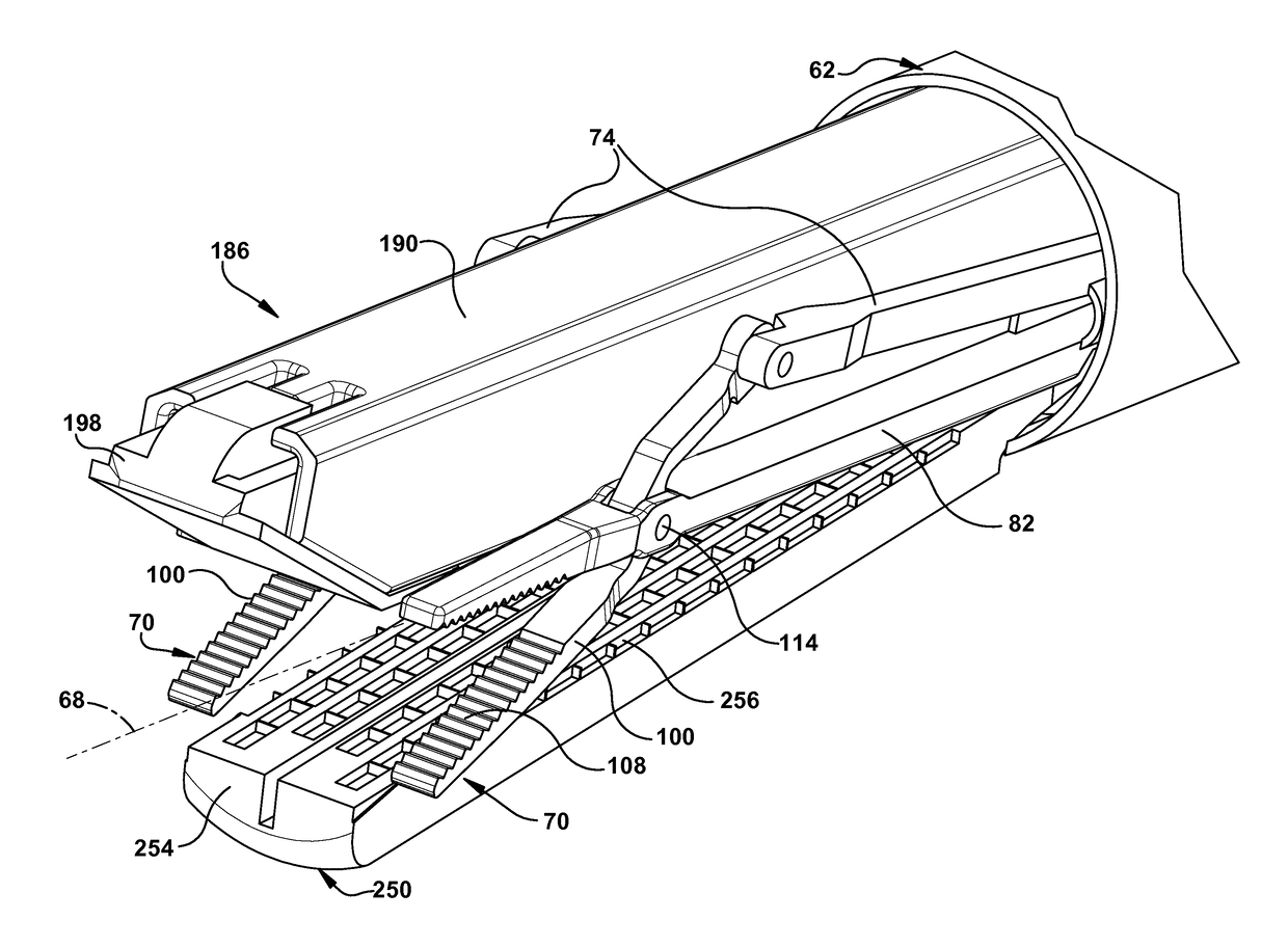
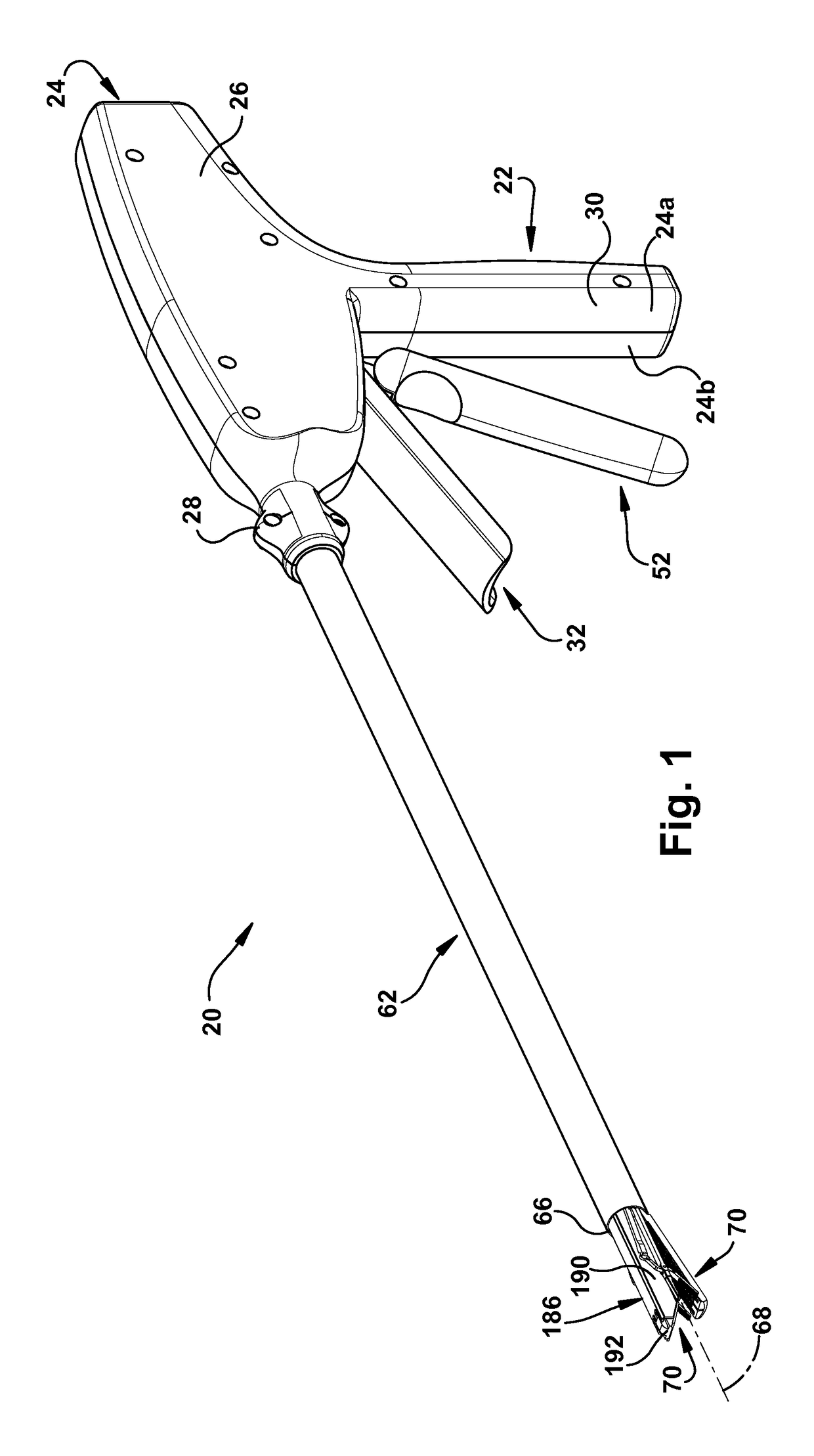
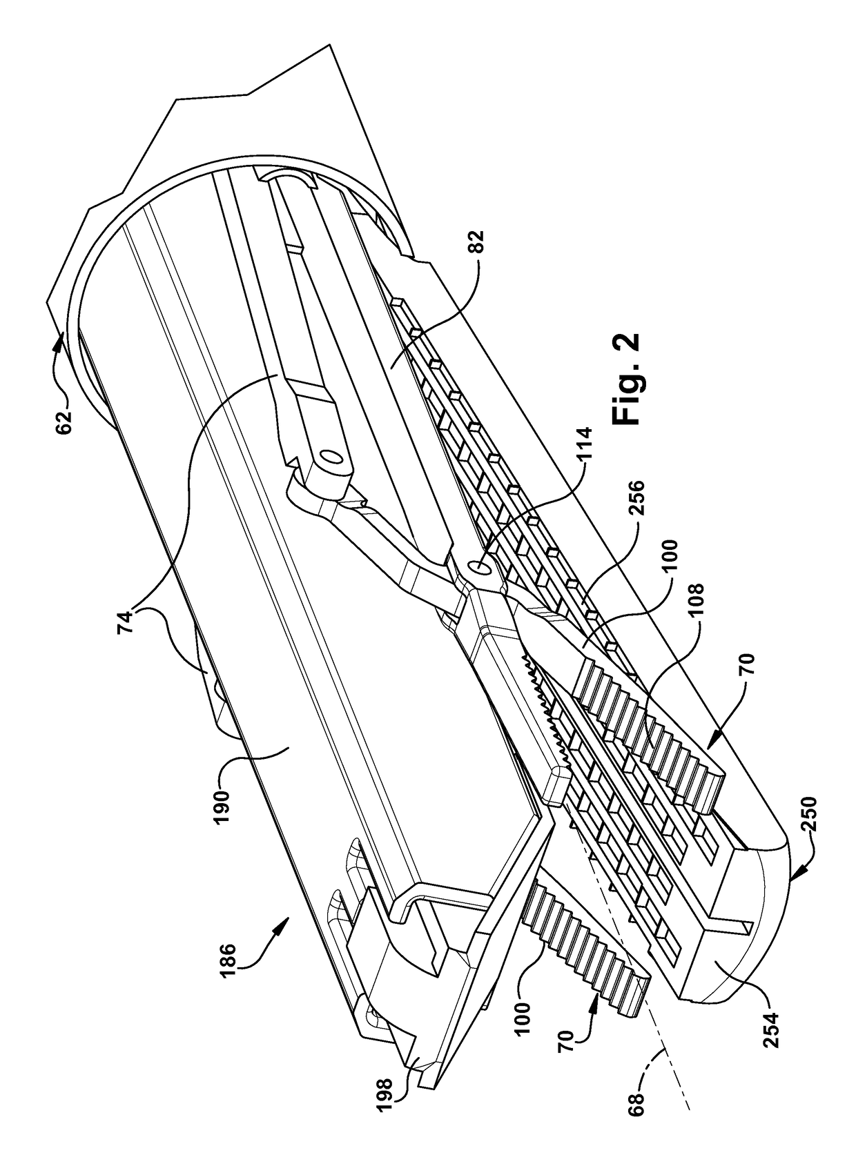
[0023]The present invention relates to an endoscopic stapler and, in particular, relates to a stapler that pulls tissue inward before stapling. FIGS. 1-10 illustrate a stapling device 20 in accordance with the present invention. The stapling device 20 includes a grip assembly 22, a pair of laterally spaced-apart clamp members 70, and a stapler 186 positioned laterally between the clamp members.

[0024]The grip assembly 22 includes a housing 24, a first handle 32, a second handle 52, and a tubular member 62. As can be seen in FIG. 1, for example, the housing 24 is shaped like a pistol with an enlarged body portion 26, a tubular barrel portion 28, and a grip portion 30. The tubular barrel portion 28 projects to the left, as viewed in FIG. 1, from one end of the body portion 26. The grip portion 30 projects downwardly, as viewed in FIG. 1, from the body portion 26 at an intermediate position along the length of the body portion. The housing 24 is formed in two halves 24a and 24b that, in...


PUM


Abstract

An endoscopic stapler comprises a first jaw and a second jaw. The first jaw includes structure for retaining a plurality of staples. The first jaw and second jaw are mounted to permit pivotal movement of the first and second jaws relative to one another. The endoscopic stapler also comprises at least one clamp member for clamping tissue and pulling the tissue into a position between the first jaw and the second jaw to facilitate inserting at least one staple of the plurality of staples into the tissue. A first handle is operably coupled with the first jaw such that movement of the first handle effects delivery of the at least one staple of the plurality of staples to the tissue. A second handle is operably coupled with the second jaw and the at least one clamp member such that movement of the second handle effects movement of the second jaw and movement of the at least one clamp member.

Description

CROSS-REFERENCE TO RELATED APPLICATION[0001]This application claims the benefit of U.S. Provisional Patent Application No. 61 / 701,852 filed on Sep. 17, 2012, and entitled ENDOSCOPIC STAPLER, which is incorporated herein by reference in its entirety.TECHNICAL FIELD[0002]The present invention relates to an endoscopic stapler and, in particular, relates to a stapler that clamps and pulls tissue before stapling the tissue.BACKGROUND OF THE INVENTION[0003]Tissue stapling is used in a variety of clinical applications to treat injury, correct deformities, and promote healing. One application that requires tissue stapling is Zenker's Diverticulum (ZD). ZD is a condition in which excessive pressure within the lower pharynx causes the weakest portion of the pharyngeal wall to balloon out, forming a diverticulum which may reach several centimeters in diameter. Symptomatic cases of ZD have been traditionally treated by neck surgery to either resect the diverticulum and / or incise the cricopharyn...

Claims


Application Information

Patent Timeline
Patent Type & Authority Patents(United States)
IPC IPC(8): A61B17/04A61B17/08A61B17/072A61B17/29
CPCA61B17/07207A61B17/29A61B2017/2906
Inventor LORENZ, ROBERT R.
Owner THE CLEVELAND CLINIC FOUND
Why Eureka
Try Eureka