

Method for preparing microscopic image of holographic digitalized sliced sheet
A technology of microscopic images and production methods, applied in the direction of digital output to display equipment, microscopes, image communication, etc., can solve long-term problems, achieve clarity, optimize consultation operating procedures, increase storage and repeatability Effect
- Summary
- Abstract
- Description
- Claims
- Application Information
AI Technical Summary
Problems solved by technology
Method used
Image

Examples
Embodiment 1
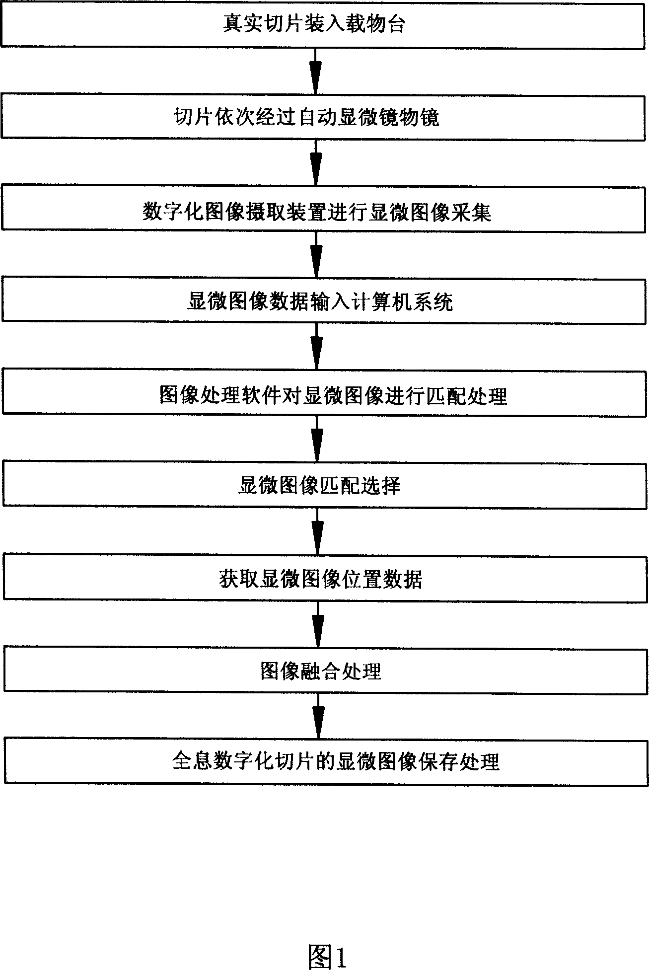
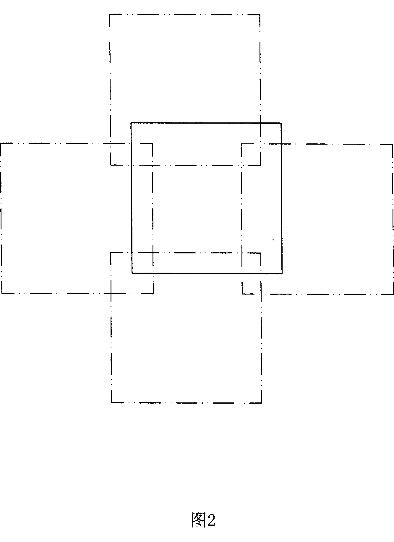
[0023] Referring to Fig. 1 and Fig. 2, the method for making a microscopic image of a holographic digital slice, the specific steps are as follows:
[0024] A. Use an automatic microscope to collect the microscopic image information of a slice, and control the stage of the automatic microscope through the set control program to carry the slice through the objective lens of the automatic microscope in sequence according to the specified collection range, collection order and step length, The microscopic image units displayed on the objective lens are photographed one by one by a digital image capture device connected to the automatic microscope, and the data conversion is completed to generate serialized multiple microscopic image unit data; The micro image unit data includes at least all pixel information of the microscopic image of the slice, the magnification information of the objective lens, the sequence information of the micro image unit, the coordinate information of the...
Embodiment 2
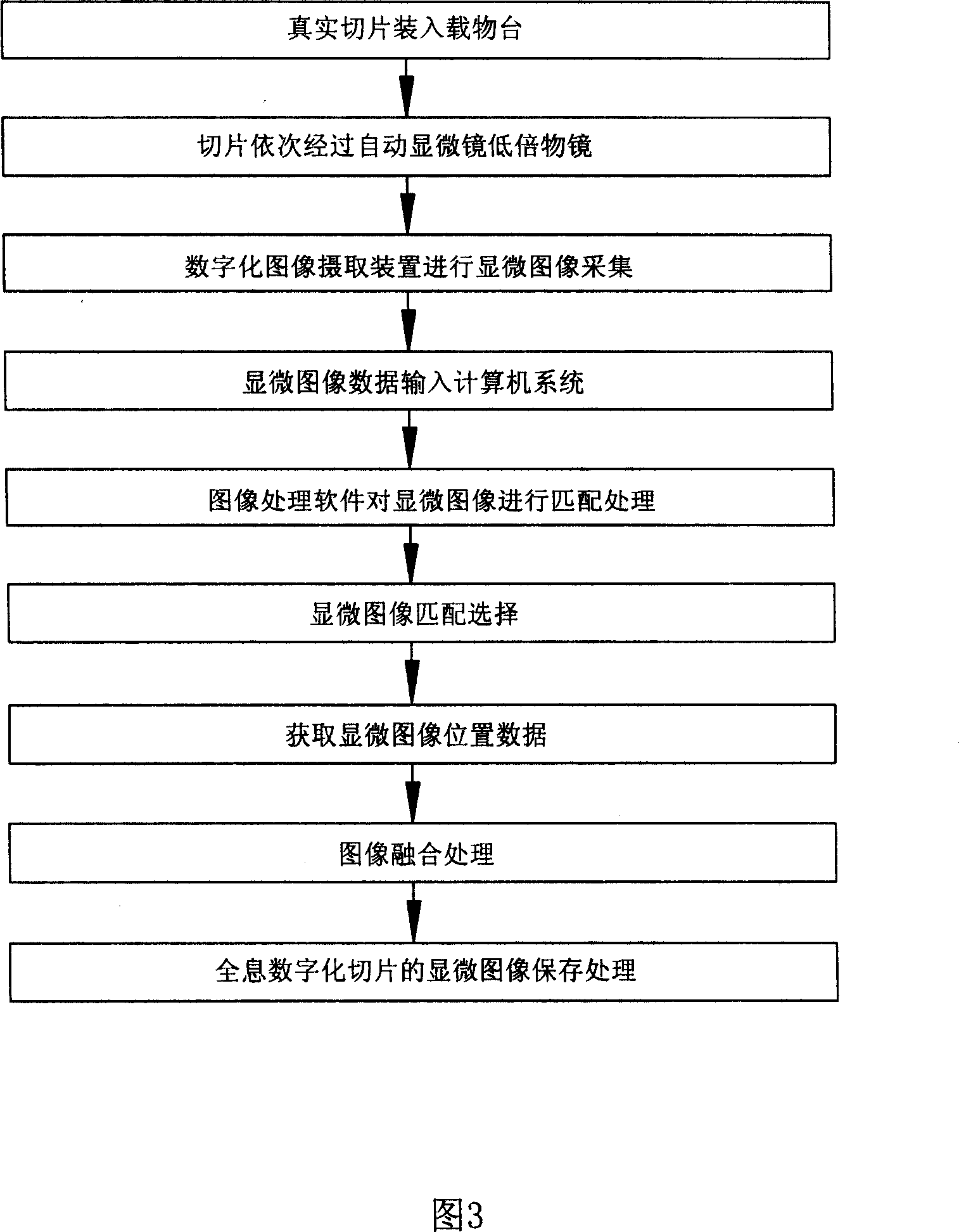
[0048] Referring to Fig. 3, the present embodiment is an improvement on the basis of Embodiment 1. When the automatic microscope collects microscopic image information of a slice, the used objective lens is a low power objective lens (the magnification of the objective lens is 4, referred to as low magnification objective lens), according to the steps described in Embodiment 1, collect and make, the microscopic image of the generated holographic digital slice is a low magnification image, adopt the JPEG algorithm to compress the microscopic image, and classify and divide the microscopic image according to the compression format Blocks are stored in an image file. The low magnification objective lens refers to 4 times or 10 times. This method collects fewer images, has faster image processing speed, saves computer resources, and is suitable for fast panorama browsing.
Embodiment 3
[0050] Referring to Fig. 4, the present embodiment is an improvement on the basis of Embodiment 1. When the automatic microscope collects microscopic image information of a slice, the used objective lens is a high-magnification objective lens (the magnification of the objective lens is 40, referred to as High magnification objective lens), collect and make according to the steps described in Embodiment 1, the microscopic image of the generated holographic digitized slice is a high magnification image, adopt the JPEG algorithm to compress the microscopic image, and classify and divide the microscopic image according to the compression format Blocks are stored in an image file. The grading refers to converting the high-magnification microscopic image of the holographic digitized slice into microscopic images of several magnification levels, for example: 4 times, 10 times, 20 times, 40 times. The microscopic images of the holographic digitized slices of the four magnification lev...
PUM
 Abstract
Description
Claims
Application Information