Supercharge Your Innovation With Domain-Expert AI Agents!

Digital X-ray pictures-taking diagnosis device and the encapsulation method thereof

A technology of a diagnostic device and a packaging method, applied in the directions of diagnosis, instruments for radiological diagnosis, X-ray equipment, etc.

Inactive Publication Date: 2008-01-30
徐敏
View PDF0 Cites 0 Cited by
  • Summary
  • Abstract
  • Description
  • Claims
  • Application Information

AI Technical Summary

Problems solved by technology

[0003] However, since the aperture of the scintillation plate is 43 × 43 centimeters, the aperture of the charge-coupled device is 40 × 40 millimeters, and the ratio of the focal length of the lens to the aperture is generally set at 1: 1.2, like this, in order to make the scintillation plate produce Fluorescence can fully enter the lens for charge-coupled devices to generate clear digital images. In digital X-ray imaging diagnostic systems, there are strict requirements for the positions of charge-coupled devices, lenses and scintillation plates, that is, charge-coupled devices are generally required The conjugate matching with the scintillation plate must reach the order of 10 microns, which is an extremely difficult technical problem in the current technical field

Method used


Image

  • Digital X-ray pictures-taking diagnosis device and the encapsulation method thereof
  • Digital X-ray pictures-taking diagnosis device and the encapsulation method thereof
  • Digital X-ray pictures-taking diagnosis device and the encapsulation method thereof

Examples


Embodiment Construction

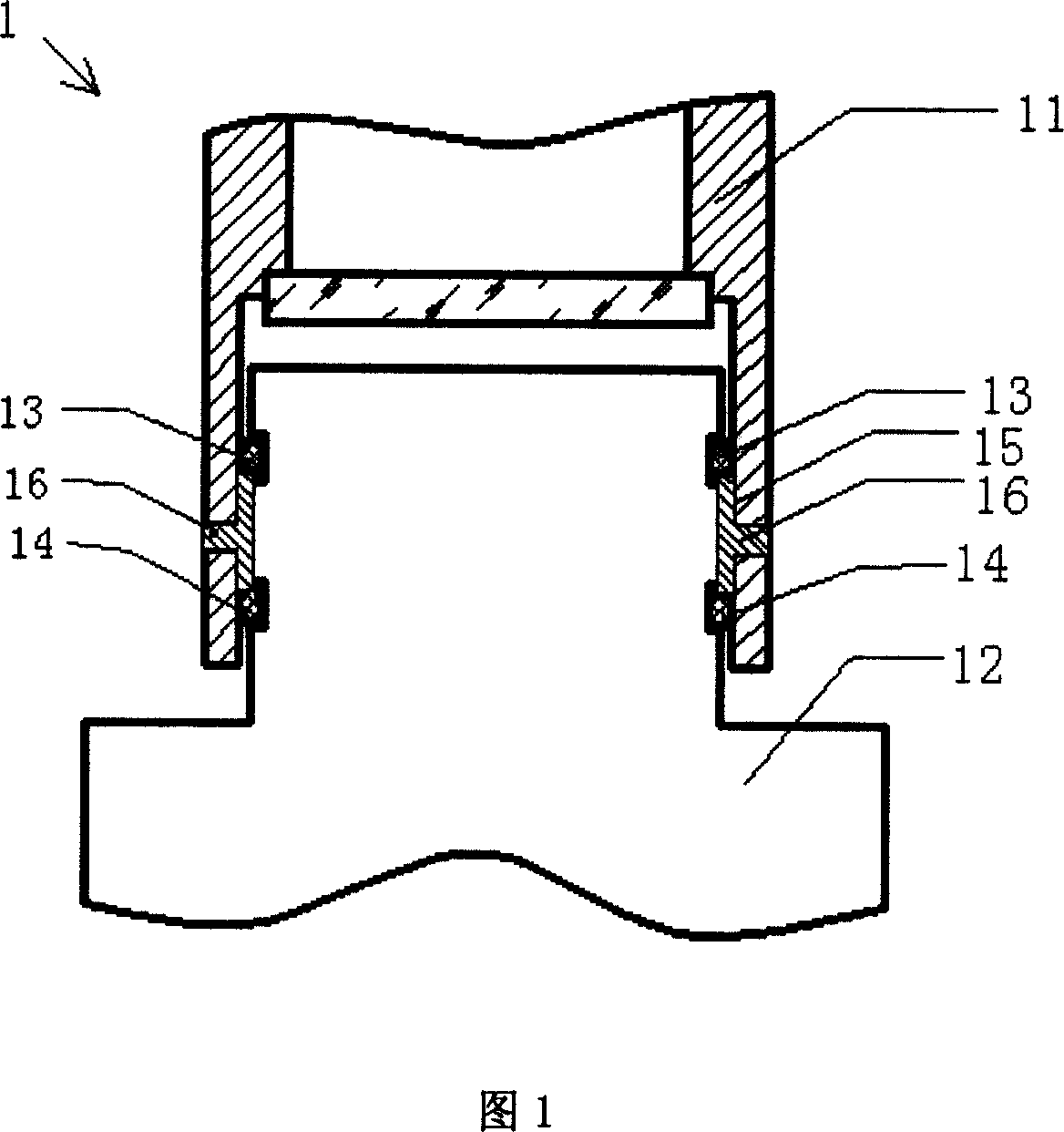
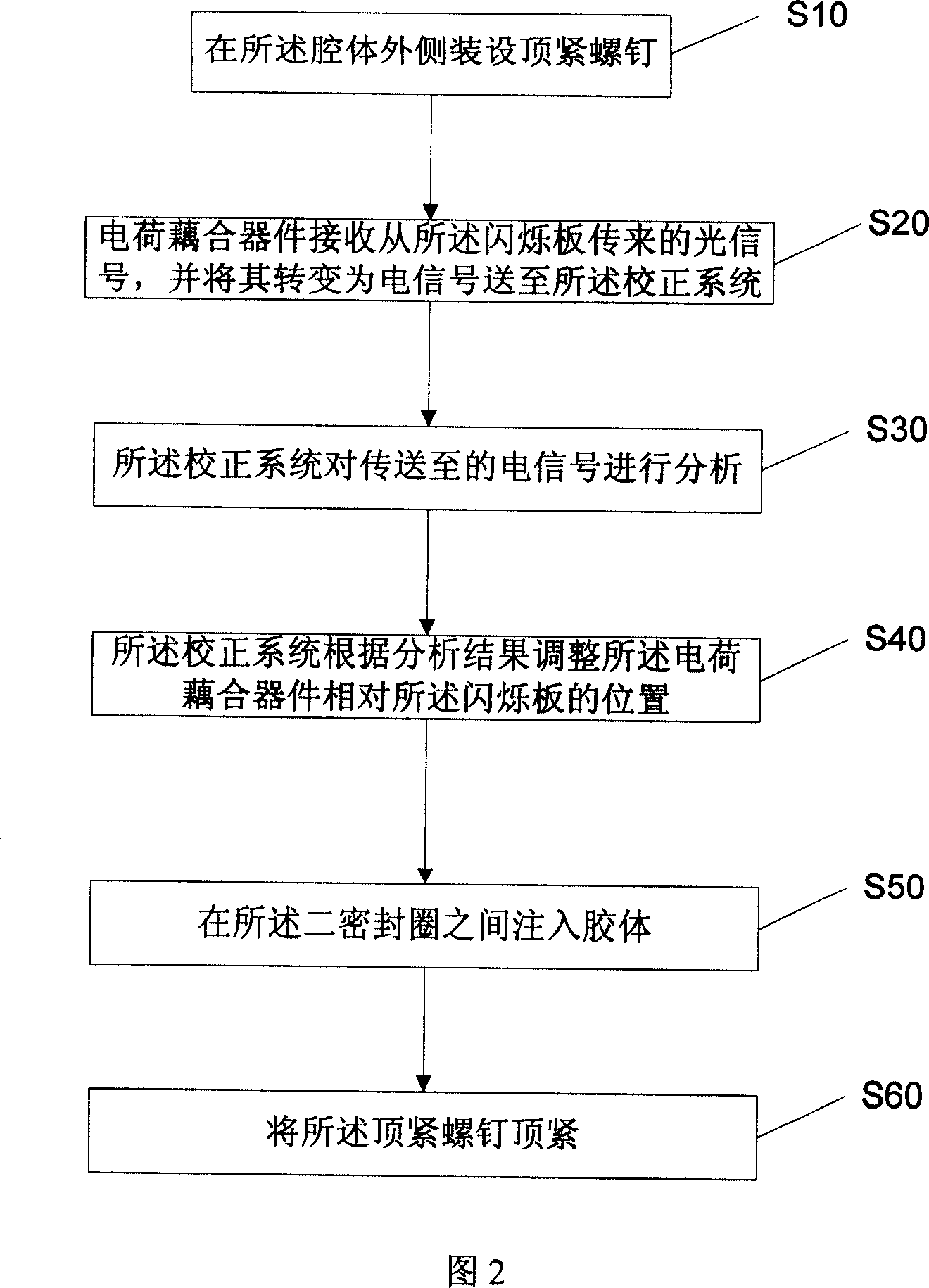
[0014] Please refer to FIG. 1 , the digital X-ray diagnostic device 1 of the present invention at least includes: a cavity 11 , an accommodating space, two sealing rings 13 and 14 and a colloid layer 15 .

[0015] The cavity 11 is used for accommodating the charge-coupled device and the lens 12, and 6 small holes are arranged on the outside thereof, and the accommodating space is provided between the cavity 11 and the lens 12, wherein the accommodating The space can be implemented as a groove extending circumferentially on the inner surface of the cavity 11 (as shown in FIG. 1 ), or as a groove extending circumferentially on the inner surface of the lens 12 (not shown). , the two sealing rings 13 and 14 are used to snap into the accommodating space, and the colloidal layer 15 is connected between the two sealing rings 13 and 14, wherein the colloidal layer 15 is made of epoxy resin .

[0016] When installing, first evenly smear some vacuum lubricating oil on the two sealing r...


PUM


Abstract

The invention provides a digital X-ray photographic diagnostic device and an enclosing method, wherein, the digital X-ray photographic diagnostic device comprises a cavity for containing a charge-coupled device and a lens, a containing space kept between the cavity and the lens, two sealing loops connected and arranged in the containing space, and a colloid layer arranged between the two sealing loops. The enclosing method of the digital X-ray photographic diagnostic device is that the charge-coupled device is positioned and regulated at first, the colloid is injected between the two sealing loops to fix the charge-coupled device, therefore, the conjugate match of 10 micrometres of the charge-coupled device and the flash plated comprised by the digital X-ray photographic diagnostic device is achieved.

Description

technical field [0001] The utility model of the invention relates to an X-ray photographic diagnostic device and a packaging method thereof, in particular to a digital X-ray photographic diagnostic device and a packaging method thereof. Background technique [0002] Although the Digital X Radiographic diagnostic system (Digital X Radiographic, DR) is currently the most advanced digital X-ray diagnostic system in the world, its working principle is that when the X-ray source emits The X-rays irradiated by the object under inspection on the scintillation plate will cause the scintillation plate to produce fluorescence, and the distribution of the fluorescent light intensity on the surface of the scintillation plate will reflect the internal characteristics of the object under inspection. The fluorescence of the internal characteristics of the object is sent to the photosensitive surface of the charge coupled device (CCD), and the charge coupled device converts the received flu...

Claims


Application Information

Patent Timeline
Patent Type & Authority Applications(China)
IPC IPC(8): A61B6/00H05G1/02
Inventor 徐敏
Owner 徐敏
Features
  • R&D
  • Intellectual Property
  • Life Sciences
  • Materials
  • Tech Scout
  • Unparalleled Data Quality
  • Higher Quality Content
  • 60% Fewer Hallucinations
Learn More