Method for detecting immune body affinity
A technology for detecting antibodies and affinity, applied in the field of medicine and biology, can solve problems such as long time required, and achieve the effects of good repeatability, stable and reliable detection results, and high sensitivity
- Summary
- Abstract
- Description
- Claims
- Application Information
AI Technical Summary
Problems solved by technology
Method used
Image
Examples
Embodiment 1
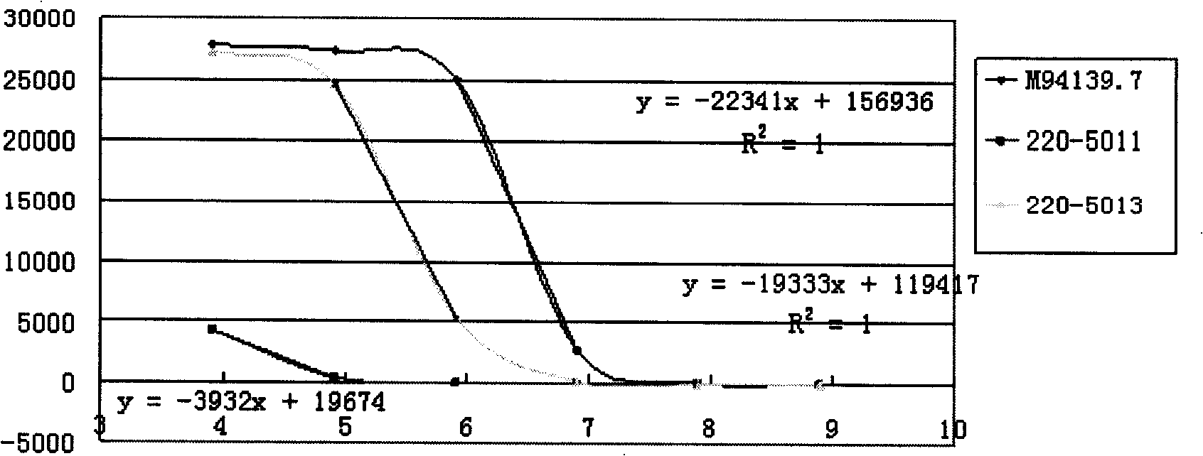
[0065] Take the affinity detection of three different antibodies (cloning numbers M94139.7, 220-5011, and 220-5013) to β-HCG (β-subunit human chorionic gonadotropin) as an example.
[0066] The steps of the antibody affinity detection method are as follows:
[0067] Antigen-coated microspheres:
[0068] -Use a vortex oscillator or ultrasonic to suspend the microspheres (Luminex, USA), about 20s;
[0069] - Take 50μL of microspheres in a 1.5ml centrifuge tube and centrifuge at a speed of ≥8,000g for 1-2min;
[0070] -Remove the supernatant, resuspend the microspheres in 100 μL double distilled water, suspend the microspheres with a vortex shaker or ultrasonic wave for about 20 seconds, and centrifuge at a speed of ≥8000g for 1-2 minutes;
[0071] - Discard the supernatant, add 80 μL of phosphate buffer (pH 6.2), vortex for about 20 seconds, and ultrasonically suspend the microspheres for about 20 seconds;
[0072] -Add 10μL 50mg / mL Sulfo-NHS and mix gently with a vortex shak...
Embodiment 2
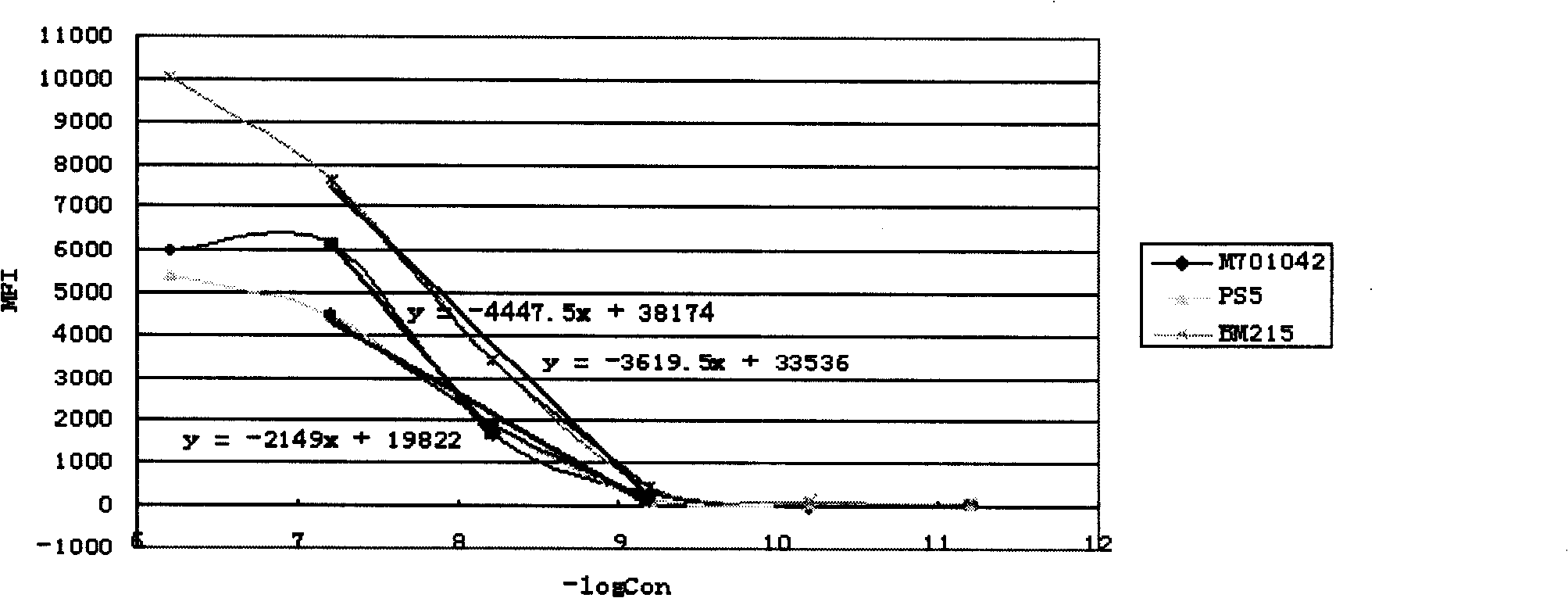
[0119] Affinity detection of three antibodies (cloning numbers: M701042, PS5, BM215) of PSA (Prostate Specific Antigen).
[0120] The method used is the same as that of Example 1. The result is as follows,
[0121] Table 2
[0122] μg / ml mol / L Negative logarithm of concentration M701042(MFI value) PS5(MFI value) BM215(MFI value)
[0123] 100 6.25E-07 6.20412 6323.5 5369 10054.5
[0124] 10 6.25E-08 7.20412 6479 4481.5 7657
[0125] 1 6.25E-09 8.20412 2031.5 1910.5 3447.5
[0126] 0.1 6.25E-10 9.20412 640 183.5 418
[0127] 0.01 6.25E-11 10.20412 336.5 97 125
[0128] 0.001 6.25E-12 11.20412 438.5 52.5 57
[0129] The result is calculated as follows:
[0130] Calculation of the affinity constant of the M701042 antibody:
[0131] y=(6323.5-438.5) / 2=2942.5, substitute into the formula y=-4447.5x+38174, and get x=7.92164
[0132] Affinity constant K=1 / (10 -7.92164 )=8.35E+07
[0133] Similarly, the affinity constants of PS5 and BM215 antibodies can be calculated, which...
Embodiment 3
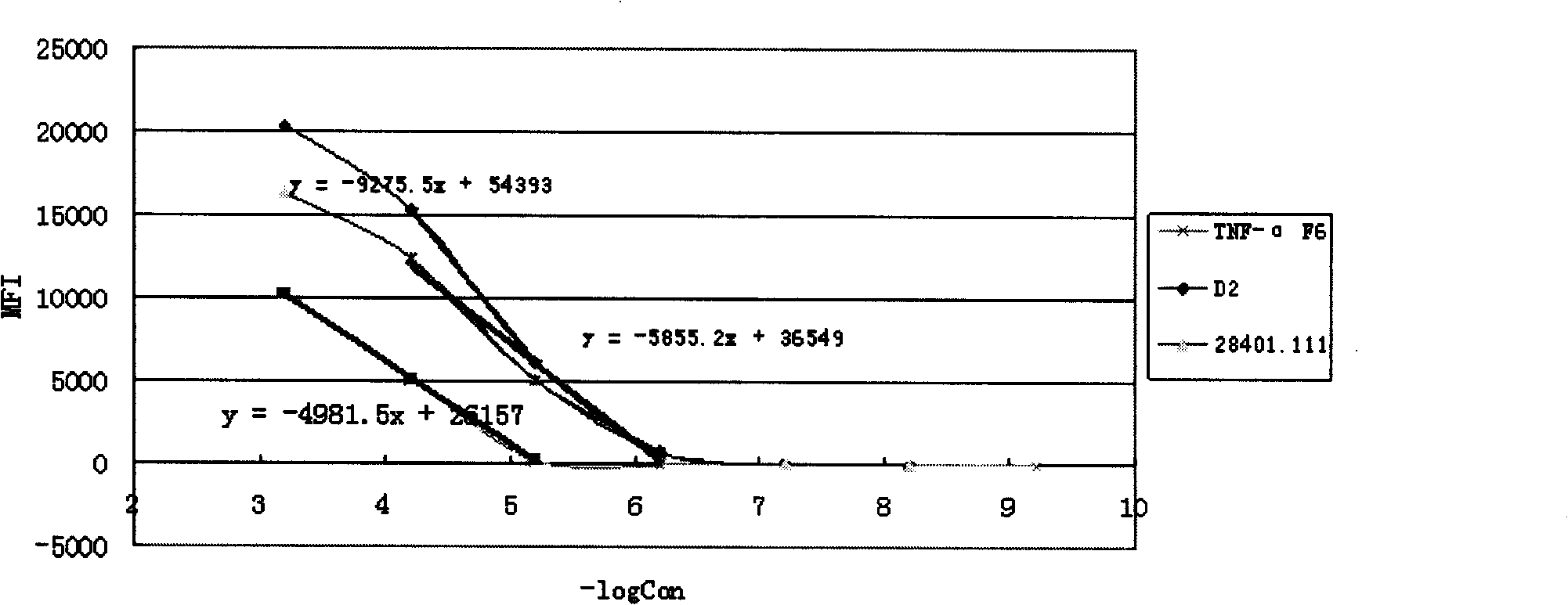
[0135] Affinity detection of three antibodies to TNF-α (Tumor necrosis factor-alpha, tumor necrosis factor α) (cloning numbers are respectively TNF-αF6, D2, 28401.111).
[0136] The method used is the same as that of Example 1. The result is as follows,
[0137] table 3
[0138] Negative logarithm of μg / ml mol / L concentration TNF-αF6 (MFI value) D2 (MFI value) 28401.111 (MFI value)
[0139] 100 0.000625 3.204119983 10233.5 20352 16397
[0140] 10 6.25E-05 4.204119983 5137.5 15397.5 12433
[0141] 1 6.25E-06 5.204119983 270.5 6122 5077
[0142] 0.1 6.25E-07 6.204119983 0 779 722.5
[0143] 0.01 6.25E-08 7.204119983 0 70.5 73
[0144] 0.001 6.25E-09 8.204119983 2 15.5 16
[0145] 0.0001 6.25E-10 9.204119983 16
[0146] The result is calculated as follows:
[0147] Calculation of TNF-αF6 antibody affinity constant:
[0148] y=(10233.5-2) / 2=5116.75, substitute into the formula y=-4981.5x+26157, and get x=4.2237
[0149] Affinity constant K=1 / (10 -4.2237 )=1.67E+04
[...
PUM
 Abstract
Description
Claims
Application Information
 - R&D
- Intellectual Property
- Life Sciences
- Materials
- Tech Scout
- Unparalleled Data Quality
- Higher Quality Content
- 60% Fewer Hallucinations
Browse by: Latest US Patents, China's latest patents, Technical Efficacy Thesaurus, Application Domain, Technology Topic, Popular Technical Reports.
© 2025 PatSnap. All rights reserved.Legal|Privacy policy|Modern Slavery Act Transparency Statement|Sitemap|About US| Contact US: help@patsnap.com


