Method for testing instantaneous speed and accelerated speed of dissection type M type kinetocardiogram

An instantaneous acceleration, instantaneous velocity technology, applied in the direction of organ motion/change detection, ultrasonic/sonic/infrasound image/data processing, ultrasonic/sonic/infrasonic Permian technology, etc. Problems such as the detection of instantaneous velocity and acceleration of cardiogram

Inactive Publication Date: 2008-11-05
FUZHOU UNIV
View PDF2 Cites 4 Cited by
  • Summary
  • Abstract
  • Description
  • Claims
  • Application Information

AI Technical Summary

Problems solved by technology

Although the invention patent: Velocity Field and Acceleration Field Detection Method and Device of Omnidirectional M-mode Cardiogram (its patent number is ZL200310104284.0) solves the detection of the instantaneous velocity field and acceleration field of omnidirectional M-mode Cardiogram, it has not It can solve the detection of instantaneous velocity and acceleration of anatomical M-mode cardiogram

Method used


Image

  • Method for testing instantaneous speed and accelerated speed of dissection type M type kinetocardiogram

Examples


Embodiment Construction

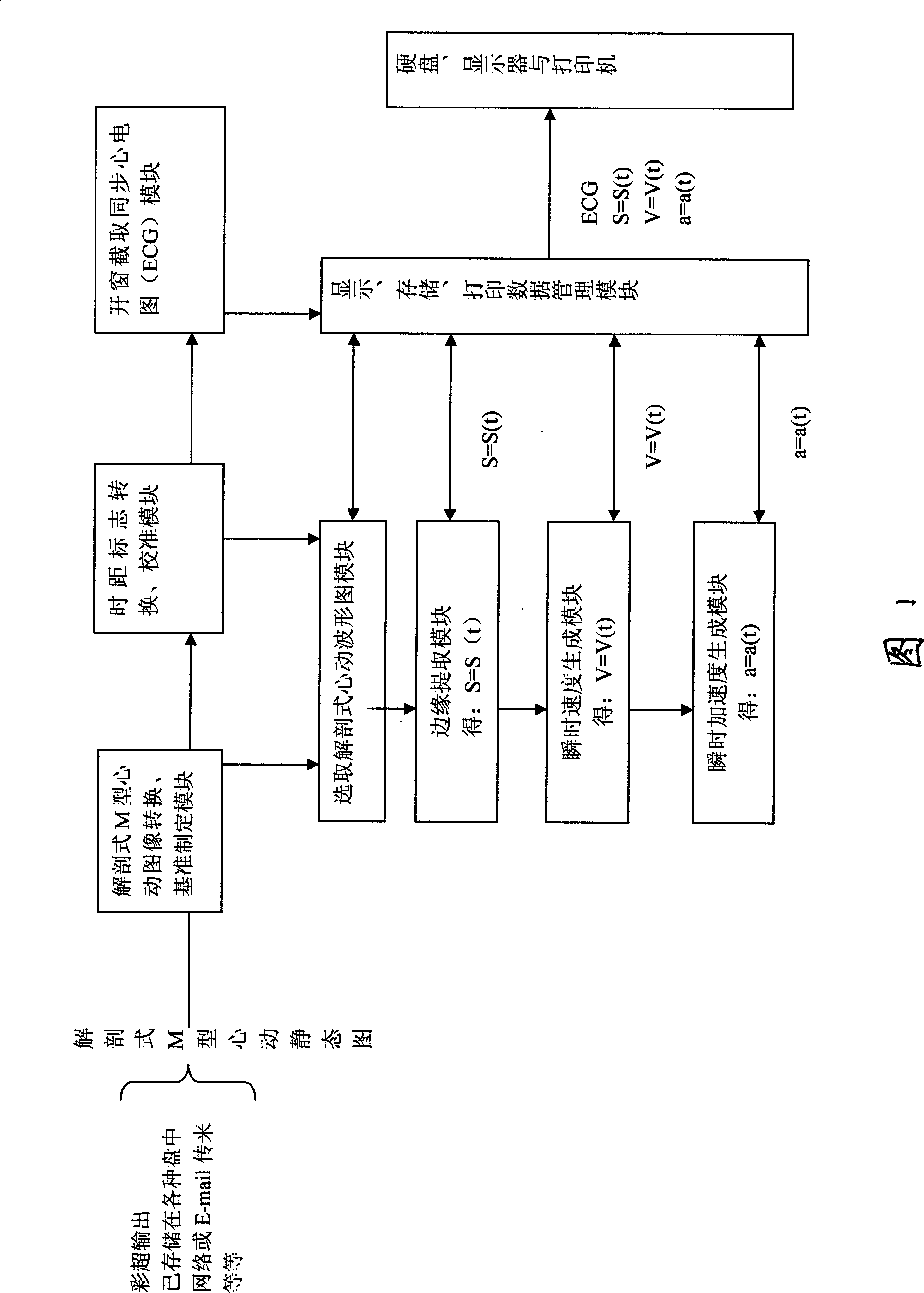
[0021] As shown in the figure, the present invention comprises the following steps:

[0022] 1. Convert anatomical M-mode cardiac still images (in JPG, BMP, etc. formats) output from color ultrasound or stored in various disks or from the Internet or E-mail, etc. into anatomical M-mode cardiac image conversion, The baseline formulation module converts the incoming anatomical M-mode static images in various formats into BMP format static images, and determines the baseline of the time axis (starting point) and the extracted anatomical M-mode static images.

[0023] 2. The time distance mark and the calibration module convert the time and distance scales on the extracted anatomical M-type heartbeat static map into the X and Y direction pixel point interval time distance values ​​of the current map, so as to achieve the (y, The conversion of the actual displacement reference unit of measurement (cm, etc.) and the time reference measurement unit (second, etc.) of the measurable pi...


PUM


Abstract

This invention relates to a detection method for an instantaneous speed and an instantaneous acceleration of an anatomic M-typed kinetocardiogram, which is characterized by comprising an edge extracting module, a speed generating module and an acceleration generating module, wherein, the edge extracting module comprises a linear template, the invention chooses waveform of the anatomic M-typed kinetocardiogram of a certain part of a certain structure randomly in the anatomic M-typed kinetocardiogram, time discrete function differentiation is made for the waveform after the edge extracting is made so as to obtain the motion speed of every moment of the part, and then the time discrete function differentiation is made for the waveform again, thus obtaining the motion acceleration of every moment of the part (a repeating part). The invention can detect the motion speed and the motion acceleration of of every moment of every part of every heart structure accurately, thus disclosing the motion information of every part of the heart further in a noninvasive mode and providing important scientific evidences for the study of the diagnosis of the heart disease and the hemodynamics.

Description

technical field [0001] The invention relates to a detection method of instantaneous velocity and acceleration, in particular to a detection method of instantaneous velocity and acceleration of anatomical M-mode cardiogram. Background technique [0002] A large number of devices in the currently imported color Doppler ultrasound (i.e. color Doppler echocardiography) have the function of anatomical M-mode echocardiography. It is based on the two-dimensional ultrasonic heart cross-sectional image to reconstruct the movement of each structural tissue of the heart on a certain direction line versus time waveform. [0003] In addition to the Doppler detection of the original blood velocity, the currently imported color Doppler ultrasound also realizes the detection of the component of the instantaneous motion velocity of a certain position of the heart tissue in the direction of the ultrasonic beam, that is, the tissue Doppler technology. The tissue Doppler function of the existi...

Claims


Application Information

Patent Timeline
Patent Type & Authority Applications(China)
IPC IPC(8): A61B8/08
Inventor 林强
Owner FUZHOU UNIV
Why Eureka
Try Eureka