Imaging system and method

An imaging system and imaging technology, applied in the field of biomedical imaging, can solve problems such as deformation and motion artifacts, and achieve the effect of eliminating motion artifacts

Inactive Publication Date: 2009-06-17
HUAZHONG UNIV OF SCI & TECH
View PDF1 Cites 11 Cited by
  • Summary
  • Abstract
  • Description
  • Claims
  • Application Information

AI Technical Summary

Problems solved by technology

[0004] In view of this, the object of the present invention is to provide an imaging system and method for sol...

Method used


Image

  • Imaging system and method
  • Imaging system and method
  • Imaging system and method

Examples


Embodiment Construction

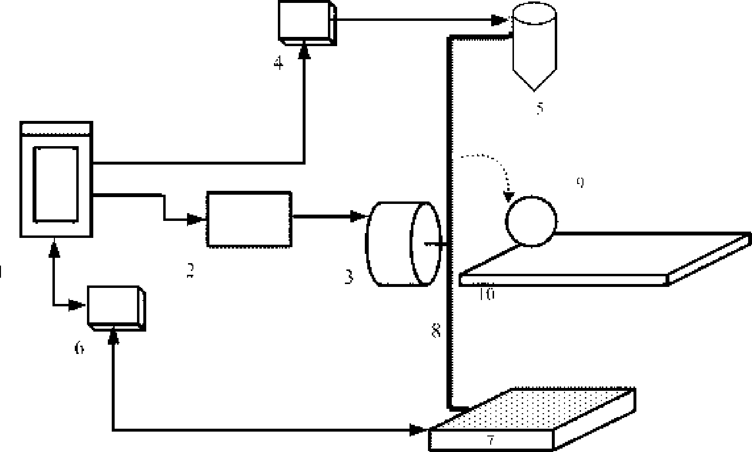
[0024] The embodiment of the present invention provides a system and method for imaging small animals, which uses the rotation of the light source and the detector to replace the sample rotation, keeps the sample still, and the light source and the detector rotate around it, which solves the problem of deformation caused by the rotation of the sample. Motion artifact problem.

[0025] In order to make the object, technical solution and advantages of the present invention clearer, the present invention will be further described in detail below in conjunction with the accompanying drawings.

[0026] Such as figure 1 As shown, the imaging system provided by the embodiment of the present invention specifically includes:

[0027] Computer 1 , motor driver 2 , electric rotating mechanism 3 , radiation source controller 4 , microfocus X-ray source 5 , data acquisition device 6 , flat panel detector 7 , rotating frame 8 , and sample stage 10 .

[0028] The micro-focus X-ray source 5...


PUM


Abstract

The present invention discloses an imaging system which comprises the following components: a sample carrying table which is placed with a sample to be imaged; a beam source part which radiates beam; a beam detecting collecting part which receives a beam image; a rotation control part which has two ends respectively fixedly connected with the beam source part and the beam detecting collecting part and drives the beam source part and beam collecting part to rotate; and a computer which transmits control instruction to the beam source part, the rotation control part and the detecting collecting part and receives the image received by the beam detecting collecting part for imaging processing. The invention also correspondingly provides an imaging method. According to the imaging method, an expected imaging requirement is input. The related parameters are calculated. A control instruction is transmitted. The beam source part and the beam detecting collecting part are fixed at two ends of the rotation control part. According to the control instruction, the beam source part radiates beam, the beam detecting collecting part collects the projection image of beam after transmitting the sample. The projection image is executed with imaging. The invention settles the problems of deformation and motion artifact caused by sample rotation.

Description

technical field [0001] The invention relates to biomedical imaging technology, in particular to an imaging system and method for digital X-ray transmission imaging, tomography imaging and three-dimensional imaging of small animals. Background technique [0002] Digital X-ray transmission imaging technology is a relatively mature medical imaging technology and has been successfully applied in clinical practice. This technique plays an important role in the study of bone tissue and can provide detailed structural information of small animals. Micro-CT, also known as micro-CT, differs from hospital-used CT mainly in spatial resolution. The spatial resolution of CT used in hospitals is generally on the order of millimeters, while the spatial resolution of micro-CT can reach the order of microns. [0003] The Chinese invention patent application with the application number 200510135935.1 discloses an X-CT scanning system, including a main control and data processing computer, a...

Claims


Application Information

Patent Timeline
IPC IPC(8): A61B6/03
Inventor 骆清铭杨孝全龚辉方正邓子林
Owner HUAZHONG UNIV OF SCI & TECH
Why Eureka
Try Eureka