Method for acquiring three-view drawing of medical image

A medical image and acquisition method technology, applied in the field of image processing, can solve the problem that the three-view does not provide a direct and effective acquisition method

Inactive Publication Date: 2010-04-07
SHANGHAI JIAO TONG UNIV
View PDF0 Cites 28 Cited by
  • Summary
  • Abstract
  • Description
  • Claims
  • Application Information

AI Technical Summary

Problems solved by technology

[0004] After searching the existing technology, it was found that Yang Zhiqiang's paper "Acquisition Method of CT Slices in Any Orientation" published in the Journal of Shandong Normal University in 2006 realized the rendering and display of CT slices in 3D volume da

Method used


Image

  • Method for acquiring three-view drawing of medical image
  • Method for acquiring three-view drawing of medical image
  • Method for acquiring three-view drawing of medical image

Examples


Embodiment Construction

[0046] The embodiments of the present invention are described in detail below. This embodiment is implemented on the premise of the technical solution of the present invention, and detailed implementation methods and specific operating procedures are provided, but the protection scope of the present invention is not limited to the following implementation example.

[0047] The specific implementation process of this embodiment is as follows:

[0048] 1. Firstly, perform a neck CT scan on the patient to obtain a neck CT picture. The picture is in DICOM format, a 12-bit grayscale image, and the picture pixels are 512*512.

[0049] The DICOM is a digital imaging and communication standard.

[0050] 2. Perform 3D reconstruction of the collected medical images:

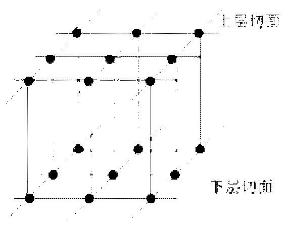
[0051] 2.1) Read two slices each time to form a layer, such as figure 1 As shown in , each dot represents a pixel of the slice, and 8 vertices of a cube are composed of 4 pixels on adjacent layers;

[0052] 2.2) Class...


PUM


Abstract

The invention provides a method for acquiring a three-view drawing of medical image, which belongs to the technical field of image processing. The method comprises: firstly, acquiring a CT medical image and performing the three-dimensional reconstruction of the medical image to obtain a three-dimensional image; reading the three-dimensional image and obtaining a three-dimensional reconstructed image by reducing the triangular plates in the three-dimensional image by using triangular plate simplification technology to reduce the image drawing time and the occupation of a memory; computing a three-view drawing normal vector and a three-view drawing position coordinate of a point according to the three-dimensional coordinate of the point in the three-dimensional reconstructed image and acquiring a cross section view, a coronal plane view and a sagittal plane view which pass through the point by conversion matrix processing, namely, acquiring the grey values of all points in the cross section view, the coronal plane view and the sagittal plane view by using a trilinear interpolation method to acquire the cross section view, the coronal plane view and the sagittal plane view. In the method, the triangles in the three-dimensional image are reduced to reduce the image drawing time and the occupation of the memory and improve interaction.

Description

technical field [0001] The invention relates to a method in the technical field of image processing, in particular to a method for acquiring three views of a medical image. Background technique [0002] In the current imaging medical diagnosis, doctors mainly find lesions by observing a group of two-dimensional slice images of CT (Computed X-ray Tomography) and MRI (Magnetic Resonance Imaging). film experience. Using medical visualization technology to reconstruct three-dimensional human organs from a series of continuous two-dimensional slices can truly reproduce the original appearance of objects and assist medical staff in quantitative and qualitative analysis of diseased bodies or regions of interest, which can greatly improve medical diagnosis. accuracy. [0003] Using computer image processing technology to obtain virtual slices in other directions (such as cross-section, coronal view and sagittal view) from the tomographic image sequence can help doctors observe and...

Claims


Application Information

Patent Timeline
IPC IPC(8): G06T11/00G06T15/00G06T15/08
Inventor 王宸昊黄茂祥马文娟刘允才
Owner SHANGHAI JIAO TONG UNIV
Why Eureka
Try Eureka